• Welcome to Phoenix Rising!

    Created in 2008, Phoenix Rising is the largest and oldest forum dedicated to furthering the understanding of and finding treatments for complex chronic illnesses such as chronic fatigue syndrome (ME/CFS), fibromyalgia (FM), long COVID, postural orthostatic tachycardia syndrome (POTS), mast cell activation syndrome (MCAS), and allied diseases.

    To become a member, simply click the Register button at the top right.

My MRI images

sb4

Senior Member
Messages
1,654
Location
United Kingdom
@Hip Very interesting, thank you. I hear you on the low CCI symptoms, that combined with no response to traction is quite significant in terms of thinking it is not this. However I do have some symptoms which make me think maybe, my neck is quite (uncomfortable?) at night time in most sleeping positions. I have weird yet slight neck ache and back ache that appears in the evening and coincides with the time of day I feel best. Perhaps that is from disc bulges as it feels in a similar place (lower neck). Then there is the TMJ, receded gums, EDS in imediate family, and now, what seems to be significantly past the norm neck extension, even though P Smith thinks this isn't causing issues.

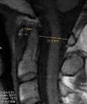
@pattismith I have not measured translational BAI, could you give me a quick run down on what it is and I will look at it tomorrow morning.
 

Hip

Senior Member
Messages
17,824
I have not measured translational BAI, could you give me a quick run down on what it is and I will look at it tomorrow morning.

Translational BAI is the maximum change in basion-axis interval (BAI) value as the head moves from flexion to neutral to extension. A normal translational BAI is less than 1 mm (ie, the BAI should not change much when the head nods up or down). Greater than 2 mm is pathological.

So first you need to measure the BAI in flexion, neutral and extension, and if these BAI values vary by more than 2 mm, then you have a pathological translational BAI.

This video shows how to measure the BAI.

This diagram also shows the BAI and BDI:

1568844302293.png
 

sb4

Senior Member
Messages
1,654
Location
United Kingdom
@Hip I had a go at the measurement but my confidence is super low in it's accuracy, I couldn't even get it to match up with P Smiths measurements and I had it as a direct reference:

bd1.png

bd2.png


The first 3 are my measurements and the last one is Smiths.
 

pattismith

Senior Member
Messages
3,930
@sb4
do you know how much is your ADI?
Mine is big (about 5 mm if i am correct).
This would correlate with a dental pannus and a potential Atlanto-Axial Subluxation, but many people with AAS are non symptomatic, so it doesn't say if my symptoms are related to it or not....What a mess...
 

pattismith

Senior Member
Messages
3,930
@sb4 it looks good in neutral, and hypermobile in flexion-extension, but I don't know how it should be in F and E...
 

pattismith

Senior Member
Messages
3,930
@sb4 , I don't know how severe are your symptoms, nor if they are related to your spine hypermobility/instability. However, the neurosurgeons who perform cranio-cervical fusion only cast severe patients.
Physio is offered for others, with the goal of reinforcing cervical muscles and to correct posture.
Hormonal optimisation probably has some importance, as lack of testo and T3 can alter muscle strength.
 

sb4

Senior Member
Messages
1,654
Location
United Kingdom
@pattismith Yeah surgery is out of the question for me, it is more about knowing whether spine is causing sypmtoms are not so that I can rule it out, or focus my energies on other forms of treatment for spine. Speaking of which, what neck exercises are recomended?

I have been working all year to improve my posture with some mild success however I am still not in good posture most of the day though have stopped a decent amount of forward head posture.

As for severity I reached my pit 3 years ago, I would classify as severe. Since then I have been steadily improving however if a moderate stressor comes along my body goes right back to how it was 3 years ago but only temporary. I would classify myself as moderate now.
 

pattismith

Senior Member
Messages
3,930
@sb4
We need a good physiotherapist to help us for posture and strenghtening, I am looking for one at the moment.
Also it's important not to push your spine too much (not going too far in flexion, extension or rotation).
Maybe the problem is elsewhere, there is so much we don't know yet, but working on our spine health will always be rewarding. If we don't, our spine problems will just come on top of our previous issue.
 

valentinelynx

Senior Member
Messages
1,310
Location
Tucson
Thanks, I had a go at measuring. Let me know what you think.

Just a quick comment: according to the slide you copied, the "ADI is seen on lateral plain films or CT scan." This may mean that it should NOT be measured on MRI. From a video of a talk by Dr. Bolognese I learned that MRI only show the marrow of bones. This is also why he uses a "soft" measurement of CXA on MRI rather than a "hard" one, which I discussed in a previous post (because the cortex of bone is dark on MRI). Both CT scans and plain films are X-rays and show the true borders of bones.
 

pattismith

Senior Member
Messages
3,930
@sb4
There is another measurement that can be done on MRI, that can detect anormal bent of the brainstem.
it is the skull bas angle, explained here with it's modification for MRI
Angle formed by:

"line extending across the anterior cranial fossa to the tip to the dorsum sellae line drawn along the posterior margin of the clivus
children: 114° +/- 5°
adults: 117° +/- 6°"


1569302434513.png
 

sb4

Senior Member
Messages
1,654
Location
United Kingdom
@pattismith It's super hard to tell from my MRI but here is my attempt.

zz.png


You think that is at the right spot?

Just looking at my clivus it does seem to be at a sharp angle and judging by the power ratio my opisthion seems raised, almost like the skull is rotated forwards relative to the start of the cervical spine. Could this be putting a little too much pressure on my brain-stem causing dysautonomia?

Is laxity on the space between the opisthion and C1 the reason I can extend my head back significantly above range?
 

pattismith

Senior Member
Messages
3,930
Yes, your clivus/odontoid angle doesn't look like a perfect angle.
But you have to choose another picture to measure the skull base angle, with a better view of the cord and the dens so that you will be just in the middle of your skull.
 

sb4

Senior Member
Messages
1,654
Location
United Kingdom
Also if you take the wild inaccuracies of my measurements in to account then I probably fall in range. One measurement significantly over, the other under, meaning I am definitely not measuring it correctly.
 

pattismith

Senior Member
Messages
3,930
I took one picture in your first post in this thread to show you the one that would work for this measure (I just change luminosity/contrast so you can see better)

sb2.jpg
 

sb4

Senior Member
Messages
1,654
Location
United Kingdom
asd.png


Problem is @pattismith I have no idea where to place the lines, my image looks much different to the one posted earlier and I cannot see the bones. Have you an example of this measurement done on yourself?