pattismith
Senior Member
- Messages
- 3,990
I can't do it either, as I don't have proper image of my brain/nose...

In fact, reading your measurements, I thought 145 was fine because the consensus says that pathological in under 135. But Henderson seems not to follow this strict rule.@pattismith Yeah Prof Smith puts it at 145.3. He noted it was out of normal range however nothing more.
Yeah, I can sometimes feel better (neck discomfort lessening) in flexion briefly but cant hold it. I figure if I properly sort my posture I will be able to hold my head in the correct position without forward head posture. This will take time though as I think my gastroparesis and thus stomach bloating is pulling the rest of my spine out of position.In fact, reading your measurements, I thought 145 was fine because the consensus says that pathological in under 135. But Henderson seems not to follow this strict rule.
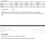
It is strange to notice that your CXA improves in flexion, which is not expected, did you check that?
In this article you can read that some atlanto-occipital horizontal posterior instability resolves in flexion, so I wonder if this could be your case.
edit:
According to traumatology articles, you need a CT multiplanar reconstruction of atlanto occipital joint to access instability inthat joint, I wonder why the EDS-fusion specialists doesn't use it...
yes, radiologist said everything is fine, and spine surgeon said "why are you coming to me, neurosurgeons are not supposed to deal with painful neck, you should go to a rheumatologist"...Did you get a report with your imaging?

Interestingly he finds for the CXA Neutral 137 (145.3), Flexion 141 (147.2), and extension 150 (154.5).
@Hip He hasn't diagnosed me with AAI he said it is possible but I would definitely need more imaging.
It's strange how there is such a discrepancy between smith and gillettes neutral CXA reading (137 vs 145.3). Perhaps G is doing soft tissue measurements? Perhaps this is why he dx far more people than S. For what its worth, my amatuer attempts at measuring CXA came closer to Smiths.
@Hip It appears you have mistaken somebody elses report for mine.