- Forums
- Related Conditions
- Comorbid Conditions, Differential Diagnoses, etc.
- Connective Tissue Disorders: Collagen, EDS, CCI
You are using an out of date browser. It may not display this or other websites correctly.
You should upgrade or use an alternative browser.
You should upgrade or use an alternative browser.
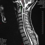
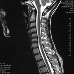
My MRI images
- Thread starter sb4
- Start date
sb4
Senior Member
- Messages
- 1,910
- Location
- United Kingdom
Thanks @JenB I have requested to join the group.
pattismith
Senior Member
- Messages
- 3,990
hello @sb4
I just had my cervical supine RMI done today, and it looks rather similar to your, with a small hernia( C5-C6 for me).
I'd like to do some pictures and mesures, what software should I use?
Any advise on this would be welcome, thanks!
I just had my cervical supine RMI done today, and it looks rather similar to your, with a small hernia( C5-C6 for me).
I'd like to do some pictures and mesures, what software should I use?
Any advise on this would be welcome, thanks!
sb4
Senior Member
- Messages
- 1,910
- Location
- United Kingdom
Did you have it done at medserena or elsewhere? Did the images come with software or just raw images?hello @sb4
I just had my cervical supine RMI done today, and it looks rather similar to your, with a small hernia( C5-C6 for me).
I'd like to do some pictures and mesures, what software should I use?
Any advise on this would be welcome, thanks!
sb4
Senior Member
- Messages
- 1,910
- Location
- United Kingdom
@JenB I rarely use facebook so I am having some difficulty at understanding the layout. I click on discusions tab and I see the top posts I guess but I would have no idea where to post my images or how to view similar threads of other peoples images.
pattismith
Senior Member
- Messages
- 3,990
I had it done in my local clinic in my country. I think the images are raw (DICOM)Did you have it done at medserena or elsewhere? Did the images come with software or just raw images?
sb4
Senior Member
- Messages
- 1,910
- Location
- United Kingdom
https://www.visus.com/en/downloads/jivex-dicom-viewer.htmlI had it done in my local clinic in my country. I think the images are raw (DICOM)
This appears to be software that came with mine. Try to install it and see if you can get it to load your images.
sb4
Senior Member
- Messages
- 1,910
- Location
- United Kingdom
@pattismith You have any luck with that software?
pattismith
Senior Member
- Messages
- 3,990
Hello @sb4 , thank you for your help, i finally managed to open the pics with the Software in the mri disc ! I will try to do some measures when I will back home. The radiologist said nothing significant, but the hernia leaves a free space for the cord that is about 10 mm or less so it seems consistant with a moderate stenosis. My transverse ligament seems similar to yours .@pattismith You have any luck with that software?
hello @sb4
I just had my cervical supine RMI done today, and it looks rather similar to your, with a small hernia( C5-C6 for me).
I'd like to do some pictures and mesures, what software should I use?
Any advise on this would be welcome, thanks!
Have you decided whether to treat it or not? I know thats a big choice to make, the cost / benefit is very difficult to estimate considering there's no guarantee that such a surgery will help you. That being said there's still a chance that it might.
sb4
Senior Member
- Messages
- 1,910
- Location
- United Kingdom
There is no way I can afford surgery, especially considering how at the moment, I think based on my MRI that it could be causing or at least significantly contributing to my autonomic dysfunction but it could also not, or only be having a mild effect. I am really not sure at this point but at the moment leaning slightly in the direction of it being significant.Have you decided whether to treat it or not? I know thats a big choice to make, the cost / benefit is very difficult to estimate considering there's no guarantee that such a surgery will help you. That being said there's still a chance that it might.
Either way my treatment plan would be non surgery.
sb4
Senior Member
- Messages
- 1,910
- Location
- United Kingdom
Interesting. When you makes some measurements will you post them in this thread? I will be interested in comparing.Hello @sb4 , thank you for your help, i finally managed to open the pics with the Software in the mri disc ! I will try to do some measures when I will back home. The radiologist said nothing significant, but the hernia leaves a free space for the cord that is about 10 mm or less so it seems consistant with a moderate stenosis. My transverse ligament seems similar to yours .
pattismith
Senior Member
- Messages
- 3,990
I don't Know yet but there is very little chance that I might be offered any surgery for such benign stenosis. Surgery here seems to be proposed to patients who have motor or reflexe deficit only.Have you decided whether to treat it or not? I know thats a big choice to make, the cost / benefit is very difficult to estimate considering there's no guarantee that such a surgery will help you. That being said there's still a chance that it might.
pattismith
Senior Member
- Messages
- 3,990
@sb4
My grabb oakes measure is below 9 mm so still correct, even though my transverse ligament seems a bit thickened; Did you measure your grabb?
My stenosis is about 10 mm at C5-C6 and C6-C7.
I didn't expected much more from this MRI, the question now is wether or not the stenosis and concomittant instability of these 3 vertebras are potentially involved in my symptoms or not....


sb4
Senior Member
- Messages
- 1,910
- Location
- United Kingdom
@pattismith I measured my grabb-oakes however it wasn't pathological. I think something like 7mm. Although comparing our images my upper spinal cord seems to bend more at this point.
I get about the same readings at you for the stenosis lower down.
Yeah the million dollar question is, is it causing symptoms. I wonder why your images look at lot clearer than mine? Perhaps I was moving too much.
I get about the same readings at you for the stenosis lower down.
Yeah the million dollar question is, is it causing symptoms. I wonder why your images look at lot clearer than mine? Perhaps I was moving too much.
pattismith
Senior Member
- Messages
- 3,990
Yes, I think you were sitting, whereas I was lying supine with a special pillow under my neck.@pattismith I measured my grabb-oakes however it wasn't pathological. I think something like 7mm. Although comparing our images my upper spinal cord seems to bend more at this point.
I get about the same readings at you for the stenosis lower down.
Yeah the million dollar question is, is it causing symptoms. I wonder why your images look at lot clearer than mine? Perhaps I was moving too much.
I learned from Small Fiber Neuropathy that polymorphism in the voltage gated sodium channel in the nerve can produce gain of function involved in this disease...So I wonder if this kind of polymorphism could be involved for some of us, making us more sensitive to any strain on our spinal cord...just a wild hypothesis...
pattismith
Senior Member
- Messages
- 3,990
I forgot to say that I had a previous Xray in 2017 that showed arthrosis in C4-C5.
When i went to the osteopath in july this year, he told me my neck is deviated to the left, and took a photo to show me.
I had a look at my 2017 xray, and yes the deviation was already there. I can find it also on the last MRI.
I would like to know what is producing this deviation...
When i went to the osteopath in july this year, he told me my neck is deviated to the left, and took a photo to show me.
I had a look at my 2017 xray, and yes the deviation was already there. I can find it also on the last MRI.
I would like to know what is producing this deviation...
sb4
Senior Member
- Messages
- 1,910
- Location
- United Kingdom
I have gotten my report back from Professor Smith and on the whole everything seems normal. The only abnormal things brought up were:
-Mild posterior annular bulges at C4/5 and C5/6 however this doesn't impinge on the nerve root or the spinal cord.
-Cervical spine angle measurement of extension was 72.1 degrees with normal mean values being 32 and upper limit 55 degrees. This may suggest increased flexibility however no element of ligament laxity, spinal instability, or fluctuating spinal stenosis.
-In neutral, the clivo-vertebral angle is estimated at 145.3 degrees (normal range 150-180).
-On turning to the right there are 35 degrees of rotation of the atlas over the axis which is at the upper limits of normal. No evidence of subluxation of the facets of the atlantoaxial joint to suggest atlantoaxial instability.
On the whole I am disappointed. I still don't know whether or not my symptoms are likely to be coming from my neck or not. The report notes the same things we noted but the way it was written seems to suggest they are of no concern. They being C4/5 5/6 bulges and clivo-vertebral angle being a little below range.
The fact that my neck extension was 72 deg with the upper limit being 55 and mean 32 indicates I do have connective tissue problems when taken in the wider context of things like TMJ, etc. It is annoying as well because for both flexion and rotations I could have gone a lot further however the head brace they put you in wouldn't let me. Makes me wonder if they would have shown up as increased flexibility.
I notice on @Hip survey (which I will now fill out) Smith dx the majority (53%) as okay yet Gillette has around 90%. Perhaps I would get some kind of dx from him but is he over-diagnosing or is Smith missing more subtle ques?
@pattismith @jeff_w @JenB @borko2100 @valentinelynx
Last edited:
Hip
Senior Member
- Messages
- 18,305
I notice on @Hip survey (which I will now fill out) Smith dx the majority (53%) as okay yet Gillette has around 90%. Perhaps I would get some kind of dx from him but is he over-diagnosing or is Smith missing more subtle ques?
Looking at the 15 patients (so far) in the CCI survey who had their scans viewed by Prof Smith:
8 of these 15 were diagnosed negative fro CCI by Prof Smith. And 6 out of these 8 negative cases were diagnosed positive by Dr Gilette. So there are 6 discrepancy cases where Prof Smith diagnosed CCI negative, but Dr G diagnosed CCI positive.
In 5 of these 6 discrepancy cases, the pathological measurement detected by Dr G was the translational BAI, which is a measurement Dr G utilizes, and I believe Dr H does also, but it is not usually taken into consideration by Dr B (as Dr B does not usually look at images in flexion and extension, which are required to measure the translational BAI).
2 out of the 5 also had a pathological CXA as well as a translational BAI.
All 6 discrepancy cases were also positive for atlantoaxial instability (AAI).
You have a low number of cervical medullary syndrome (CMS) symptoms, just 3 out of 32. CMS symptoms are usually present in CCI, AAI, Chiari and cervical spinal stenosis. Most of the people in the survey diagnosed positive for CCI, AAI or related conditions had in the range of 12 to 28 CMS symptoms. Although there are a handful of positive cases in the survey where the number of CMS symptoms is low, in the range of 5 to 8.
It is annoying as well because for both flexion and rotations I could have gone a lot further however the head brace they put you in wouldn't let me. Makes me wonder if they would have shown up as increased flexibility.
Dr B points out that one issue with flexion and extension images is that the angle of head tilt is not standardized, so you get different amounts of tilt from different MRI setups.
Last edited:
pattismith
Senior Member
- Messages
- 3,990
@sb4
did you try to measure your translational BAI?
did you try to measure your translational BAI?
