Human Herpesvirus-6 Reactivation, Mitochondrial Fragmentation, Paper Pub. 4/1/20 - Dr. Prusty

Hip

Senior Member
Messages
18,150
Just so I am on the same page, does the virus induce/produce MFF, FIS1, and DYM1L directly which goes on to drop transmembrane potential, or does it drop transmembrane potential another way and only after it's dropped the cell induces MFF, etc

The way I read it was that the virus induces or produces the proteins, which cause a collapse in the mitochondrial transmembrane potential, which in turn triggers mitochondrial fragmentation. But the paper is not very clear on this.
 

Learner1

Senior Member
Messages
6,311
Location
Pacific Northwest
Agree that the fragmentation/fission happens a lot. It may be new to our audience, but being able to manipulate it, encourage greater recycling of better mitochondria than we had as well as stimulating the proliferation of more good mitochondria seems useful.
Some years back I was in contact with David Whitlock, who has a low nitric oxide theory of autism and ME/CFS.

It was Whitlock who told me that if basal levels of NO are low, then you get less turnover of mitochondria in the cells. He says that the modern culture of daily bathing with strong soaps has killed off the natural ammonia-oxidizing bacteria (AOB) that once lived on our skin, and generated lots of NO which entered into the bloodstream.

Whitlock therefore thinks that modern humans are suffering from low NO, leading to diseases like autism and ME/CFS. His company (AOBiome) sells a spray which replenishes the AOB on the skin.
I think this deserves more press than it's gotten around here. Can you connect it to Prustys paper or should we start a new thread?
 

Hip

Senior Member
Messages
18,150

Learner1

Senior Member
Messages
6,311
Location
Pacific Northwest
I think we should start a new thread. Those two had done content, but they were detailed by a whole bunch of other stuff.

A couple of points are pertinent:

- low NO may prevent mitochondrial biogenesis, the making of new mitochondria

- low NO leads to more superoxide production and more peroxynitrites which damage mitochondria

- this can deplete glutathione, and a cascade of other bad things, none of which are good for mitochondria.

- depleted BH4 makes all this worse.
 

ZeroGravitas

Senior Member
Messages
141
Location
UK
What is interesting about Prusty's work is the finding of "something in the serum" of ME/CFS patients that appears to trigger the mitochondrial alterations.
Yes.:) More so that they were able to make this signal (or one with identical effects) in a modified standard laboratory cell line. Able to turn the signal on and off on demand.

Plus documented many distinctive changes to the secondary cultures, which could be used by others to detect the serum factor without needed a fancy "nano-needle".

I believe Dr Prusty is proposing that some factor (such as an immune signaling molecule) may be transmitted from HHV-6-infected cells to uninfected cells
I believe they ruled out the recognised immune signalling molecules as part of the factor...? (It's a part of the paper I've digested the least and molecules I know little about.)
No significant differences were seen for IL-1 and IL-5. These results ruled out a potential role of IFN response in the mitochondrial fragmentation and antiviral response in ME/CFS patients.

But as far as I know, the potential fatigue factors he's previously talked about are still on the table:
Fatigue factor molecule types Prusty has talked about looking for specifically [YouTube]:
• Mitochondrial metabolites (analysing in conjunction with Naviaux).
• Exosomes containing small non-coding RNAs or proteins. Are these the "cryptic peptides" [Twitter]?
• RNA - Cellular or pathogenic (including U14, which has been found in many other viruses and bacteria).
• Antibodies - auto (against self), against viruses.
• Calcium flux - altered inside cells so also outside.


If T3 or NO are low, then you get reduced turnover of mitochondria.
Ah, cool. Both important molecules we're commonly low in (I think).

Myhill, for example, usually pushes thyroid supplementation to a high level. Although I've seen that I have high-normal "reverse T3", which is made in place of regular T3 to reduce use of limited energy. So am am assuming that some other block on energy is more limiting (for me, currently).

So, reduced mito-genesis could mean lower numbers of mitos, or less frequently replaced mitos with more damage that run slower. Don't see any particular link to Prusty's work (as you guys say :)).
 

ZeroGravitas

Senior Member
Messages
141
Location
UK
I thought of a few more (and modified some of my) questions, above:

(3) Was Naviaux's contribution to the paper purely advice and/or interpretation (no lab work)?
(9) ATP production:

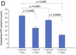
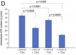
(a) Was this shown halved in the U2-OS cells, upon transactivation or with transfered supernatant. But a much more marginal reduction in the A549 cells (am I'm reading the paper right)?(b) Does [Fig.2D] show halved ATP production purely due to application of TSA? (If so, is that a big distorting factor on results?)(c) Are the measured ATP concentrations indicate a matching scaling of flux (in production rate)?(d) Would you expect to see a similar contrast in effect between different tissues within a patient? Or between patients? Or are these results not indicative at all, because they are cancer cells?(e) Why is there a factor of 10 difference between ATP concentrations graphed in fig.2d verses fig.1c...? (An error?)
(12) Personal relevance:

(a) Gradual onset - can this work on latent virus reactivation fit with (very) gradual onset of ME/CFS?
(b) Predisposing factors - could deleterious SNPs of SOD2 (or methylation enzymes, upregulated COMT, etc) make viral activation more likely? Or its effects more pronounced?
(c) Delayed sleep - Could the CDR state (or viral proteins) directly slow down the 24h clock gene expression pattern? Specifically, the astrocytes in the SCN seem to be the body's master time keepers. Because circadian rhythm is so commonly delayed in the illness - probably more of a high level neurological issue (but it was my first symptom, worsening with very gradual onset).


Finally time to barrage Prusty with them on Twitter!o_O Heh.:lol:
 
Last edited:
Messages
73
Location
Richmond, VA
I think we are sleeping on the whole SOD2 / PDP1 down-regulation that Prusty found. Understanding these deficiencies may lead to better interim treatments as research continues to occur around "what's in the blood".

PDP1 down-regulation would lead to deficiencies in the PDH pathway (converting pyruvate into Acetyl-COA), which means problems with excess lactate buildup. PDP1 is most expressed in the skeletal muscle and liver - knockout of this enzyme leads to fatal exercise intolerance. It is also expressed in immune cells as well as the brain - excess lactate in these systems leads to fascinating downstream effects that would explain a majority of our symptoms.

SOD2 down-regulation causes an increase in ROS, which explains the increase in oxidative stress in ME/CFS. This breaks a whole ton of stuff when left unchecked. Increased oxidative stress in ME/CFS has been widely researched but never explained. Increased ROS will also cause a decrease in PDH activity, which further exacerbates hypo-metabolism issues caused by inhibited PDP1.

The interesting thing is the interplay between these two systems - this probably explains why there are gradations in ME/CFS severity, as well as the concept of "crashes".

Treatments-wise: these two deficiencies explain why a lot of ME/CFS patients benefit from higher fat, lower carb diets (either through caloric restriction or going on a keto diet) - since this largely bypasses the PDP1 deficiency. This combined with a heavy antioxidant treatment protocol (like @Learner1 does) would help make up for deficiencies in SOD2. If the "something in the blood" winds up being HHV6 related, then it also validates Montoya's research around using Valcyte for extended periods of time as a treatment course.

I might dive a bit deeper on this and post a more in-depth look into these pathways + validating our understanding based on these pathways.
 
Messages
73
Location
Richmond, VA
For completeness and for those reading these posts I just want to point out that Prustys pSILAC experiment that saw down regulated PDP1 and SOD2 were on reactivated HHV6A cells. pSILAC analysis was not performed on the ME/CFS plasma swap experiments.

Yep, this is a very important call-out. We need someone to do a fast-follow experiment to confirm these two pathways are inhibited in ME/CFS cells specifically. This seems like a natural research path for Naviaux to explore, since it would tie his ME/CFS metabolomics studies together nicely.

PDP1:
  1. Unexplained build ups of lactic acid in the muscles of ME/CFS patients after exercise has been reported in literature, which is consistent with PDP1 enzyme inhibition.
  2. Fluge found that ME/CFS patients have a weird buildup of certain amino acids that point to a deficiency somewhere in the PDH complex, however he could not find exactly where the deficiency occurred.
  3. Ohba replicated this study successfully and also found deficiencies in the PDH complex after exercise for a mouse model of ME/CFS.
SOD2:
 

wigglethemouse

Senior Member
Messages
776
@ShepherdK I'm not sure how feasible it is to do the experiment since you are looking at proteins inside the cell you would have to remove the serum first, and that might affect the results..... Prusty said pSILAC is ~EUR1000 per sample just for materials, which is why he only did the one experiment with acivated vs non activated cells.
 

Learner1

Senior Member
Messages
6,311
Location
Pacific Northwest
I think we are sleeping on the whole SOD2 / PDP1 down-regulation that Prusty found. Understanding these deficiencies may lead to better interim treatments
I think SOD problems are important for many of us - SNPs are pretty common and if there's excessive oxidative stress and superoxide production, lack of adequate SOD is a big problem. Lack of BH4, NADPH, or heme can lead to increased superoxide production, which can lead to peroxynitrite production with too little SOD, particularly MnSOD. And peroxynitrites lead to increased hydrogen peroxide, impaired complex 1, and damaged membranes.
PDP1 down-regulation would lead to deficiencies in the PDH pathway (converting pyruvate into Acetyl-COA), which means problems with excess lactate buildup. PDP1 is most expressed in the skeletal muscle and liver - knockout of this enzyme leads to fatal exercise intolerance. It is also expressed in immune cells as well as the brain - excess lactate in these systems leads to fascinating downstream effects that would explain a majority of our symptoms.
I never gave high lactate and I know several other patients who don't either. High lactate can be reduced by increasing thiamine, by increasing exercise, and other strategies.
SOD2 down-regulation causes an increase in ROS, which explains the increase in oxidative stress in ME/CFS. This breaks a whole ton of stuff when left unchecked.
Yes, it does.
Treatments-wise: these two deficiencies explain why a lot of ME/CFS patients benefit from higher fat, lower carb diets (either through caloric restriction or going on a keto diet) - since this largely bypasses the PDP1 deficiency.
While there can be benefits to ketogenic diets, they can increase oxidative stress. Additionally, many if us have low amino acids and need to increase amino acid intake, making it challenging to keep fat intake at a high enough percentage to stay in ketosis.

And, as my recent metabolic and lab testing showed, I have some sort of problem burning fat - my body prefers to burn glucose. I do t know how common this is among ME/CFS patients (the testing was 55 minutes and pretty arduous) but I have found a keto diet us not appropriate for me.
  1. Not all of us have high lactate
  2. Fluge and Mella found group 2 and 3 amino acids to be reduced in ME/CFS patients. This pattern was the same in my lab results - all those aminos were low, which lead to my needing to increase protein intake to 1.6-1.8g a day, far above normal, and making it impossible to be on a ketogenic diet.
In his conclusion at last year's NIH Accelerating Research in ME/CFS Conference, Ton Tomkins made a special point of calling out increased oxidative and nitrosative stress as a key finding of the researchers. This was striking, because every other item in his list had been discussed during the 2 day conference, and this one had not, but he apparently felt it was significant enough to call it out.
 

raghav

Senior Member
Messages
818
Location
India
We are on the cover page of Immunohorizons.

1589008712067.png
 

ZeroGravitas

Senior Member
Messages
141
Location
UK
a lot of ME/CFS patients benefit from higher fat, lower carb diets
Or is it higher protein (lower carb) that is the most important aspect? AA or BCAAs supplements helping many (right @Mary, think I've seen you saying some place?). As Learner1 says, a common serum metabolite deficiency finding, there:
Additionally, many if us have low amino acids and need to increase amino acid intake, making it challenging to keep fat intake at a high enough percentage to stay in ketosis.
I didn't realise protein could break ketosis... (Thought only carbs over, like 50g ish, whatever.) Have you got the decimal point in the wrong place here?:
protein intake to 1.6-1.8g a day


the interplay between these two systems - this probably explains why there are gradations in ME/CFS severity, as well as the concept of "crashes".
So, you hypothesise that regular amounts of ROS from muscle exertion adds to an already saturated degradation capability, impacting energy production further via impact on PDH...?
PDP1 down-regulation would lead to deficiencies in the PDH pathway (converting pyruvate into Acetyl-COA), which means problems with excess lactate buildup.
OK, first, I think it can be important to be very clear about the location of specific metabolic differences. I.e. inside muscle cells, or in the serum, or are these closely linked in practice, and will these changes also be relevant to neurons/brain, etc...?

Second, the Australians have repeatedly found *lower* serum lactate levels (and raised glucose). This is in PEM when sampled after overnight fasting [HealthRising 2019].

We discussed this above, also with regard to...:
So hypo/hyper are two sides of the same shift in energy metabolism:
Reduced (hypo) use of oxygen in mitochondria for oxidative phosphorylation (electron transport chain, etc with lactic acid as an end-point).
Increased (hyper) anaerobic glycolysis, which is far less efficient, way more resource intensive and so catabolic (muscle breakdown).
Am I talking sense, here...? I *really* need to go find a very nice clear energy metabolism diagram that includes all the relevant metabolites and enzymes (without being too big). It confusing because "glycolysis" is used for both aerobic and anaerobic ATP metabolism, but with different end-points and inputs, I think... :xeyes:


Unexplained build ups of lactic acid in the muscles of ME/CFS patients after exercise has been reported in literature, which is consistent with PDP1 enzyme inhibition.
I've not read it carefully, but wasn't the lactate measured in the blood? (Taken from arteries *during* exercise!:jaw-drop:) The key graph of the study:

PHY2-7-e14138-g004.jpg


So patients immediately have higher lactic acid, ramping up sooner and then marginally enhanced lactate next day, during PEM. But change is in opposite direction to controls.


We need someone to do a fast-follow experiment to confirm these two pathways are inhibited in ME/CFS cells specifically.
And do you think it's possible to confirm Prusty's finding, of reduced expression of PDH (and SOD), in patient cells, in practice?

The accessible cells that will grow in lab conditions are the PBMC (serum immune cells). Those were used in Xinnan Wang's experiments that showed greatly increased (non)mitochondrial ATP production, without patient serum. Could one do a 50/50 mix of serum/standard supernatant, like Prusty did, and get meaningful results?
So, greatly increased glycolysis, which Cort's write up talked more about. With slightly less than normal ATP from mitochondria, but double the normal from outside the mitos (glycolytically):

cropper2020-05-03-07-40-02-8158201-jpg.37164
Wang's results also showed slightly *reduced* mito ATP production (not sure if statistically significant), despite lack of serum factor (and an increase in mitochondrial cristae). But presumably that's because they were in an activated state and deliberately suppressing it.

So patient immune cells are difficult, what cells should be used for (a lab controlled) confirmation?
 
Last edited:

ZeroGravitas

Senior Member
Messages
141
Location
UK
Questions for Prusty (paper summarised above).
Questions asked and replied to on this tweet. Prusty's responses in quotes:


(1) Does time frame of transfer effect suggest mechanism for patient's 1-2 day delayed PEM, crashes, etc? Naviaux's previously said [2018] that "Mitochondria change their function rapidly under stress. Within minutes[...]". But you cultured the HHV-6 transactivated cells for 2 days and then the responder cells for 2 days in the transferred supernatant.
(a) Were these durations necessary for the phenotype transfer to work? (I.e. to accumulate or respond to the molecular factor.) Did you try shorter times to find a minimum?​
(b) Or was the duration part of technical requirements of the analysis methods?​

Ql: PEM related crashes and fatigue is a complex physiological process and can involve many factors. We are in the process of understanding this complex phenomenon. Mitochondrial fragmentation is a very quick process under experimental conditions. With a sufficient stimulus, one can induce mitochondrial fragmentations within 2-3 h. But this is not how it works in nature. The CDR response is initiated from a limited number of cells and it requires sufficient time to reach every cell and completely shut down every cell. This is usually in the range of 2-4 days depending upon the number of original events. Yes, we can see effects starting to show up within 24 h. But the statistically significant effect that we want to show takes a longer time. In short, the metabolically and energetically crunch time is the hardest time that the cell or the body faces, when every other alternative is exhausted. [Twitter]


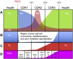
(2) Did this paper show cells in a CDR1 state? "M1" mitochondria and anti-viral protection are characteristic of Naviaux's CDR1 state. But he categorises ME/CFS as being a CDR3 disease [2019, Fig.2]:
1-s2.0-S1567724918301053-gr2.jpg
(a) Was he possibly mistaken about ME/CFS being CDR3 associated?​

(b) Or are the majority of our cells stuck in CDR3 as a result of a smaller population of cells in CDR1 (e.g. with HHV-6 transactivation) preventing completion of the healing cycle?​
Q2: This is what I also asked Bob when I had my results and he explained me very nicely, which is not possible to do it here. Just to tell you that M1 mitochondria is the shape of mitochondria when they are under stress like PEM. But if the mitochondria remain in M1 form forever, the cells will simply die (mitophagy). This possibly happens in neurons causing neuronal damage in long run. The M2 form is probably preferred under resting conditions. But unfortunately, we cannot test this in the body. [Twitter]
(c) Or are the your observations not sufficient to draw conclusions on this because they are in cancer cells?​
These cells are treated with both control & patient serum. So cell line does not make any difference here. [Twitter]
Q2: Our work is done in cancer cell lines as it is necessary for creating the HHV-6 reactivation system. All the other work are in cancer cell lines because we need to have a uniform system to work. [Twitter]
(d) Are the lab cancer cells naturally in a proliferative CDR2 state...?​


(3) Was Naviaux's contribution to the paper purely advice and/or interpretation (no lab work)?
Q3: Bob has helped in me in making a lot of sense out of my work. He has not done any actual experiment for this paper. But we are continuing on some of the cool stuffs now [Twitter]


(4) Which cells in patients are (or aren't) affected?:
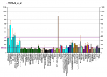
(a) Are initial HHV-6A infections mostly limited to cells with higher CD46 expression (chart below)? And HHV-6B to cells with CD134, primarily CD4 T-Cells, etc?​
PBB_GE_CD46_207549_x_at_fs.png
Q4: CD46 is expressed in every nucleated human cell. Hence HHV-6 can infect virtually every nucleated human cell. Same is also true for HHV-6B. T-cells only allow productive virus infection, which is clinically not that relevant in my personal opinion. [Twitter]
(b) Was HHV-6A virus chosen, instead of 6B or 7, too be able to infect U2-OS and A549​
We wanted to have a unique fluorophore-based system for our study. As HHV-6A is the only virus out of the three (HHV-6A, HHV-6B and HHV-7), whose genome is available in the form of a BAC, we chose this one for our work. We will continue working with the rest of the two in future. The cell type in which the virus is integrated and where (chromosomal site) it is integrated, might influence the rate of reactivation and hence the severity of the disease. [Twitter]
(c) Could the extent of the initial (latent) infection determine the severity of ME/CFS, after the reactivation triggering event has passed?​
(d) Or is infection and reactivation in specific locations more likely to be key? E.g. You've found infections all over the brain and brainstem [YouTube].​
(e) Could worsening of ME/CFS severity come from a spread of HHV-6 infection to more cells?​
I do not think that these viral reactivation spreads from cell to cell as there is literally no virus particle formation. Virus particle formation would be the simplest infection scenario possible as the host immune system would then recognize it and will eliminate it. [Twitter]

(5) Why do most people *not* get ME/CFS after acute infections, surgery, etc? If everyone has HHV-6 (and other viruses) latent in their bodies. (Some factors in 3, genetic and/or metabolic status?)
Q5: People do reactivate these viruses from time to time. But most of the times, our own immune system takes care of it. We do not even realize it. But in some of us, there must be a genetic component that makes us susceptible to conditions like ME/CFS. We are trying to understand that. [Twitter]


(6) How does HHV-6 transactivation become chronic in patients?

(a) The U2-OS and A549 cells are incapable of late stage viral replication. Is this also true for some cells in the human body (that can be infected)?​
(b) Does the virus deliberately prevent itself from complete replication? What's the evolutionary advantage, if so? Is it related to blocking competing viruses from getting its host cell destroy by the immune system...?​
(c) Is another mechanism needed for chronic HHV-6 activation? e.g. another infection, accumulation of toxic elements or other researcher's hypothesis (below)?​

Q6: In our body, most of the cell types does not allow productive HHV-6 infection. Only CD4+ cells allow productive HHV-6 infection. HHV-6 would never want to get fully eliminated from the cell. So, it would prefer to stop itself being detected as soon as it starts reactivating. The Immediate early (IE) viral RNA would immediately be detected in cell cytoplasm by host innate immune machinery. Hence it starts producing small non-coding RNAs first, which helps in preparing the stage for further virus reactivation. It is a constant battle between the cells and the virus. Sometimes the cell wins and sometimes the virus.

An ideal cellular environment is the most important requirement for fully productive virus infection. [Twitter]


(7) Do you suspect interactions with any other proposed mechanisms? I.e. as them causing sustained HHV-6 transactivation, or vice versa? Specifically:

(a) Robert Phair's IDO2 mutation tryptophan trap [2019]?​
(b) Nuno Sepúlveda's "Hyper-Regulated Immune System" [2019]?​
(c) Michael VanElzakker's "vagus nerve infection" hypothesis [2013]?​
(d) Any others you have an eye on...? (E.g. research on ROS/oxidative stress.)​


(8) Any hints of patient symptom phenotypes being separable by something you've measured...?:

(a) Detectable U14 (in 40% of sampled patients)?​
(b) Inherited ciHHV-6?​
(c) What about the never sick patients verses those who catch everything (or at least have repeatedly infection symptops).​


(9) ATP production:

(a) Was this shown halved in the U2-OS cells, upon transactivation or with transfered supernatant. But a much more marginal reduction in the A549 cells (am I'm reading the paper right)?​
(b) Does [Fig.2D] show halved ATP production purely due to application of TSA? (If so, is that a big distorting factor on results?)​
2020-05-07 Fig2.d ATP in U2-OS cells.jpg
Q9: The figure 2D is the ATP content in U2-OS cells after adoptive transfer of the culture supernatant. The results are more or less the same in A549 cells. TSA do have some effect, but it is very clear that virus reactivation itself has a stronger effect. If one normalizes the data to the TSA added sample, the difference still stands clear and significant.
The A549 ATP assay that you see in figure 5 is from patient serum treated cells. I guess we need to grow the cells for longer time to see a more significant effect on cellular ATP content. But I do not think that we can see a dramatic decrease in ATP content under any condition. Minor changes in Physiological levels of ATP can have a drastic effect on cells. [Twitter]
(c) Are the measured ATP concentrations indicate a matching scaling of flux (in production rate)?​
No, we did not measure the production rate of ATP. [Twitter]
(d) Would you expect to see a similar contrast in effect between different tissues within a patient? Or between patients? Or are these results not indicative at all, because they are cancer cells?​
Again, cancer cells have a different physiology. But until we have another system, we have to depend upon these cells for our work. In future, we will try utilizing primary cells for such work.
Of course, different tissues or cells will show different levels of effect. [Twitter]
(e) Why is there a factor of 10 difference between ATP concentrations graphed in fig.2d verses fig.1c...? (An error?)
Good observation. This depends upon the number of cells being used for the study. We did not show ATP content per cell. We take similar number of cells per experiment. The figure 1C was from experiments done a long time back. Figure 2D was done during the revision. As we have used more cells for 2D, we have almost 10 times more ATP content. [Twitter]


(10) Issue with the paper - Does the last sentence of your abstract seems to claim too much? You clearly showed strong similarities but no direct evidence of causation by HHV-6 in patients; it was an in vitro study. Is there unpublished work that bridges this gap?
Q10: I do not think that it will ever be possible to show HHV-6 infection in ME/CFS patients causing the disease. It is not a localized disease like cancer, and it is not an overnight disease. So, it is up to you to decide whether we claim too much or not. We do not claim that we solved the mystery. [Twitter]


(11) Serum factor...:

(a) Can you give us any more hints about what molecules you are currently honing in on, or the nature of the tests you've mentioned?​
Q11: We are trying everything that we can think about. I am not in a position to tell you anything. It is a very competitive field. If I tell you anything, someone will come out and tell that it is too early, and we do not have enough data to support. So let us work and come to stronger results. [Twitter]
(b) Could the U14 protein be the factor? Seeing as it turns up in so many pathogens. But you only found it in 40% patient serum, so would that make it one of multiple factors? Or could it be pressent in all, but below your detection threshold?​
No, I do not think that U14 protein is a factor. I guess you are talking about U14 small non-coding RNA. It is very much possible that other viruses have similar RNA. But our probes are extremely stringent and do not detect anything having more than one nucleotide difference. So, we are definitely not detecting any similar RNA from other herpesviruses. It needs tons of money to test all the other herpesviruses. We will do it systematically as and when possible. [Twitter]
(c) Have you been able to rule out any of the types of molecule you mentioned previously [YouTube]? I.e. Mitochondrial metabolites, exosomes containing small non-coding RNAs or proteins, cellular RNA, antibodies, calcium flux...? And what about purinergic signalling (ATP, etc)?​
Yes, we are ruling out some of them. But once again, I cannot tell anything about it at this moment. [Twitter]


(12) Personal relevance:

(a) Gradual onset - can this work on latent virus reactivation fit with (very) gradual onset of ME/CFS?​
Q12: Yes, I think so. Virus reactivation and its consequential effects are slow process. It matches well with the slow onset process of the disease. [Twitter]

(b) Predisposing factors - could deleterious SNPs of SOD2 (or methylation enzymes, upregulated COMT, etc) make viral activation more likely? Or its effects more pronounced?​
Yes, it can have consequences. [Twitter]

(c) Delayed sleep - Could the CDR state (or viral proteins) directly slow down the 24h clock gene expression pattern? Specifically, the astrocytes in the SCN seem to be the body's master time keepers. Because circadian rhythm is so commonly delayed in the illness - probably more of a high level neurological issue (but it was my first symptom, worsening with very gradual onset).​
The link to circadian clock is fascinating. Some of the ME/CFS patients have spoken to me on this. I am very much fascinated with the idea and the literature do support an association between circadian rhythm and mitochondrial morphology. I think hormonal changes, metabolism and other factor that has a link to circadian rhythm can make the situation more interestink. I would love to test this in future if time and finances permits. [Twitter]


(13) General curiosities:

(a) Can HHV-6 DNA show up sometimes in whole genome sequencing? I assume this is deliberately filtered out of the final data, even if its chromosomally integrated...?​
Yes, one can see HHV-6 in whole genome data. But an extremely high sequencing depth is required to see these sequences. Yes, most of the times these sequences are discarded during initial data filtration. [Twitter]
(b) How close are we to having a working CRISPR system that could remove HHV-6 from humans (in vivo)? Would have to do this by providing immunity, blacklisting the key viral proteins, or something?​
Some of my colleagues are working on CRISPR technology to remove integrated HHV-6 from the genome. But I guess we are still very far away from any successful results. [Twitter]
(c) Are there any exciting new laboratory technologies or equipment you expect to have access to in the near future?​
We keep developing exciting technologies and also keep looking for new ones developed by others. It is hard to say here what we really want. [Twitter]
 
Last edited:

ZeroGravitas

Senior Member
Messages
141
Location
UK
As you can see above, Prusty made a valiant effort to work through the litany of questions, above. :woot: We missed a couple, between us, so I've pinged him back with those. But he certainly cleared up a few things for me! :) And I've thrown in these follow-up questions:


(14) So, following on for question 6, incomplete (HHV-6) reactivation is actually the more common, by cell type. Even amongst nucleated cells, the interior is more limiting of viral replication than the surface proteins needed for virus ingress...
(a) Do most cells lack certain molecular machinery/enzymes/gene expression? What is it?​
(b) Or are some/most cell types just better at actively suppressing complete replication?​
Q14: Still not fully understood. It is hard to say whether it is virus or the host who decides the fate of virus reactivation. I would say it's the host cell. [Twitter]
(15) How do anti-virals suppress transactivation (or its effect)? If indeed they do? (Sorry, I've no idea of their mechanisms of action.)
Q15: Most of the antivirals against herpesviruses inhibit virus DNA replication. [Twitter]


(16) Do you think it could be possible to (quickly) confirm the finding of reduced PDH, SOD2, etc, activity directly in cells taken from patients?
(a) What cell type(s) might be suitable for this, if any? (Keeping in mind the lab methods required, too.)​
Q16: Yes, it should be possible to test PDH and SOD2 levels in patients. Blood cells would be ideal as it will be difficult to get any other cells. But two factors are key to success. First, A simple western blot or mass spec would not tell you anything as it measures everything in the cells. One has to pSlLAC like experiments that we did where you measure the proteins as they are synthesized avoiding the proteins that have a longer half like and they are there in cell without much of changes. We plan to follow another very fascinating approach to check this in future. [Twitter]
(b) Or is there little value in this? As you've said, above, that the serum/supernatant factor is all important?​
It is hard to say what is important and what is not. Every result at this stage is valuable. We will see how to utilize the data at a later time. [Twitter]
 
Last edited:

Wishful

Senior Member
Messages
6,126
Location
Alberta
I think we are sleeping on the whole SOD2 / PDP1 down-regulation that Prusty found.

I don't feel that these are problems for some PWME. I don't have any observations that seems like excess lactate, and antioxidants and peroxynitrite scavengers make me feel worse. I would guess that lactate and ROS problems are downstream of the core problem, and affect only some people.
 
Messages
65
So patient immune cells are difficult, what cells should be used for (a lab controlled) confirmation?
I think the type of cell is extremely important. Warren Tate, who's published a notable review article on ME/CFS, tried placing PBMCs in ME/CFS serum and control serum. He found that both inhibited ATP production, which is very strange. That's why you need to either use healthy grown muscle cells or U2-OS cells to replicate Fluge's and Prusty's studies.

Another important point is energetic stress. Lower ATP will be more apparent under energetic stress. From Fluge & Mella:
. . . exposure to ME/CFS serum led to increased rates of mitochondrial respiration. . . This effect was particularly evident under conditions of energy depletion, when mitochondrial respiration works at a maximum rate (condition IV).

And Paul Fisher:
. . . this may leave the cells less able to respond to further acute increases in ATP demand, because the signaling and metabolic pathways involved are already chronically upregulated.
 

Wishful

Senior Member
Messages
6,126
Location
Alberta
Prusty's reply: "The CDR response is initiated from a limited number of cells and it requires sufficient time to reach every cell and completely shut down every cell. This is usually in the range of 2-4 days depending upon the number of original events. Yes, we can see effects starting to show up within 24 h. But the statistically significant effect that we want to show takes a longer time."

That's outside of the timeframe for my PEM. My physically-induced PEM would flare up abruptly 24 hrs after the exertion, and usually be gone the next day. My cerebrally-induced PEM (same set of symptoms) would flare up within an hour, and be gone the next day. When I was sensitive to tryptophan, my ME symptoms would flare up dramatically 20 minutes after starting the meal, and decline in less than an hour. These responses don't seem to fit the CDR response as he describes it.

Does anyone else's ME not fit that time frame?
 
Back