Human Herpesvirus-6 Reactivation, Mitochondrial Fragmentation, Paper Pub. 4/1/20 - Dr. Prusty

wigglethemouse

Senior Member
Messages
776
Also, as others were saying above (I think) the pSILAC analysis was performed only on ciHHV-6 U"-OS cells, I think(?). Did they analyses the levels of all proteins in the cells and then only show the ones associated with mitos? I don't clearly see all the elements they state were most notably altered, in the abstract (but random protein/enzyme names, eh?):
Bhupesh Prusty said that pSILAC is very expensive, so I suspect that they didn't have enough data to make conclusions with any weight on individual proteins. Yes, looks like it was only that one pSILAC analysis on U2-OS cells.
 

Learner1

Senior Member
Messages
6,311
Location
Pacific Northwest
No idea of relative numbers, sorry. Just know that Naviaux describes the M1 (fragmented) mitochondria as set up do deliberately produce ROS, with reduced use of oxygen almost as a side effect (to free of the oxygen for making the toxic defensive oxidants).
How can one test this?
Or does one really need to test? (I took one pass at testing *everything*, 6 years back,
YES!! I have fine a NutrEval almost every 9 months, fir 7 years, through my battle with stage 3 cancer and my subsequent battle with ME/CFS. Dramatically changing results over time have guided me toward answers and treatment that works. What I took in 2012 and 2016 were vastly different from each other and from now.

Genova has a London office.
makes me wonder about MyHill pushing vit-C megadosing (to bowel tolerance). Other's too, specifically for (covid) viral infection, etc. Too much of a good thing...?
I've done high dose C, with good and bad effect. If I get COVID, it's my first strategy. It killed my cancer as it's been shown to in trials. But, too much can cause endogenous oxalate production, sharp crystals that damage mitos and everything else. The mechanism is to up superoxide production and then create hydrogen peroxide, which must be gotten rid of. If one has lousy SOD SNPs, peroxynitrites can form, which damage mito membranes and impair complex I, and if one doesn't produce adequate catalase, one can't get rid of damaging hydrogen peroxide. The good news is that these damage infectious agents and cancer, too, so it's a double edged sword and must be managed.:nerd:
 

ZeroGravitas

Senior Member
Messages
141
Location
UK
How can one test this?
Measure what state all your mitochondria are in...? Or how much ROS your cells are making...? Is either possible? (You seem to have measured overall oxidative stress, though... Or have indications that it's too high, from metabolic disturbances....?)

Genova has a London office.
Yeah... So I have to get a new nutritionist (parted ways with the first one as I'm an awkard sod and she wanted me to just be the patient), order the tests through her and probably through another intermediate company, then last time I had to physically travel 110 miles to London for a blood draw at the Biobank, then get the results back though nutritionist.

Added bonus of coronavirus now and less physical energy. If there were a home finger-prick test I'd hop on it, but not even bothered looking into it again, in the last 5 years. Buying, trying and reacting to boxes full of supplements wore me out emotionally, too. Anyway....

If one has lousy SOD SNPs
Like these of mine with SOD2? (That I've not looked at in a long time.):
Cropper2020-04-24-16-25-19-6646863.jpg


It was a question I had in mind for Prusty, too - what SNPs (or nutritative deficiencies/excesses) might enhance the likelihood of HHV-6 transactivation. E.g. if SOD is itself part of the serum factor signal.
 

Wishful

Senior Member
Messages
6,120
Location
Alberta
I've not read it through, yet, but this table of information seems quite relevant, here, it describing which cells they think HHV-6 (and the other viruses) infect and then are latent in (mostly immune system cells... :wide-eyed:):

I hope researchers do the same tests on the other herpes viruses, to see whether they create the same 'something in the blood' as HHV-6. I didn't notice my ME getting worse when I developed cold sores.
 

Slushiefan

Senior Member
Messages
121
I hope researchers do the same tests on the other herpes viruses, to see whether they create the same 'something in the blood' as HHV-6. I didn't notice my ME getting worse when I developed cold sores.

Interesting observation here. Possible the effect is more of an on/off situation?
 

Learner1

Senior Member
Messages
6,311
Location
Pacific Northwest
Measure what state all your mitochondria are in...? Or how much ROS your cells are making...? Is either possible? (You seem to have measured overall oxidative stress, though... Or have indications that it's too high, from metabolic disturbances....?)
Determine if one's mitos are fragmented and, if so, to what degree? And a test that can test if this situation is improving as treatments are applied,?

Measure how many ROS my mitos are making. I do have a sense of overall sense if oxidative stress. And that it's too high.

My goal is to figure out what's driving it, deranged mitochondria, lack of SOD or catalase, NOS uncoupling, Fenton reactions, or an imbalance of antioxidants. It done combination of all, to figure out what can be changed to improve the situation.;)

Like these of mine with SOD2?
Yes, exactly those. I have come to believe those have greatly contributed to my inability to deal with oxidative and nitrosative stress. Without adequate SOD, superoxide radicals created by mitochondria immediately react with No to become peroxynitrites, which damage mito membranes and impair complex I. The classic pattern for ME/CFS patients on the MitoSwsb test is low complex I and high complex IV activity. And, those SNPs are not too rare, so they may be playing a part for at least some of us. They also lead to NOS uncoupling.

It was a question I had in mind for Prusty, too - what SNPs (or nutritative deficiencies/excesses) might enhance the likelihood of HHV-6 transactivation. E.g. if SOD is itself part of the serum factor signal.
Yes. What mito nutrients, SNPs and environmental factors (mold, toxins, it other CDR danger) may make our mitos and us more vulnerable? What processes are weakened)impacted by the fragmentation?
 

wigglethemouse

Senior Member
Messages
776
Some rambling for @ZeroGravitas and @Learner1 to ponder. Just wondering how the different ME studies on mitochondria tie together.

I went back and looked at the Wang Stanford study on mitochondria of PBMC's free of serum to see if there were any other clues.
https://www.healthrising.org/blog/2...duction-found-chronic-fatigue-syndrome-mecfs/
ME/CFS patients were actually producing normal levels of mitochondrial ATP but over double the levels of non-mitochondrial ATP. That finding lead us right back to the glycolysis question, and put the study findings in line with the results from the Armstrong/McGregor and Naviaux papers.
Finally, the study found increased density of folds in the inner mitochondrial membranes called “cristae” in ME/CFS. Wang noted that the cristae are “exquisitely” regulated in reaction to the mitochondria’s need to produce ATP: the more folds present the greater the mitochondrial need to produce energy. That suggested that even though the glycolytic production of ATP was through the roof in the ME/CFS patients, their mitochondria were still under pressure to produce more ATP.
Paul Fisher presented at the Emerge 2019 conference that mitochondria in lymphoblasts were working overtime to produce ATP due to proton leak. This ties in with Wang's findings of increased cristae and increased non-mitochondrial ATP generation to meet the energy demand.

Another finding I remember about mitochondria is that Jonas Berquist presented at the 2018 Stanford Symposium that pregnenolone which is made in the mitochondria from cholesterol is reduced in ME/CFS. Pregnenolone is a precusor for many other steroid hormones and is thought to provide some neuroprotective effects. It also seems to have an effect on the fission/fusion transition.

Somehow these studies must tie together...........
 

ZeroGravitas

Senior Member
Messages
141
Location
UK
I hope researchers do the same tests on the other herpes viruses, to see whether they create the same 'something in the blood' as HHV-6.
Sure, I think that's just a matter of funding, from what Prusty has said. But given that we know so many of these ecode the U14, too [Wiggle's Tweet to Prusty] it's expected that they would cause the same kind of CDR situation as in this HHV-6 paper... If they are in a transactivated, 'smouldering' state of infection. Which is the more interesting thing to investigate - what mechanisms can cause this state to be sustained? (E.g. the Sepúlveda Hyper-Regulated immunity hypothesis.)

didn't notice my ME getting worse when I developed cold sores.
Would one expect surface legions to cause a profound effect throughout the body? I think cellular context of reactivation is probably essential. So activation in the brain stem, hypothalamus or certain immune blood cells could have a strong affect. Unless one is thinking of the coldsores as a litmus test for a changed state in the body, overall (metabolic, nutritative, immune, etc), that will also be triggering reactivation in more important tissues...?

Also, cold-sores mean there full viral replication, right? So a more advanced/complete stage than needed for the CDR cell phenotype.


My goal is to figure out what's driving it
That's the point of this thread and the studies discussed, is looking at higher level, overarching processes and cause for cellular promotion of ROS and all the othe CDR changes. Looking in detail at each of one's deranged metabolite levels, and such, certainly can allow you to put out the worst of the fires. But much better to find the arsonist....
 

ZeroGravitas

Senior Member
Messages
141
Location
UK
the Wang Stanford study
ME/CFS patients were actually producing normal levels of mitochondrial ATP but over double the levels of non-mitochondrial ATP.
As I was saying about this [above], this raised production was in the context of being cultured in clean supernatant for days, without any patient fatigue factor.

The seemingly big finding, of apparently doubled ATP by glycolysis, mostly meant that the specific immune cells being examined had been activated, for some reason (I assume this effect is sustained for many days). What this likely means (from the thread on the Wang paper)...:
Proliferating cells (immune cells) use glycolysis to proliferate, they actively stop pyruvate from glycolysis entering the mitochondria in a process called the warburg effect. [...] and this transfer expands the variety of non-essential amino acids available in the cell. You need a variety of amino acids to build new proteins for replication an glutaminolysis provides this.

So the authors of the paper should be aware that this is exactly what one would expect to be occurring in an immune cell.

So how does this relate to the hypometabolism in the blood? Well the hypometabolism may actually be caused by these immune cells functioning this way because proliferation takes a lot of metabolites, cofactors, vitamins and minerals to occur. Blood is very different to the cell and individual cell types are different from each other. Chronic immune activation may be what leads to the hypometabolism state.

Why should there be changes in pathways involved in synthesis of nucleotides and/or proteins? One strong reason would be to fight pathogens within the cell [...]
...So, the cells *want* to make energy less efficiently, without (so much) glucose feeding into the mitochondria, so that more non-essential amino acids are created as a by-product of the earlier glycolysis stage, which requires input of proteins. Aiming to use this resource for cell/DNA replication. The extra ATP itself might be more of a by-product...

It looks like past me knew more about these aspects of mitochondria back in that thread [1, 2, 3, etc] (and in the Naviaux thread before that). And @Hip probably still does, having related all that to the Myhill/McLarren work, etc.

Maybe the PBMC immune cells were activated, as off the leash from the kind of hyper-regulation Sepúlveda described... But I've no idea if things even work vaguely like that; the immune system is a total maze of differentiated cell types which each have radically different purposes and contrasting metabolic profiles.

Another finding I remember about mitochondria is
The specifics are all fascinating, and often seem to have a useful bearing on treatments to improve function, somewhat. But there's a virtually endless number of them...
Somehow these studies must tie together...........
Sure all the evidence all ties together. Each in-vitro study is a cropped close-up shot of the same elephant (or elephants, maybe). ATP is literally the most central, widely connected molecule in metabolism, affecting everything! So mitochondria are even more central, in being the main site of producing ATP and doing a whole load of other things beside...

I think we've sidetracked too much into discussing specifics of mitochondrial function. It's not the point of the HHV-6 study, and I feel that looking too closely at them is obscuring the forest by doing a bark rubbing... Or like scrutinising a momentary snapshot of only the tip of a helicopter rotor-blade to see which way it's going - which fails to tell us anything about how the helicopter overall is moving, what it's doing (let alone why - a higher level of abstraction). The metaphors... I know... :rolleyes:

I'm also aware that I'm mincing the science with my vaguries (quite possibly getting the odd specific backwards, even, in trying to reconstruct my understanding). And I've not baited anyone with more definitive understanding to correct me, at all, so I think I should maybe stop speculating. (Going to see if I can throw up some questions for Prusty, though...)
 
Last edited:

ZeroGravitas

Senior Member
Messages
141
Location
UK
... Wait. I just realiseded something: This figure I clipped earlier, with only modest ATP differences, was from the A549 cells:
Prusty's new study saw only a modestly lower overall ATP production (below). Different tissue types of the cells, of course. I think they decided (unless I'm misinterpreting) the overall ~2x rise (above), entirely from glycolysis, might be due to patient's immune cell's ongoing activation. Changes previously hidden, in vivo, suppressed by the serum factor...?

cropper2020-05-03-07-38-00-5635442-jpg.37165

[Fig.5C - A549 cells cultured in respective serum]

... The U2-OS cells saw a *much* stronger reduction in ATP. But during transactivation of the virus within them [Fig.1C] (annotated by me):

2020-05-07 Prusty 2020 cellular ATP.jpg


So seems like the serum factor doesn't cause nearly as strong of an effect as direct viral transactivation within the same cell... But it's an apples and orange comparison between A549 and U2-OS cells; have they measured ATP in U2-OS cells cultured in transferred supernatant...?

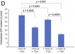
Edit: looks like ATP production in U2-OS cells cultured in supernatant from transactivated U2-OS cells is what's shown in [Fig.2d]. And it looks like very similar numbers as with the directly infected cells (except there's a decimal point missing from the numbers on the x-axis scale?!):

2020-05-07 Fig2.d ATP in U2-OS cells.jpg
 
Last edited:

ZeroGravitas

Senior Member
Messages
141
Location
UK
[Answers here! In-line with these questions re-posted + some bonus ones.]

Questions for Prusty (paper summarised above):


(1) Does time frame of transfer effect suggest mechanism for patient's 1-2 day delayed PEM, crashes, etc? Naviaux's previously said [2018] that "Mitochondria change their function rapidly under stress. Within minutes[...]". But you cultured the HHV-6 transactivated cells for 2 days and then the responder cells for 2 days in the transferred supernatant.
(a) Were these durations necessary for the phenotype transfer to work? (I.e. to accumulate or respond to the molecular factor.) Did you try shorter times to find a minimum?​
(b) Or was the duration part of technical requirements of the analysis methods?​


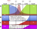
(2) Did this paper show cells in a CDR1 state? "M1" mitochondria and anti-viral protection are characteristic of Naviaux's CDR1 state. But he categorises ME/CFS as being a CDR3 disease [2019, Fig.2]:
1-s2.0-S1567724918301053-gr2.jpg
(a) Was he possibly mistaken about ME/CFS being CDR3 associated?​

(b) Or are the majority of our cells stuck in CDR3 as a result of a smaller population of cells in CDR1 (e.g. with HHV-6 transactivation) preventing completion of the healing cycle?​
(c) Or are the your observations not sufficient to draw conclusions on this because they are in cancer cells?​
(d) Are the lab cancer cells naturally in a proliferative CDR2 state...?​

(3) Was Naviaux's contribution to the paper purely advice and/or interpretation (no lab work)?

(4) Which cells in patients are (or aren't) affected?:
(a) Are initial HHV-6A infections mostly limited to cells with higher CD46 expression (chart below)? And HHV-6B to cells with CD134, primarily CD4 T-Cells, etc?​
PBB_GE_CD46_207549_x_at_fs.png
(b) Was HHV-6A virus chosen, instead of 6B or 7, too be able to infect U2-OS and A549​
(c) Could the extent of the initial (latent) infection determine the severity of ME/CFS, after the reactivation triggering event has passed?​
(d) Or is infection and reactivation in specific locations more likely to be key? E.g. You've found infections all over the brain and brainstem [YouTube].​
(e) Could worsening of ME/CFS severity come from a spread of HHV-6 infection to more cells?​

(5) Why do most people *not* get ME/CFS after acute infections, surgery, etc? If everyone has HHV-6 (and other viruses) latent in their bodies. (Some factors in 3, genetic and/or metabolic status?)


(6) How does HHV-6 transactivation become chronic in patients?

(a) The U2-OS and A549 cells are incapable of late stage viral replication. Is this also true for some cells in the human body (that can be infected)?​
(b) Does the virus deliberately prevent itself from complete replication? What's the evolutionary advantage, if so? Is it related to blocking competing viruses from getting its host cell destroy by the immune system...?​
(c) Is another mechanism needed for chronic HHV-6 activation? e.g. another infection, accumulation of toxic elements or other researcher's hypothesis (below)?​


(7) Do you suspect interactions with any other proposed mechanisms? I.e. as them causing sustained HHV-6 transactivation, or vice versa? Specifically:

(a) Robert Phair's IDO2 mutation tryptophan trap [2019]?​
(b) Nuno Sepúlveda's "Hyper-Regulated Immune System" [2019]?​


(8) Any hints of patient symptom phenotypes being separable by something you've measured...?:

(a) Detectable U14 (in 40% of sampled patients)?​
(b) Inherited ciHHV-6?​
(c) What about the never sick patients verses those who catch everything (or at least have repeatedly infection symptops).​


(9) ATP production:

(a) Was this shown halved in the U2-OS cells, upon transactivation or with transfered supernatant. But a much more marginal reduction in the A549 cells (am I'm reading the paper right)?​
(b) Does [Fig.2D] show halved ATP production purely due to application of TSA? (If so, is that a big distorting factor on results?)​
2020-05-07 Fig2.d ATP in U2-OS cells.jpg
(c) Are the measured ATP concentrations indicate a matching scaling of flux (in production rate)?​
(d) Would you expect to see a similar contrast in effect between different tissues within a patient? Or between patients? Or are these results not indicative at all, because they are cancer cells?​
(e) Why is there a factor of 10 difference between ATP concentrations graphed in fig.2d verses fig.1c...? (An error?)​


(10) Issue with the paper - Does the last sentence of your abstract seems to claim too much? You clearly showed strong similarities but no direct evidence of causation by HHV-6 in patients; it was an in vitro study. Is there unpublished work that bridges this gap?


(11) Serum factor...:

(a) Can you give us any more hints about what molecules you are currently honing in on, or the nature of the tests you've mentioned?​
(b) Could the U14 protein be the factor? Seeing as it turns up in so many pathogens. But you only found it in 40% patient serum, so would that make it one of multiple factors? Or could it be pressent in all, but below your detection threshold?​
(c) Have you been able to rule out any of the types of molecule you mentioned previously [YouTube]? I.e. Mitochondrial metabolites, exosomes containing small non-coding RNAs or proteins, cellular RNA, antibodies, calcium flux...? And what about purinergic signalling (ATP, etc)?​


(12) Personal relevance:

(a) Gradual onset - can this work on latent virus reactivation fit with (very) gradual onset of ME/CFS?​

(b) Predisposing factors - could deleterious SNPs of SOD2 (or methylation enzymes, upregulated COMT, etc) make viral activation more likely? Or its effects more pronounced?​

(c) Delayed sleep - Could the CDR state (or viral proteins) directly slow down the 24h clock gene expression pattern? Specifically, the astrocytes in the SCN seem to be the body's master time keepers. Because circadian rhythm is so commonly delayed in the illness - probably more of a high level neurological issue (but it was my first symptom, worsening with very gradual onset).​


(13) General curiosities:

(a) Can HHV-6 DNA show up sometimes in whole genome sequencing? I assume this is deliberately filtered out of the final data, even if its chromosomally integrated...?​
(b) How close are we to having a working CRISPR system that could remove HHV-6 from humans (in vivo)? Would have to do this by providing immunity, blacklisting the key viral proteins, or something?​
(c) Are there any exciting new laboratory technologies or equipment you expect to have access to in the near future?​


[Answers here! In-line with these questions re-posted + some bonus ones.]
 
Last edited:

Hip

Senior Member
Messages
18,150
A couple of replies here have mentioned how mitochondria are constantly repairing & recycling themselves, and have a lifespan of 2-4 weeks. Has anyone here come across mention of precise and consistent timing for mitochondria (or other cellular components) that would fit 21 days, +/- hours rather than days?

The lifespan of mitochondria is often quoted as 2 to 4 weeks, but there's no definitive evidence for this, and other sources suggest a lifespan of only 2 days:

It is believed that mitochondria are normally replaced every 2–4 weeks in rat brain, heart, liver, and kidneys, although recent studies have shown that the turnover rate might be considerably higher.
Source: here

the estimated median half life of liver mitochondria is 1.83 days
Source: here
 
Last edited:

Hip

Senior Member
Messages
18,150
I came across a paper recently which mentions some very interesting things about viruses and mitochondrial fission (aka mitochondria fragmentation), and also says that menthol can inhibit mitochondria fragmentation.

The paper focuses on coxsackievirus B infection, which induces mitochondrial fragmentation as a means to promote infection; but the paper says that several other viruses also trigger mitochondrial fragmentation, including cytomegalovirus and hepatitis B virus:

It was reported that certain viruses induce mitochondrial fission to support infection. We documented that CVB triggers mitochondrial fission and blocking mitochondrial fission limits infection.

Our preliminary findings suggest that CVB relies on TRPV1-mediated mitochondrial fragmentation

Other viruses have also been shown to rely on mitochondrial fission and mitophagy to further infection. Hepatitis B virus induces fission and mitophagy as a method to impair antiviral apoptosis

Cytomegalovirus has been shown to induce mitochondrial fission via the viral antiapoptotic protein vMIA

Menthol treatment reduces mitochondrial fission basally and during CVB infection- HeLa cells were treated with 1 mM menthol and then examined after 6 h.

The paper is discussed in this thread.
 

Hip

Senior Member
Messages
18,150
This paper describes the mechanism of virally-triggered mitochondrial fission/fragmentation:
Mitochondrial fission can be induced via a collapse of mitochondrial transmembrane potential (∆Ψm) and is modulated by outer mitochondrial membrane proteins, including FIS1 (fission, mitochondrial 1) and MFF (mitochondria fission factor).

So it seems that as soon as the voltage across the mitochondrial membrane (transmembrane potential) drops down, the mitochondria may fragment.


This excerpt explains how hepatitis B and C viruses purposely induce mitochondrial fission, which in turn is followed by the mitochondria being broken down and removed (mitophagy).
In the context of virus-induced mitophagy, during the early process of viral infection, some viruses induce the collapse of mitochondrial transmembrane potential, which results in depolarized mitochondria by promoting mitochondrial fission/fragmentation.

These viruses may modulate virus-induced mitochondrial fission as a precursor to the host mitophagic process, which can be triggered by PRKN or PRKN-independent mediators for persistent infection.

Perhaps the earliest described example of this concept was seen with hepatitis viruses. Hepatitis B virus (HBV) and hepatitis C virus (HCV) stimulate the gene expression of DNM1L and MFF and promote DNM1L recruitment to mitochondria by stimulating the phosphorylation of DNM1L (Ser616), leading to mitochondrial fission.

Virus-induced DNM1L-mediated mitochondrial fission is then followed by PRKN-dependent mitophagy. These findings implicate mitophagy as a potential therapeutic target against HBV- and HCV-associated liver diseases.

DNM1L = Dynamin-1-like protein.
MFF = mitochondria fission factor.


And the paper explains how in cells infected by coxsackievirus B, mitochondrial fragmentation is observed:
Likewise, in Venezuelan equine encephalitis virus (VEEV)-infected cells, DNM1L, PRKN, and PINK1 are enriched in mitochondrial fractions as compared with uninfected cells, and mitophagy ensues in infected cells [38]. Similar findings were reported for coxsackievirus B (CVB), classical swine fever virus (CSFV), and porcine reproductive and respiratory syndrome virus (PRRSV).


This paper explains the rationale for why a hepatitis C virus triggers mitochondrial fission: amongst other things, mitochondrial fission helps prevent apoptosis (killing the virally-infected cell) and inhibits interferon secretion which would normally fight the viral infection.
 

bread.

Senior Member
Messages
499
I've edited/updated my paper summary post a bunch [above]. I hadn't realised that the green florescent mitochondria images were taken in a second batch of U2-OS cells without ciHHV-6 in them, just activated by the supernatant signal.

Also, as others were saying above (I think) the pSILAC analysis was performed only on ciHHV-6 U"-OS cells, I think(?). Did they analyses the levels of all proteins in the cells and then only show the ones associated with mitos? I don't clearly see all the elements they state were most notably altered, in the abstract (but random protein/enzyme names, eh?):


Well, not *just* that, for sure. But factors that cause damage (toxins, etc) could also trigger ME/CFS. And damage (of at least some varieties) is more likely to accrue while in an ME/CFS state. This will pressumably limit one's maximum (good day) capabilities, when not crashing, or whatever.

I don't think there's anything so precise... Turn-over's going to vary a lot by tissue, as Learner1 says, by an order of magnitude (fast in liver, for example). And also by one's activity and food intake, etc. Might be interesting to read up on if mitos have any kind of expiration time mechanism... I'd think that damage would be more likely a determining factor.


So, basically *all* the anti-oxidants then? Lol!:) We're kinda going well off-topic, here, but have you ever noted a negative effect of these on muscle mass? They're supposed to reduce/block the hormesis effect of muscle damage from exercise/exertion that builds them up. Vit-E I know's been associated with raised cancer risk (in people with smokng DNA damaged lungs). But perhaps your body's producing so much more ROS, neither of these are likely to be an issue?

Where's this image from...? :wide-eyed:


Could you explain to me why antioxidants could lead to muscle wasting? Ty!
 

ZeroGravitas

Senior Member
Messages
141
Location
UK
Could you explain to me why antioxidants could lead to muscle wasting?
I don't know that they can. Almostly certainly not on their own.

But it seems fairly accepted that, by counteracting the ROS (and RNS - reactive nitgoren spieces), anti-oxidants can blunt the benefits of high intensity workouts. In this case, hormeisis (restoration of enhanced function in compensation for mild damage/stress) is dependant on ROS as a signal. [2015 - NCBI]


Thanks for coming to post on the thread, Hip.:) Have you been looking into this HHV-6 work much? Any observations/questions to pass on?
This paper explains the rationale for why a hepatitis C virus triggers mitochondrial fission: amongst other things, mitochondrial fission helps prevent apoptosis (killing the virally-infected cell) and inhibits interferon secretion which would normally fight the viral infection.
Thanks for this link, I'll have to take a look later. Its something not explored in the paper - why do virus proteins appear to (paradoxically) deliberately induce an anti-viral cell state?


And could you sanity check me a bit? I was saying that:
I believe mito recycling can be sped up in various ways, too, like fasting, which will trigger cells to start breaking down some of their machinery to burn it for fuel.
Am I getting that wrong? Because this diagram from a 2012 paper shows starvation (and stress) leading to hyper-fused mitochondria... Is that right...?
1-s2-0-s0005272812000692-gr1-jpg.33541

Mitochondrial fragmentation is a response to cell danger (also pre-empts apoptosis, etc).
And isn't what I've said there correct? Fragmentation preempts apoptosis? But you say it prevents it...? o_O
 

Wishful

Senior Member
Messages
6,120
Location
Alberta
The lifespan of mitochondria is often quoted as 2 to 4 weeks, but there's no definitive evidence for this, and other sources suggest a lifespan of only 2 days:

Some papers I came across yesterday imply that there's a wide range of differences in mitochondria in different parts of the body, even in different cell types in the same part of the body. So, I guess there is no standard lifespan for mitochondria, and there could be mitos somewhere in the body with a 21-day lifespan, or there might not be.

My question was more for networking, in case someone had encountered some research that mentioned body systems with a precise, consistent timing mechanism that could have a period of 21 days. It seems like an unusual system, since I expect most biological systems change gradually over time, rather than having this alarm clock (calendar) timing. Something seems to be counting circadian cycles or cell replications or some such thing, and T2 triggers a new counting cycle.
 

sb4

Senior Member
Messages
1,809
Location
United Kingdom
So it seems that as soon as the voltage across the mitochondrial membrane (transmembrane potential) drops down, the mitochondria may fragment.
Just so I am on the same page, does the virus induce/produce MFF, FIS1, and DYM1L directly which goes on to drop transmembrane potential, or does it drop transmembrane potential another way and only after it's dropped the cell induces MFF, etc
 

mariovitali

Senior Member
Messages
1,216
@Hip

Thank you for mentioning menthol.

Just wanted to add that i was lucky in the sense that i could feel the smallest changes to my symptoms and menthol was one of the foods that would have a very positive effect on me, perhaps for a reason.

Unfortunately it may act as a 5AR inhibitor (if originated by peppermint oil) which -according to the techniques i use- suggest that 5AR inhibition could lead to ME or ME-like symptoms.
 

Hip

Senior Member
Messages
18,150
Thanks for coming to post on the thread, Hip.:) Have you been looking into this HHV-6 work much? Any observations/questions to pass on?

I have not spent that much time looking at the Prusty research, so have not really got much insight into it as yet.

But it does seem from the papers I posted above that quite a few viruses will induce mitochondrial fragmentation for their own purposes of survival and replication. So viral effects on mitochondria are not unique to HHV-6.

What is interesting about Prusty's work is the finding of "something in the serum" of ME/CFS patients that appears to trigger the mitochondrial alterations.

I believe Dr Prusty is proposing that some factor (such as an immune signaling molecule) may be transmitted from HHV-6-infected cells to uninfected cells, and it is this factor which he thinks may be causing the uninfected cells to also develop mitochondrial alterations.


And could you sanity check me a bit? I was saying that:
I believe mito recycling can be sped up in various ways, too, like fasting, which will trigger cells to start breaking down some of their machinery to burn it for fuel.

I know that the rate of mitochondrial biogenesis (the creation of new mitochondria in the cell) is dependent on certain factors. Potent stimulators of mitochondrial biogenesis are T3 (triiodothyronine, the thyroid hormone) and nitric oxide. If T3 or NO are low, then you get reduced turnover of mitochondria.

Some years back I was in contact with David Whitlock, who has a low nitric oxide theory of autism and ME/CFS.

It was Whitlock who told me that if basal levels of NO are low, then you get less turnover of mitochondria in the cells. He says that the modern culture of daily bathing with strong soaps has killed off the natural ammonia-oxidizing bacteria (AOB) that once lived on our skin, and generated lots of NO which entered into the bloodstream.

Whitlock therefore thinks that modern humans are suffering from low NO, leading to diseases like autism and ME/CFS. His company (AOBiome) sells a spray which replenishes the AOB on the skin.


And isn't what I've said there correct? Fragmentation preempts apoptosis? But you say it prevents it...?

I don't really know enough about the subject to answer this. The paper I quoted says some viruses will fragment mitochondria in order to prevent the cell from self-destructing by apoptosis.

But this paper says "Fission machinery plays a critical role in programmed cell death — Mitochondrial fission is an early event during apoptosis".
 
Back