• Welcome to Phoenix Rising!

    Created in 2008, Phoenix Rising is the largest and oldest forum dedicated to furthering the understanding of and finding treatments for complex chronic illnesses such as chronic fatigue syndrome (ME/CFS), fibromyalgia (FM), long COVID, postural orthostatic tachycardia syndrome (POTS), mast cell activation syndrome (MCAS), and allied diseases.

    To become a member, simply click the Register button at the top right.

A systematic review of neurological impairments in myalgic encephalomyelitis/chronic fatigue syndrome using neuroimaging techniques

leokitten

Senior Member
Messages
1,542
Location
U.S.
This is a pretty well done and thorough review paper, makes for a good reference to find papers you might've read long ago on particular neuroimaging studies of ME.

While most, if not all, the neuroimaging studies in ME had fairly small sample sizes (due to abysmal ME funding!) and most used Fukuda criteria because CCC and ICC didn't become mainstream for ME research until after many of these studies were published, seeing all the papers again in one document shows that there were indeed multiple replicated results, with some being replicated in a number of papers. Though of course there were also inconsistent results between some papers, so hopefully with CCC criteria selected patients and larger sample sizes (increased funding for ME research) that future neuroimaging studies can produce more highly statistically significant findings and possible markers of ME.

My first pleasant surprise about this review, and I didn't realize this about ME until I saw this with all the results in one place, is wow the cingulate cortex brain region appears to very implicated in ME. It was found to have abnormalities/impairments in cerebral blood flow studies, neuroinflammation and other PET studies (i.e. lower SERT transporter density, glucose hypometabolism), functional connectivity studies, EEG studies, and cognitive function fMRI and SPECT studies (and possibly a couple types I missed). And many of these cingulate cortex findings are replicated by multiple papers. Of course there are other brain regions with abnormalities discussed in the review that are important, but the cingulate cortex appeared to me to stand out as it has been implicated in almost all the different study modalities.

The cingulate cortex is a key brain region that has a lot of important and coordinating functions.

From Neuroanatomy, Cingulate Cortex:
As seen from the extensive neural pathways it shares with other brain regions, the cingulate cortex can be considered, in a sense, a connecting hub of emotions, sensation, and action. Some of these pathways are those involved in motivational processing, which is apparent through the connections with the orbitofrontal cortex, basal ganglia, and insula, which together make up the reward centers of the brain. Also, the cingulate cortex projects pathways to the lateral prefrontal cortex which is involved in executive control, working memory, and learning. Pathways between cingulate cortex and motor areas like the primary and supplementary motor cortices, spinal cord, and frontal eye fields, suggest an important role in motor control. Moreover, the cingulate cortex, frontal and parietal lobes comprise a neural network for orienting attention, and expectedly, injury to any of these areas is known to cause hemineglect. The neural circuits cingulate cortex shares with the hippocampus and amygdala suggest a role in consolidating long-term memories and processing emotionally-relevant stimuli, respectively.

The cingulate cortex is also involved in regulation of autonomic and neuroendocrine responses and pain perception.


A systematic review of neurological impairments in myalgic encephalomyelitis/chronic fatigue syndrome using neuroimaging techniques
Maksoud et al. PLOS One April 2020

Abstract

Background
Myalgic encephalomyelitis/ Chronic Fatigue Syndrome (ME/CFS) is a multi-system illness characterised by a diverse range of debilitating symptoms including autonomic and cognitive dysfunction. The pathomechanism remains elusive, however, neurological and cognitive aberrations are consistently described. This systematic review is the first to collect and appraise the literature related to the structural and functional neurological changes in ME/CFS patients as measured by neuroimaging techniques and to investigate how these changes may influence onset, symptom presentation and severity of the illness.

Methods
A systematic search of databases Pubmed, Embase, MEDLINE (via EBSCOhost) and Web of Science (via Clarivate Analytics) was performed for articles dating between December 1994 and August 2019. Included publications report on neurological differences in ME/CFS patients compared with healthy controls identified using neuroimaging techniques such as magnetic resonance imaging, positron emission tomography and electroencephalography. Article selection was further refined based on specific inclusion and exclusion criteria. A quality assessment of included publications was completed using the Joanna Briggs Institute checklist.

Results
A total of 55 studies were included in this review. All papers assessed neurological or cognitive differences in adult ME/CFS patients compared with healthy controls using neuroimaging techniques. The outcomes from the articles include changes in gray and white matter volumes, cerebral blood flow, brain structure, sleep, EEG activity, functional connectivity and cognitive function. Secondary measures including symptom severity were also reported in most studies.

Conclusions
The results suggest widespread disruption of the autonomic nervous system network including morphological changes, white matter abnormalities and aberrations in functional connectivity. However, these findings are not consistent across studies and the origins of these anomalies remain unknown. Future studies are required confirm the potential neurological contribution to the pathology of ME/CFS.
 
Last edited:

Wishful

Senior Member
Messages
5,684
Location
Alberta
I wonder if any of the ME research organizations are keeping track of research that has shown significant results and which need replication and/or larger studies. Completely novel research isn't necessarily going to find the one true answer, brightly lit with banners flying and a marching brass band. An important part of ME may already be found, but is sitting on a shelf gathering dust, because it lacked the waving banners. It might just need a bit of cleaning and polishing.
 

leokitten

Senior Member
Messages
1,542
Location
U.S.
Unfortunately in ME research you also see a lot of one-offs by labs which do a very good first study and then you never see another ME publication from them again. It's quite uncommon in other research areas and I believe it's due to the lack of money in ME research. Labs can usually find enough money for one project on their own, but the issue is that technical and other knowledge about protocols etc doesn't build.
 
Last edited:

Pyrrhus

Senior Member
Messages
4,172
Location
U.S., Earth
My first pleasant surprise about this review, and I didn't realize this about ME until I saw this with all the results in one place, is wow the cingulate cortex brain region appears to very implicated in ME.

Interestingly, the Anterior Cingulate Cortex (ACC) in the cortical brain also figures prominently in research studies on Attention Deficit Disorder (ADD).[1] One reason why the ACC may be critical for maintaining attention is its connections with the thalamus in the sub-cortical brain.[2] The thalamus is the part of the subcortical (autonomic) brain that is most closely connected to the cortical (conscious) brain.

Although the thalamus is best known as a relay station that provides all the body's sensory information to the cortical brain, the cortical brain also provides "feedback" to the thalamus, allowing the thalamus to suppress certain sensory information. It is thought that this cortical "feedback" is how the conscious brain decides which sensory information to pay attention to, and which sensory information to ignore, a phenomenon called "sensory gating". If there is a problem with this "feedback", then the conscious brain may be inundated with too much sensory information.

References
[1] https://pubmed.ncbi.nlm.nih.gov/23713508/
[2] https://pubmed.ncbi.nlm.nih.gov/23371564/
 

Pyrrhus

Senior Member
Messages
4,172
Location
U.S., Earth
Interestingly, the Anterior Cingulate Cortex (ACC) in the cortical brain also figures prominently in research studies on Attention Deficit Disorder (ADD).[1] One reason why the ACC may be critical for maintaining attention is its connections with the thalamus in the sub-cortical brain.[2] The thalamus is the part of the subcortical (autonomic) brain that is most closely connected to the cortical (conscious) brain.

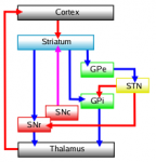
I forgot to mention that, in addition to the thalamo-cortical circuits, there are also basal ganglia-thalamus-cortical circuits:
https://en.m.wikipedia.org/wiki/Cortico-basal_ganglia-thalamo-cortical_loop

A rough diagram:
E56D65BD-5560-4EF7-9E00-E4BF41755BA6.png
Connectivity diagram showing excitatory glutamatergicpathways as red, inhibitory GABAergic pathways as blue, and modulatory dopaminergicpathways as magenta. (Abbreviations: GPe: globus pallidus external; GPi: globus pallidus internal; STN: subthalamic nucleus; SNc: substantia nigra pars compacta; SNr: substantia nigra pars reticulata)

Hope this helps.
 

Pyrrhus

Senior Member
Messages
4,172
Location
U.S., Earth
It might be interesting to compare the conclusions from this review of neuroimaging by Staines & Marshall-Gradisnik with this other review of neuroimaging, by Michael Van Elzakker:
https://forums.phoenixrising.me/thr...-a-critical-review-of-research-methods.76890/

Donald Staines & Sonya Marshall-Gradisnik's review:
  • "A total of 55 studies were included in this review."
  • "Out of the 55 papers, 43 relied on the Fukuda criteria to classify ME/CFS patients."
  • "Out of the 55 studies: 16 studies utilised MRI, 17 used functional MRI (fMRI), five used PET scans and 11 used EEG. The remaining studies used magnetic resonance spectrometry (MRS)."
  • "The articles reported on multiple outcomes including brain structure, cerebral blood flow (CBF), gray and [white matter (WM)] volumes, sleep and EEG, and [functional connectivity (FC)]."
  • "The results suggest widespread disruption of the autonomic nervous system network including morphological changes, white matter abnormalities and aberrations in functional connectivity."
  • "However, these findings are not consistent across studies and the origins of these anomalies remain unknown."
  • "The search for definitive cerebral signatures for ME/CFS remains difficult. The lack of reproducibility of the studies and low sample size also contribute to the inability to arrive at a consensus regarding potential neurological markers for ME/CFS. There are, however, consistent examples of altered brain health, e.g. an increase in neuroinflammation suggestive of a neuroimmune mechanism and reduced functional efficiency."

Michael Van Elzakker's review:
  • "The name “myalgic encephalomyelitis” essentially means “muscle pain related to central nervous system inflammation” and many efforts to find diagnostic biomarkers have focused on one or more aspects of neuroinflammation, from periphery to brain."
  • "The current review focuses on three methods used to study putative neuroinflammation in ME/CFS: (1) positron emission tomography (PET) neuroimaging using translocator protein (TSPO) binding radioligand (2) magnetic resonance spectroscopy (MRS) neuroimaging and (3) assays of cytokines circulating in blood and cerebrospinal fluid."
  • "We argue that the vast majority of ME/CFS neuroimaging has failed to use optimal techniques for studying brainstem, despite its probable centrality to any neuroinflammatory causes or autonomic effects."
  • "We argue that both the biological mechanisms of cytokines and the innumerable sources of potential variance in their measurement make it unlikely that a consistent and replicable diagnostic cytokine profile will ever be discovered."
  • "Kraynak et al. (19) conducted a useful meta-analysis of this basic neuroimmunology research. They synthesized results from studies that performed neuroimaging during peripheral immune activation by either an immune stimulating antigen (e.g., lipopolysaccharide [LPS]) or proinflammatory cytokines (e.g., interferon alpha [IFN-α]). [...] Consistent activation occurred in basal ganglia (bilateral striatum), limbic structures (right amygdala, bilateral hippocampus, and hypothalamus), brainstem/pons, and neocortex (right anterior insular cortex, right temporal and left parahippocampal gyri, subgenual and dorsal anterior cingulate cortex [sgACC and dACC], and dorsomedial and ventromedial prefrontal cortex [dmPFC and vmPFC])."
  • "The dACC (which would be considered anterior midcingulate cortex [aMCC] by some anatomists) [...] was consistently activated and therefore could also be considered an a priori region of interest in neuroinflammation studies. Given the role of dACC in attention and cognitive control, we suggest that its function in ME/CFS could be considered particularly important for “brain fog” symptoms."
  • "Furthermore, Kraynak et al. (19) reported that the thalamus was also consistently detected across multiple study designs, but not in a way that demonstrated functional connectivity. However, we consider thalamus an important region of interest in ME/CFS given its detection by Nakatomi et al. (8) and given the role of thalamus in sensory filtering, a likely mechanism for the common symptom of sensory sensitivity."
  • "The current gold standard for in vivo imaging of neuroinflammation is PET scanning using a translocator protein-binding radioligand."
  • "Nakatomi et al. (8) conducted the first case-control study using PET to measure [translocator protein (TSPO)] expression in the central nervous system of ME/CFS patients vs. healthy controls. [...] Nakatomi et al. (8) remains an important, groundbreaking study that should be replicated with complementary methods."
  • "The ME/CFS research field has been stuck in a somewhat defensive posture, with a focus on demonstrating “this is a real condition” by showing significant biological differences between patients and controls. [...] The field is ready to move past proving “this is a real condition” and to start elucidating the specific relationship of ME/CFS symptoms to neuroinflammation."
 

pattismith

Senior Member
Messages
3,931
Interestingly, the Anterior Cingulate Cortex (ACC) in the cortical brain also figures prominently in research studies on Attention Deficit Disorder (ADD).[1] One reason why the ACC may be critical for maintaining attention is its connections with the thalamus in the sub-cortical brain.[2] The thalamus is the part of the subcortical (autonomic) brain that is most closely connected to the cortical (conscious) brain.

Although the thalamus is best known as a relay station that provides all the body's sensory information to the cortical brain, the cortical brain also provides "feedback" to the thalamus, allowing the thalamus to suppress certain sensory information. It is thought that this cortical "feedback" is how the conscious brain decides which sensory information to pay attention to, and which sensory information to ignore, a phenomenon called "sensory gating". If there is a problem with this "feedback", then the conscious brain may be inundated with too much sensory information.

References
[1] https://pubmed.ncbi.nlm.nih.gov/23713508/
[2] https://pubmed.ncbi.nlm.nih.gov/23371564/


Thalamic neuroinflammation as a reproducible and discriminating signature for chronic low back pain
Torrado-Carvajal, Angela,b; Toschi, Nicolaa,c; Albrecht, Daniel S.a; Chang, Kena; Akeju, Oluwaseund; Kim, Minhaea; Edwards, Robert R.e; Zhang, Yid; Hooker, Jacob M.a; Duggento, Andreaa,c; Kalpathy-Cramer, Jayashreea; Napadow, Vitalya; Loggia, Marco L.a,*


PAIN: April 2021 - Volume 162 - Issue 4 - p 1241-1249
doi: 10.1097/j.pain.0000000000002108

Using positron emission tomography, we recently demonstrated elevated brain levels of the 18 kDa translocator protein (TSPO), a glial activation marker, in chronic low back pain (cLBP) patients, compared to healthy controls (HCs).
...................................
. Our observations suggest that thalamic neuroinflammatory signal is a reproducible and discriminating feature for cLBP, further supporting a role for glial activation in human cLBP, and the exploration of neuroinflammation as a therapeutic target for chronic pain.

This work further shows that TSPO signal contains a richness of information that the simple mean might fail to capture completely.
 

Pyrrhus

Senior Member
Messages
4,172
Location
U.S., Earth
Thalamic neuroinflammation as a reproducible and discriminating signature for chronic low back pain

Thanks for sharing this new study by Marco Loggia!

Marco Loggia's team first published evidence of neuroinflammation in chronic pain a number of years ago, and then followed it up with this classic study on fibromyalgia:

Brain glial activation in fibromyalgia – A multi-site positron emission tomography investigation (Albrecht et al. 2019)
https://forums.phoenixrising.me/thr...-of-fibromyalgia-patients.61667/#post-2321470
 

pattismith

Senior Member
Messages
3,931
Interestingly, the Anterior Cingulate Cortex (ACC) in the cortical brain also figures prominently in research studies on Attention Deficit Disorder (ADD).[1] One reason why the ACC may be critical for maintaining attention is its connections with the thalamus in the sub-cortical brain.[2] The thalamus is the part of the subcortical (autonomic) brain that is most closely connected to the cortical (conscious) brain.

The first question is how many CFS/ME or Fibro patients are undiagnosed ADHA ?
My new neuropsy seems to think I may have ADHA from the start + chronic iron deficiency.
I looked back at my history and found some mental features commonly found in ADHA
- crying a lot when I was under 8
- a good amount of physical accidents
- eating disorders that vary according to age
- attention deficit that made me unable to learn only by listening or reading (I had to write things to learn it)

The fact Methylphenidate is helping me seems to favor this hypothesis...

On the other hands I don't have typical impulsivity/hyperactivity found in this disorder, Or maybe when I was very young, but it quickly disappeared (I think education can change the way the syndrome will move)
 
Last edited:

pattismith

Senior Member
Messages
3,931
Interestingly, the Anterior Cingulate Cortex (ACC) in the cortical brain also figures prominently in research studies on Attention Deficit Disorder (ADD).[1] One reason why the ACC may be critical for maintaining attention is its connections with the thalamus in the sub-cortical brain.[2]


I wish to add some input about the ACC/mesolimbic dopamine connections (mesolimbic circuit goes from VTA/midbrain to Striatum/Basal Ganglia);

it is from a 2021 article about chronic pain, but likely to be still valid for some other functionalities

1635144986737.png


Figure 1.
Interactions between the Ventral Mesostriatal Dopaminergic Pathway and Corticostriatal Glutamatergic Afferents in Chronic Pain. The scheme presents only those neuronal connections, whose participation in pain processing and involvement in chronic pain have been documented directly.

Glutamatergic, dopaminergic, and GABAergic neurons are depicted as orange, blue, and green ovals, respectively, and their projections are depicted as arrows. “+” and “−“ signs denote excitatory and inhibitory effects on the postsynaptic cell, respectively.

Left panel. “A” and “H” placed along projections denote effects of activation of the particular projection on pain responses: analgesia and hyperalgesia/pain, respectively. These signs reflect effects on both sensory and affective aspects of pain, or either (see text for details), and show general tendencies, some of which become explicit only under tonic or chronic pain.

Right panel. Changes in the activity of particular neuronal populations and their projections under chronic pain have been reflected by the “cell body” size and the arrow width. ↑ and ↓ arrows denote the final effect on pain (increase and decrease, respectively) resulting from both the function of a particular projection in pain modulation (refer to “A” and “H” on the left panel) and changes in the activity of this projection in chronic pain.

?*–In contrast to some other studies, Ren et al. [67] provide evidence of an increase in dopaminergic neurotransmission in the medial VTA–medial NAc shell projection in chronic pain. This scheme has been based on the studies by Schwartz et al. [89], Lee et al. [81], Ren et al. [63,67], and Gao et al. [85]. The nigrostriatal dopamine pathway has not been included in the figure due to the lack of corresponding studies on the dorsal striatum.

ACC—anterior cingulate cortex;
BLA—basolateral amygdala;
D1-MSN—D1 receptor-expressing medium spiny neuron;
D2-MSN—D2 receptor-expressing medium spiny neuron;
DA—dopamine;
IL—infralimbic cortex;
LTD—long-term depression;
LTP—long-term potentiation;
mNAc-sh—medial nucleus accumbens shell;
NAc-c—nucleus accumbens core;
PL—prelimbic cortex;
VTA—ventral tegmental area.

Association between Chronic Pain and Alterations in the Mesolimbic Dopaminergic System | Phoenix Rising ME/CFS Forums
 
Last edited:

Wishful

Senior Member
Messages
5,684
Location
Alberta
My new neuropsy seems to think I may have ADHA from the start

I think there may be a lot of diagnoses of common and easily treated (by popular highly-profitable pharmaceuticals) diseases based on very slim evidence. How many of us have been diagnosed with depression, and prescribed antidepressants even though we don't consider ourselves as clinically depressed? I think attention deficit and autism are also overdiagnosed. Some doctors would probably diagnose 90+% of the population as having all three disorders.
 

Alvin2

The good news is patients don't die the bad news..
Messages
2,997

Alvin2

The good news is patients don't die the bad news..
Messages
2,997

Alvin2

The good news is patients don't die the bad news..
Messages
2,997
My patience ran out so I am taking the lutein, 3 days and no effect so far
Will keep everyone posted
 

SWAlexander

Senior Member
Messages
1,898
"The superficial perception-motor white-matter network had decreased functional connectivity with the cortical perception-motor gray-matter networks."

Once, my neurosurgeon told me white matter is a pile of dead neurons, blocking connection to the gray matter.
This paper is saying basically the same: "The superficial perception-motor white-matter network had decreased functional connectivity with the cortical perception-motor gray-matter networks." https://pubmed.ncbi.nlm.nih.gov/29660513/