• Welcome to Phoenix Rising!

    Created in 2008, Phoenix Rising is the largest and oldest forum dedicated to furthering the understanding of and finding treatments for complex chronic illnesses such as chronic fatigue syndrome (ME/CFS), fibromyalgia (FM), long COVID, postural orthostatic tachycardia syndrome (POTS), mast cell activation syndrome (MCAS), and allied diseases.

    To become a member, simply click the Register button at the top right.

Second study might confirm neuroinflammation in ME subcortical brain

Pyrrhus

Senior Member
Messages
4,172
Location
U.S., Earth
some articles I read propose P2X7 as a new marker for neuroinflammation.

That's fascinating, thank you for sharing that. I have read about P2X7 in the context of neuropathic pain, but it never occurred to me that it might be used as a biomarker of neuroinflammation.

For anyone interested in P2X7:
https://pubmed.ncbi.nlm.nih.gov/21924325/
(note that, although intracellular ATP is an energy storage molecule, extracellular ATP is a danger-signalling molecule.)

I have a genetic change at P2RY2. Because it plays a role at muscle contraction and neurotransmitssion. maybe it is a reason, why I have sometimes problems with my muscles, who knows...

Possibly. Even now, 20 years after sequencing the full human genome, we still have so many problems matching genotype to phenotype...

I don't know if it was your explanation or my brain is working better today, but your two posts above re inflammation and TSPO (this one and this one) were amazingly easy to follow and comprehend.

Thanks, that's a true compliment. In the last 20 years or so, there has been a far greater emphasis placed on "communication skills" in the scientific community than in the past. Some scientists find "communicating" is the hardest part of their job. And so many brilliant discoveries have been overlooked entirely because they were never communicated fully to the rest of the scientific community.
 
Messages
48
Sure! If I use medical marijuana I become extraordinarily (100%) clear-headed for the whole day, and even extending into the next. But then after that, I pay the price: poor sleep, night after night, along with a heavy, swollen feeling in my head. And all the mental clarity I enjoyed is replaced with brain fog. It's terribly frustrating.
This also happens on healthy people.

Probably its the adjustment period of your endocannabinoid system causing foogines.
 

Pyrrhus

Senior Member
Messages
4,172
Location
U.S., Earth
So, we now have two studies that find neuroinflammation in the subcortical brain.

Interestingly, almost 100 years ago, a French doctor described a post-infectious condition that he called "Subcortical Dementia".[1] Unlike traditional (cortical) dementia, Subcortical Dementia did not impact long-term memory or conscious actions. Instead, Subcortical Dementia impacted working memory and autonomic (unconscious) actions. This post-infectious diagnosis was largely forgotten, although newer definitions of "Subcortical Dementia" survive to this day.

But what, exactly, is the subcortical brain?


First, let's start with the cortical brain:
1597547478366.png
This is what most people think of when they imagine the brain. The cortical brain has two large hemispheres that provide the basic outward appearance of a typical brain. The cortical brain includes four "lobes": the frontal, temporal, parietal, and occipital lobes. All conscious activity is coordinated in the cortical brain.


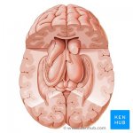
Now, let's crack open the cortical brain:
1597548172007.jpeg
Do you see those strange structures hidden inside the cortical brain, in between the two hemispheres? That's the subcortical brain. These subcortical structures handle most autonomic (unconscious) activity, and also act as a "filter" between the cortical brain and the rest of the body. As a filter, the subcortical brain determines which sensory input makes it to the cortical brain, and determines which motor output makes it from the cortical brain to the rest of the body.


Finally, let's get a closer look at the subcortical brain:
1597549095435.jpeg

In this picture, the two orange/beige areas in the middle are the left and right "thalamus". The purple area beneath the left and right thalamus is the "midbrain", and below that is the blue "pons" and the pink "medulla oblongata". Collectively, the midbrain, pons, and medulla oblongata constitute the "brainstem". The green areas on either side of the brainstem are the left and right "hippocampus".

The 2014 Japanese study found neuroinflammation in the thalamus, brainstem, and hippocampus.

In the middle of the picture, the two big red areas right behind the right and left thalamus are the right and left "basal ganglia". The new Stanford study found neuroinflammation in the basal ganglia.

Okay, that's probably enough anatomy for now. I hope you now have a better idea of what the "subcortical brain" is.


Reference
[1] L’Encephale 1922; 17:369–375.
 
Last edited:

MonkeyMan

Senior Member
Messages
405
@Pyrrhus, many thanks for this very clear and helpful explanation on the subcortical brain. Does all of this have treatment implications? So many of us are hoping for something to help relieve our symptoms. Do you think something will come along soon that may help us?
 

percyval577

nucleus caudatus et al
Messages
1,302
Location
Ik waak up
In this picture, the two orange/beige areas in the middle are the left and right "thalamus"
....

In the middle of the picture, the two big red areas right behind the right and left thalamus are the right and left "basal ganglia". The new Stanford study found neuroinflammation in the basal ganglia.

Okay, that's probably enough anatomy for now. I hope you now have a better idea of what the "subcortical brain" is.
If I recalll rightly its furthermore:
  1. The thalamus has different nuclei, which are only very little connected one to each other. They project to the cortex, and every region in the cortex is projected at from the thalamus. The different nuclei are projected at from regions in the body, and these regions project also to the cortex. These nuclei are the specific nuclei of the thalamus, and participate each exclusively in specific tasks (vision, or whatever). They also get feedback from the cortex.
  2. The different nuclei in the thalamus are though indirectly connected, via the the nucleus intralaminaris, which stretches inside the middle of the thalamus all along the thalamus. This nucleus (or its rather more than one as it as a y-junction) is part of the non-specific thalamus. Another nucleus here is the nucleus reticularis on the outer surface.
  3. The whole big of the basal ganglia projects via one part of it (globus pallidus lateralis) into one very slender specific nucleus in the thalamus (and another structure close by the thalamus).
  4. The basal ganglia project, as the thalamus does, to every region in the cortex, and they also get feedback from the cortex in the same manner.
  5. The wheel shaped basal ganglia have an upper nucleus - the nucleus caudatus - which is funnel shaped. It begins very thin near the nucleus for the memory and gets wider and wider until it ends in the nucleus accumbens, known to be important for concentration and drug addiction.

What I now find interesting is that there are two vulnerabilities:
  • The small nucleus in the thalamus where the basal ganglia are docking in. If we assume that there any any waves or rhythms in the long nucleus intralaminaris, this could well be get disturbed by irregular input from the basal ganglia.
  • The funnel shape, when it should be ever more difficult to keep up any movement arising from behind to get forward, because there is more and more action to supply.

Given that there is a wide range of symptoms, and given that they behave quite autonomously (delayed PEM), I find it quite logical that these structures are the main side of CFS symptoms in its causation. These structures may well code for the acute feeling of good or bad, and much more like short term memory, being prepared for Mondays, and things like that.

It could simply be that the action of nerves have been misguided by too much or less normal stuff, arising from any impact. So, time restricted. But although they afterwards simply make actions as usual they are in fact reproducing not normal shapes having arisen at time of impacts. Their normal algorithms may now reproduce nonsense.


So, I think this is CFS, and maybe some other badly understood diseases like depression or functional neurological disorders, or whatever. And this (re-) production of healthy or non-healthy shapes of synaptical connections cannot already be shown, nor by PET, nor by MRT, probably as well not by post mortem investigations. Or would it be possible by any of these techniques?
 
Last edited:

percyval577

nucleus caudatus et al
Messages
1,302
Location
Ik waak up
My working hypothesis in more detail is, that there are geometrical actions stretching rather over wide measures - the long nucleus intralaminaris, the funnel shaped nucleus caudatus -, and that these actions are coded by different stuff, especially by some metals and Vit B´s (not B9 and B12, which do not supply the TCA cycle).

So a hope could be to find any abnormality in the distribution of Zn, Al, Cr, Ni and Mn (my impression, with at least some possible reasoning); as well as of Vit B2, B1, B7, B5, B3 and B6.

A further hope would be, to re-erect a healthy structure by management of these atoms and molecules.

[And here I think it must be isolated application to avoid any more muddle, and even in small amounts to avoid an overreactions or a shut down of uptake into the structures. - More important, and maybe with less possible (?!) sideeffects may be simply to help canceling out the reproduced unfavaourable actions by helping with adjustment moelcules like acetate, B12, VitC and citrate under otherwise good circumstances (not this normal stuff that has been around at time of infection in exaggeration.)]

Hope it makes sense to more than simply myself. I will come back to PR later and will argue with literature (for now I have other important trouble, and chances as well!).
 
Last edited:

Pyrrhus

Senior Member
Messages
4,172
Location
U.S., Earth
@Pyrrhus, many thanks for this very clear and helpful explanation on the subcortical brain. Does all of this have treatment implications? So many of us are hoping for something to help relieve our symptoms. Do you think something will come along soon that may help us?

I'm glad you got something helpful out of it.

These studies only detect neuroinflammation. Unfortunately, they don't tell us what's causing the neuroinflammation, so they don't tell us anything about treatment. First we have to know what's causing the neuroinflammation before we can come up with a sensible approach to treatment.

Some people have proposed "treating" the neuroinflammation with anti-inflammatories. Although that approach might provide temporary relief in the short-term, it is possible that it may backfire in the long-term. Only when we identify the cause of the neuroinflammation can we move forward with a more robust plan of treatment.

Hope this helps.
 

WantedAlive

Senior Member
Messages
158
Only when we identify the cause of the neuroinflammation can we move forward with a more robust plan of treatment

Recall Fisher found mitochondrial Complex V impairment in ME/CFS? Among the plethora of symptoms associated with diseases with complex V deficiency is....brainstem degeneration. According to the publication "Syndromes: Rapid Recognition and Perioperative Implications", it states
It [complex v deficiency] is often associated with evidence of brainstem degeneration leading to coma
.

I've tried to reach out to the author here to understand why mitochondrial impairment would specifically induce brainstem degradation but his contact is difficult to access. If I ever find out I'll be sure to post.
 

Rufous McKinney

Senior Member
Messages
13,251
Among the plethora of symptoms associated with diseases with complex V deficiency is....brainstem degeneration. According to the publication "Syndromes: Rapid Recognition and Perioperative Implications", it states

Reading more at the link above about Leigh's disease/syndrome...an additional quote below- are they talking about us? Lesions on the basal ganglia, exacerbated by viruses....symptoms we often have might be stemming from some type of related issuesa:


"Most cases with mtDNA-associated LS present with symptoms between the ages of 3 and 12 months, although later childhood and adult disease onset can also occur [2]. Exacerbations are often heralded by the presence of a viral infection. The condition is progressive, and decompensation during periods of illness or over time often lead to exacerbation of the cranial lesions with associated worsening neurologic function. Due to involvement of the brain stem, cases may often present with worsening respiratory or cardiovascular function, alteration of consciousness, and apnea. Recovery after each decompensation is limited with an accumulation of symptoms over time. Cases gradually develop worsening spasticity, dystonia and movement disorders from the basal ganglia lesions, epilepsy from cortical disease, and dysphagia and respiratory insufficiency from brain-stem disease."

Do our ME researchers know this disease exists?
 

WantedAlive

Senior Member
Messages
158
Leigh's Syndrome?
Yes! and NARP amongst others. You're right many common symptoms. According to Fisher's study, this mitochondrial impairment in ME/CFS only showed up under stress test, could maybe explain PEM to some degree. Evidently the complex V impairment particularly affects the nervous system and heart, but why brainstem degradation? Maybe someone on this forum can help answer that.
 

Pyrrhus

Senior Member
Messages
4,172
Location
U.S., Earth
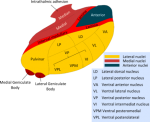
The thalamus has different nuclei, which are only very little connected one to each other. They project to the cortex, and every region in the cortex is projected at from the thalamus. The different nuclei are projected at from regions in the body, and these regions project also to the cortex. These nuclei are the specific nuclei of the thalamus, and participate each exclusively in specific tasks (vision, or whatever). They also get feedback from the cortex.

Yes, I think you're describing how the thalamus serves as a "relay station" that connects all sensory information from the body to the cortical brain. (with the sole exception of the sense of smell) Each type of sensory information is relayed through its own "nucleus" in the thalamus:
1597797840729.png
But the thalamus is more than just a relay station that provides all the body's sensory information to the cortical brain. As you mention, the cortical brain provides "feedback" nerves to the thalamus, allowing the thalamus to suppress certain sensory information. It is thought that this cortical "feedback" is how the conscious brain decides which sensory information to pay attention to, and which sensory information to ignore. If there is a problem with this "feedback", then the conscious brain may be inundated with too much sensory information. (photophobia, hyperacusis?)

There is another important aspect to the connections between the thalamus and the cortical brain- the aspect of alert consciousness. There is recurring stimulation from the thalamus to the cortical brain and then back to the thalamus, forming a loop. This recurring stimulation through the loop leads to "thalamo-cortical oscillations". Some of these oscillations can be roughly recorded using an electroencephalogram (EEG) as "alpha frequency waves". These alpha frequency waves in EEG typically represent a state of alert consciousness. If there is a problem with the thalamo-cortical oscillations, it may be hard to maintain a state of alert consciousness. (brain fog?)

EEG studies in ME have had a hard time documenting specific abnormalities. But one study noted that "In the eyes closed condition, peak alpha frequency (the frequency between 8 to 13 Hz at which the greatest amount of energy was observed) correlated negatively with the 'fatigue today' rating." This means that patients who reported more fatigue had slower alpha-frequency waves.[1] And a 2014 conference presentation "found decreased [peak alpha frequency] over 58% of the entire cortex in CFS patients when compared to controls. [...] These findings are consistent with reduced efficiency of thalamo-cortical connections in CFS participants." [2]

For those interested, here is one of the earliest papers that established a link between the speed of thalamo-cortical oscillations and attention or information processing:
EEG-alpha rhythms and memory processes (Klimesch, 1997)​
The author's later work expands on how thalamo-cortical oscillations are involved in both attention/concentration and in information processing/retrieval:

So, I think this is CFS, and maybe some other badly understood diseases like depression or functional neurological disorders, or whatever. And this (re-) production of healthy or non-healthy shapes of synaptical connections cannot already be shown, nor by PET, nor by MRT, probably as well not by post mortem investigations. Or would it be possible by any of these techniques?

It sounds like you are suggesting that there may be some "re-wiring" of the neuronal connections in ME. You are right that we can't easily detect such "re-wiring" of neuronal connections using PET or MRI. But the function of a neuronal circuit can be changed even without any "re-wiring".

To use an electrical circuit analogy, if one of the wires in the circuit experiences a large increase in resistance, the output of the circuit will change, even without any re-wiring. If the frequency of the circuit's clock/oscillator changes, then the output of the circuit will change, even without any re-wiring.


So a hope could be to find any abnormality in the distribution of Zn, Al, Cr, Ni and Mn (my impression, with at least some possible reasoning); as well as of Vit B2, B1, B7, B5, B3 and B6.

Yes, it would certainly be interesting to examine the abnormalities in the level or distribution of essential nutrients. Both neuroinflammation as well as classical inflammation trigger changes in the levels of nutrients in the area of inflammation, and sometimes in the whole body.

These metabolic changes in neuroinflammation can be different from the metabolic changes seen in classical inflammation. Furthermore, the metabolic changes seen in acute neuroinflammation may be different from the metabolic changes seen in chronic neuroinflammation. But it is indeed important to consider the "downstream" metabolic effects of neuroinflammation.

References
[1] https://www.tandfonline.com/doi/abs/10.1300/J184v02n02_04
[2] https://forums.phoenixrising.me/thr...udy-presented-at-stanford.32919/#post-2355997
 
Last edited:

Pyrrhus

Senior Member
Messages
4,172
Location
U.S., Earth
Recall Fisher found mitochondrial Complex V impairment in ME/CFS? Among the plethora of symptoms associated with diseases with complex V deficiency is....brainstem degeneration.
Leigh's Syndrome?
Evidently the complex V impairment particularly affects the nervous system and heart, but why brainstem degradation?

Yes, if there are body-wide problems with the mitochondria, symptoms tend to show up first in both muscles and in the nervous system. This is because muscles and nerve cells are the biggest consumers of mitochondrial energy in the body, and are therefore the most susceptible to mitochondrial problems.

Since the heart consumes more energy than other muscles, the heart is usually the first muscle to show problems. Since the subcortical brain is more active than the cortical brain, it consumes more energy than the cortical brain, and we tend to see mitochondrial symptoms in the subcortical brain before we see symptoms in the cortical brain. But why the brainstem in particular? I don't know.

Remember that recognized genetic problems with mitochondria, such as Leigh's Syndrome, can be detected by a genetic screen that includes mitochondrial DNA (mtDNA).

Hope this helps.
 

percyval577

nucleus caudatus et al
Messages
1,302
Location
Ik waak up
It sounds like you are suggesting that there may be some "re-wiring" of the neuronal connections in ME. You are right that we can't easily detect such "re-wiring" of neuronal connections using PET or MRI. But the function of a neuronal circuit can be changed even without any "re-wiring".

To use an electrical circuit analogy, if one of the wires in the circuit experiences a large increase in resistance, the output of the circuit will change, even without any re-wiring. If the frequency of the circuit's clock/oscillator changes, then the output of the circuit will change, even without any re-wiring.
Right, and I was not consistent in my own thoughts.

I originally didn´t want - and still don´t want - to include any necessity of a feedback from the cortex. Instead a mis-wiring in these structures should be self-sufficient, rather kind of a clou for the development from impacts. In this sense I also meant "re-production".

Another line then was, that I havn´t come so far across any wiring from the thalamus to the basal ganglia, and for the lack of such a connection within these "self-sufficient" structures (making it a bit a stronger wrong-normal) the idea surged in that via the cortex a connection exists ...

In fact this could be of importance, there is plenty of unkown enough, but the main reason to avoid it, is of course the proposed self-sufficiency which may be underlined by T-type calcium channels (I learned this from you, btw), and possibly more or more sensitive NMDAR´s (I havn´t found any paper so far, though), and a rough identification of the cortex with psychological matters having their processing or even storage there.


This last point has, as we know, lead to lots of distress. But the reliable success from psychological intervention is small, at most. Let´s assume that 5% had been reported (there is somewhere a quite prominent source for this), than the first logical question is, if these ones really had ME/CFS. If so - not the most likely guess - than an explanation could be,

that this success depends on the possibility that new cortex structures can be implemented, which will be only possible when the patient has some major psychological problems. But if this is not the case, there also cannot the bad self-sufficient and self-reproducing structures get sidestepped and new ones be implemented also in these structures.

So, having such problems would be a chance, but would not be a cause for ME/CFS. Interestingly here on the forum a member has reported to get healed over the course of two years or so from a change of sex. I would think that the new hormonal equilibrium and maybe other inputs has served as a comparable change.


Taken together I rather don´t want to rely on the feedback via cortex. Of course, there could be something really important nevertheless - in fact one of the structures should project to layer two, and gets its feedback from layer six, but my memory could be wrong -, but it may well be unlikely, I would say here.
 
Last edited:

Pyrrhus

Senior Member
Messages
4,172
Location
U.S., Earth
But what, exactly, is the subcortical brain?

I forgot to mention above that the picture of the subcortical brain that I provided is incomplete:
  • The hippocampus is an important part of the subcortical brain that is shown in the picture I provided. However, it should be noted that some neuroanatomists consider the hippocampus to be neither part of the cortical nor the subcortical brain, rather something in between.
  • The cerebellum is an important part of the subcortical brain that is not clearly shown in the picture I provided.
  • The hypothalamus is an important part of the subcortical brain that is not shown in the picture I provided.

But the thalamus is more than just a relay station that provides all the body's sensory information to the cortical brain. As you mention, the cortical brain provides "feedback" nerves to the thalamus, allowing the thalamus to suppress certain sensory information. It is thought that this cortical "feedback" is how the conscious brain decides which sensory information to pay attention to, and which sensory information to ignore. If there is a problem with this "feedback", then the conscious brain may be inundated with too much sensory information. (photophobia, hyperacusis?)

There is another important aspect to the connections between the thalamus and the cortical brain- the aspect of alert consciousness. There is recurring stimulation from the thalamus to the cortical brain and then back to the thalamus, forming a loop. This recurring stimulation through the loop leads to "thalamo-cortical oscillations". These oscillations can be roughly recorded using an electroencephalogram (EEG) as "alpha frequency waves". These alpha frequency waves in EEG typically represent a state of alert consciousness. If there is a problem with the thalamo-cortical oscillations, it may be hard to maintain a state of alert consciousness. (brain fog?)

As described above, the thalamus is the part of the subcortical brain that is most closely connected to the cortical brain. Although the subcortical brain is where most autonomic (unconscious) activity is controlled, a few parts of the cortical brain are often mentioned because they have strong connections with the thalamus:
  • The Anterior Cingulate Cortex (ACC) is a part of the cortical brain that has strong connections with the thalamus and is therefore often mentioned in relation to subcortical activity, even though it is anatomically part of the cortical brain.
  • The Insular Cortex (insula) is a part of the cortical brain that also has strong connections with the thalamus and is therefore often mentioned in relation to subcortical activity, even though it is anatomically part of the cortical brain.
  • The Prefrontal Cortex (PFC) is a part of the cortical brain that also has strong connections with the thalamus. It is well-known for its role in "executive function" and decision-making. Its connections to the thalamus are so strong that one definition of the Prefrontal Cortex is "those areas of the cortical brain that are connected to the medial dorsal nucleus of the thalamus." The Prefrontal Cortex is therefore often mentioned in relation to subcortical activity, even though it is anatomically part of the cortical brain.
 
Last edited by a moderator:

Pyrrhus

Senior Member
Messages
4,172
Location
U.S., Earth
And here's one interesting fact...

The two parts of the brain that have the highest density of mast cells are both in the subcortical brain:
  • The thalamus
  • The hypothalamus
Since mast cells are immune cells, I wonder why these two parts of the brain require higher densities of mast cells...

Also, the circumventricular organs, which lack a strong blood-brain barrier, may also show high densities of mast cells.
 
Last edited:

WantedAlive

Senior Member
Messages
158
So, I’ve been digging more into this brainstem degeneration found in Complex V deficiency syndromes (which now includes ME/CFS). It appears that both the Basal Ganglia and Brainstem are highly vulnerable to intracellular energy deprivation, and there is documented evidence of brainstem lesions in patients with acute energy deprivation syndromes such as Wernickes encephalopathy and Leighs Disease [1][2]. The big question therefore – is it the energy deprivation driving ME/CFS neuroinflammation along with the neck pain, the cranial settling and CCI issues?

If you add the evidence of cerebral hypoperfusion, ME/CFS probably fits with an ‘ischemic and energy deprivation syndrome’. There are documented cases of this. The factors in this syndrome lead to a vicious circle, leading from oxygen and glucose deprivation to potentially cell death in the basal ganglia. The basal ganglia are a high-density area for Glutamate receptors [GluN2B and GluN2A] and their physiological structure is prone to excitotoxicity and enhance inward calcium currents (recall Nimodipine has helped some PWME). The toxic elevation of calcium influx leads to increased ROS production and membrane disruption (Fisher’s study proposes a proton leak in ME/CFS), which in turn leads to necrosis, glutamate release and the vicious cycle continues. The toxic release of Cyanate, Manganese and Carbon Monoxide all target basal ganglia at the site of lesions, and of note, Cyanate and Carbon Monoxide inhibit mitochondrial Complex V.

Now carbon monoxide poisoning, endogenously, might be an interesting concept for ME/CFS that I'd never heard of. There are symptom overlaps with CO poisoning, and in fact exogenous CO poisoning has been misdiagnosed as ME/CFS (here)! Carbon monoxide competes with oxygen to bind with haemoglobin, this results in reduced respiration (reduced oxygen utilisation) and might contribute to the cerebral hypocapnia (low CO2) found in ME/CFS studies. I was surprised by this finding as I (and others) have hypercapnia in my limbs (high venous CO2), suggesting cerebral and peripheral respiration might be affected in different ways.
 

Pyrrhus

Senior Member
Messages
4,172
Location
U.S., Earth
The big question therefore – is it the energy deprivation driving ME/CFS neuroinflammation along with the neck pain, the cranial settling and CCI issues?

Wow, you're really trying to tie things together. I certainly see how energy deprivation would lead to oxidative stress, which can eventually lead to neuroinflammation. The rest, I'm not so sure what to make of it.

If you add the evidence of cerebral hypoperfusion, ME/CFS probably fits with an ‘ischemic and energy deprivation syndrome’. There are documented cases of this. The factors in this syndrome lead to a vicious circle, leading from oxygen and glucose deprivation to potentially cell death in the basal ganglia.

I believe you're talking about the vicious cycle proposed in the article you provided:
https://www.sciencedirect.com/science/article/pii/S030100821830042X

Yes, the basal ganglia are particularly vulnerable to a lack of blood flow, since the lenticulo-striate arteries that provide blood to the basal ganglia are very narrow. These arteries are common sites for blockages, and if the metabolic rate increases in the basal ganglia, these arteries may have trouble supplying enough nutrients even without any blockages. Why evolution gave the basal ganglia such narrow arteries is a mystery to me.

The basal ganglia are a high-density area for Glutamate receptors [GluN2B and GluN2A] and their physiological structure is prone to excitotoxicity and enhance inward calcium currents (recall Nimodipine has helped some PWME). The toxic elevation of calcium influx leads to increased ROS production and membrane disruption (Fisher’s study proposes a proton leak in ME/CFS), which in turn leads to necrosis, glutamate release and the vicious cycle continues.

This part of the vicious cycle seems more speculative, but might be plausible. We do know that an increase in intracellular calcium leads to mitochondrial membrane disruption and oxidative stress. But it doesn't necessarily lead to apoptosis or necrosis.

Take the example of enterovirus infection of neurons. One of the first things that enteroviruses (and many other viruses) do is to raise the intracellular calcium level. Enteroviruses do this by punching holes in membranes using a viral protein called a viroporin. This leads to mitochondrial membrane disruption and oxidative stress, but it doesn't lead to apoptosis or necrosis. The neuron probably won't function correctly, but at least the neuron is intact.

Now carbon monoxide poisoning, endogenously, might be an interesting concept for ME/CFS that I'd never heard of. There are symptom overlaps with CO poisoning, and in fact exogenous CO poisoning has been misdiagnosed as ME/CFS (here)!

Now that's a differential diagnosis that doesn't come up that often!

Carbon monoxide competes with oxygen to bind with haemoglobin, this results in reduced respiration (reduced oxygen utilisation) and might contribute to the cerebral hypocapnia (low CO2) found in ME/CFS studies. I was surprised by this finding as I (and others) have hypercapnia in my limbs (high venous CO2), suggesting cerebral and peripheral respiration might be affected in different ways.

That is interesting. I wonder if circulatory dysautonomia, as seen in orthostatic intolerance, might explain the discrepancy between cerebral and peripheral respiration. Thank you for your thoughtful and provocative ideas.
 

pattismith

Senior Member
Messages
3,932
@Pyrrhus , I think Substantia Nigra already showed up in some CFS/ME study, I don't have time to check it though...

September 2020
Regional microglial activation in the substantia nigra is linked with fatigue in MS

Results Peak voxel-based level of significance for correlation between total fatigue score and PET uptake was localized to the right substantia nigra (T-score 4.67, p = 0.001).

Similarly, SUVRs derived from atlas-based segmentation of the substantia nigra showed significant correlation with MFIS (r = 0.76, p = 0.004).

On multiple regression, the right substantia nigra was an independent predictor of total MFIS (p = 0.02) and cognitive MFIS subscale values (p = 0.007), after adjustment for age, disability, and depression.

Several additional areas of significant correlations with fatigue scores were identified, including the right parahippocampal gyrus, right precuneus, and juxtacortical white matter (all p < 0.05).

There was no correlation between fatigue scores and brain atrophy and lesion load in patients with MS.
 

Pyrrhus

Senior Member
Messages
4,172
Location
U.S., Earth
I think Substantia Nigra already showed up in some CFS/ME study, I don't have time to check it though..

Thanks, I hadn't heard about any studies with the Substantia Nigra and ME/CFS, but that study reporting neuroinflammation of the Substantia Nigra in Multiple Sclerosis is very interesting.

Note that the Substantia Nigra is physically located in the brainstem, but is anatomically considered part of the basal ganglia. Neuroanatomy can be a particularly frustrating field to wrap your head around!

Note also that the Substantia Nigra is one of the most energy-intensive parts of the subcortical brain, probably because it must manufacture and transport the dopamine for other parts of the basal ganglia, or because the Substantia Nigra must continuously fire some of its neurons roughly 25 times per second.