• Welcome to Phoenix Rising!

    Created in 2008, Phoenix Rising is the largest and oldest forum dedicated to furthering the understanding of and finding treatments for complex chronic illnesses such as chronic fatigue syndrome (ME/CFS), fibromyalgia (FM), long COVID, postural orthostatic tachycardia syndrome (POTS), mast cell activation syndrome (MCAS), and allied diseases.

    To become a member, simply click the Register button at the top right.

ME/CFS for 18 years, recently diagnosed with D-Lactic acidosis as cause of symptoms and illness.

Avenger

Senior Member
Messages
323
Hi Knackers,
an elemental diet would be great, but I was told that one was not available.
If you have any idea where one could be obtained it would be good to use when most unwell.

But it would be better to be able to reverse the illness; it is my belief that ME/CFS may be related to a number of forms of Bacterial Overgrowth that are possibly reversible through FMT in many cases. Even if the reversal was not permanent, it may be possible at some point in the future to purchase a delayed capsule with FMT product or filtered and grown versions so that you can live a normal life. But there have already been a number of successful reversals in FMT tests for ME and D-Lactic acidosis. C.Diff is just another form of Bacterial Overgrowth that is known to be caused by Antibiotics. ME may also be a side effect of Antibiotics, selecting for different Overgrowths's at least in a subset.


2018 Oct;67(4):483-487. doi: 10.1097/MPG.0000000000002043.
Intestinal Microbial and Metabolic Alterations Following Successful Fecal Microbiota Transplant for D-Lactic Acidosis.
Bulik-Sullivan EC1, Roy S2, Elliott RJ3, Kassam Z3, Lichtman SN2, Carroll IM1,4, Gulati AS2,4,5.
Author information

Abstract
Fecal microbiota transplantation (FMT) involves the transfer of stool from a healthy individual into the intestinal tract of a diseased recipient. Although used primarily for recurrent Clostridium difficile infection, FMT is increasingly being attempted as an experimental therapy for other illnesses, including metabolic disorders. D-lactic acidosis (D-LA) is a metabolic disorder that may occur in individuals with short bowel syndrome when lactate-producing bacteria in the colon overproduce D-lactate. This results in elevated systemic levels of D-lactate, metabolic acidosis, and encephalopathy. In this study, we report the successful use of FMT for the treatment of recurrent D-LA in a child who was unresponsive to conventional therapies. Importantly, we also present profiles of the enteric microbiota, as well as fecal D-/L-lactic acid metabolites, before and longitudinally after FMT. These data provide valuable insight into the putative mechanisms of D-LA pathogenesis and its treatment.
PMID: 29901551 DOI: 10.1097/MPG.0000000000002043
[Indexed for MEDLINE]
 

Avenger

Senior Member
Messages
323
Sorry if this has already been covered (I haven't been able to read all posts)

Could D-la affect PEM?

Hi Mel19,
Absolutely. The bad news is that I still get PEM after activity, even when my illness is under control. But I am not as worried about Fatigue when compared to the severity of the worst symptoms.

Fatigue fluctuates and is often better soon after using antibiotics. It is difficult to understand fully what is going on, but I was also diagnosed with autonomic dysfunction and this could be a chicken or egg situation with a number of contributing factors including antibiotics and underlying illness such as diabetes (due to Western starch based diet). But I would ideally like to trial FMT because there is already a lot of evidence that ME and a number of other diseases including D-Lactic acidosis have been fully reversed through FMT.
https://www.ncbi.nlm.nih.gov/pubmed/29901551

I have also had ME/D-La for 20 years and there may possibly be post symptoms. I can imagine that long periods of Organic poisoning, Acidosis or Neurotoxins would cause some long term damage; 'acidosis impacts the cardiovascular system in hospital settings, and chronic metabolic acidosis affects muscles, bones, kidney and cardiovascular health. Acid buffering leads to loss of bone density, resulting in an increased risk of bone fractures, renal osteodystrophy, and bone disease; as well, increased protein catabolism leads to muscle wasting'.


Sheedy et al. found that even when their CFS patients with D-Lactic Bacteria were treated, they still suffered Fatigue. They postulate that there may be other forms of Bacterial Overgrowth involved, as well as D-Lactic. But I will say that the worst symptoms have been controlled through the use of antibiotics, diet and probiotics, but it is easy to make mistakes with the diet.

Paul.
 

Avenger

Senior Member
Messages
323
Hi Sam,
I am getting ready for the Coronavirus because the fools in Cheltenham UK are holding the Cheltenham Races which regularly brings in 250.000 people to my town, which will host one of the biggest Coronavirus incubation's in history when the punters hit the pubs and clubs. Many punters are coming from Ireland and abroad! This is all about money again! Anyway...


After reading the articles below, I am going to try to hit my gut and lung with both antivirals and antibiotics as well as using things like Quinine Sulphate that can be used for both Bacterial and Viral Infections, High Vit C, Zinc, Brewers Yeast (Saccharomyces cerevisiae, which is a fungus containing Virus,that is highly beneficial to the Gut and has the potential to destroy Clostridium Difficile and other dangerous Bacteria such as Streprococcus and seems to have Phage properties as Antiviral and Antibacterial) and I will possibly add part of a protocol for destroying Biofilms in D-Lacic acidosis and Bacterial Overgrowth (Interfase Plus, NAC and Serraptase) hopefully rotating these during the incubation period experimentally.

There may be some similarities with Coronavirus related SARS in that the body has to deal initially with two major infection sites in Gut and Lungs. The Virus needs host Bacteria to reproduce, so hitting Bacteria in the Gut and Lungs may help reduce infection by destroying possible hosts.

In Aids the Virus uses Gut Bacteria as a vehicle to break down the mucosal lining to cause permeability...... although diarrhea may not be caused by the Coronavirus, it may still cause Gastrointestinal symptoms where Gut Bacteria host the Coronavirus and compound the assault (I have often wondered if D-Lactic acidosis and other Bacterial Overgrowth's may be due not only to selection through Antibiotic Resistance but also possible Overgrowth due to Viral causes in reproducing within the Host Bacteria which usually remain as part of a balanced symbiosis).

Reports below;

1. Diarrhea is a rare symptom of COVID-19, but gastrointestinal symptoms like nausea and diarrhea could be early clues of infection, a growing body of preliminary research has found. It's also a symptom mostly unique to people with COVID-19 and some children with the flu.

Still, the overlap between symptoms of COVID-19 and those of other common conditions is in part why widespread testing is necessary.

2. Crossing barriers: infections of the lung and the gut
Mucosal Immunology volume 2, pages100–102(2009)Cite this article
Abstract
Although known as respiratory pathogens, severe acute respiratory syndrome (SARS) and its sister coronaviruses frequently cause enteric symptoms. In addition, other classically non-enteric viruses (such as HIV and influenza) may also have enteric effects that are crucial in their pathogeneses. These effects can be due to direct infection of the gut mucosa, but can also be because of decreased antibacterial defenses, increased mucosal permeability, bacterial translocation, and systemic leak of endotoxin.
Coronavirus Colds and Enteritis
Molecular detection methods show that picornaviruses (rhinovirus and enterovirus) cause approximately 60% of common colds in older children and adults. The next most common are the coronaviruses, causing about 15% of colds. Human coronaviruses are classified genetically into three groups. One of the group 2 viruses, OC43, shows remarkable antigenic and genetic similarities to a common bovine coronavirus that probably first mutated and transmitted to man in the 1890s.1 Although now transmitted from person to person via the respiratory tract, OC43 causes gastrointestinal symptoms in up to 57% of infected people, along with various combinations of rhinitis (36.6%), pharyngitis (30%), and bronchitis or bronchiolitis (26.6%).2 Therefore, gastrointestinal symptoms can be as prominent as respiratory symptoms in coronavirus colds, often labeled “gastric flu”
In veterinary practice, coronaviruses are also notorious for causing infection of either the gut or lung and for sometimes moving between sites. Porcine transmissible gastroenteritis virus (TGEV) is a coronavirus related to the 229E strain of coronavirus (another cause of common colds in man). TGEV was a major cause of severe gastroenteritis in domesticated pigs, causing significant morbidity and mortality throughout worldwide.
However, in 1984 spontaneous deletions caused a new strain to emerge transmitted via the respiratory route and causing predominately upper respiratory symptoms, and often mild or inapparent infection. This new virus was sufficiently antigenetically similar to TGEV to cause cross-protection,3 so that the new strain virtually wiped out the parental strain. Therefore, the respiratory version of the coronavirus acted as a natural vaccine, eliminating TGEV as a significant veterinary problem.
SARS
When SARS broke out in the winter of 2002–2003, the world was gripped by a well-founded fear that it would become a lethal pandemic. The acronym resonated with the public, helping to focus attention not only on the virus but also on its transmissibility and global potential. The original animal reservoir of the SARS coronavirus appears to be wild bats, although it probably adapted to infect nocturnal pine civet cats before moving in to man.4 Although SARS was dubbed “respiratory” the virus was clearly not just a lung pathogen—it also affected the gut.
The emerging epidemic came to the attention of virologists when it began to spread in Hong Kong in early March 2002. One of the first major outbreaks was in a hospital, triggered by a “superspreading” event following the admission of a doctor who had acquired the infection while working with patients with atypical pneumonia in Guangdong Province of mainland China. In all, 70 hospital staff became infected in this one outbreak; this same index case apparently infected visitors to the hotel where the doctor stayed, one of whom flew to Hanoi, was admitted to hospital and there led to an outbreak in which 63 hospital staff were infected. Events in the same hotel probably led to the transmission of infection to Toronto, where a major outbreak also occurred.
The major community transmission in Hong Kong occurred in a tower block complex named Amoy Gardens. In this case, the evidence points to transmission from soil pipes and sewage which appears to have led to aerosolization of SARS coronavirus, and to inhalation and transmission to approximately 331 new individuals. This alarming ability to swop between being a respiratory and a gastrointestinal pathogen was one of the features made SARS so potentially devastating.
SARS was characterized by intense systemic symptoms and triggering of exuberant host immune responses.5 Detailed pathological studies showed that SARS coronavirus could infect not only the respiratory epithelium, but also surface enterocytes in the small bowel. Although infection caused diffuse alveolar damage, the changes in the gut are more subtle6 and might include an increase in intestinal permeability to lipopolysaccharide (LPS) and transmigration of intestinal bacteria (Figure 1c).
Figure 1


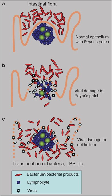
Alternative mechanisms of mucosal viral–bacterial interactions. (a) In the normal gut, intestinal flora are kept from invasion by the intact mucosa, even in the follicle-associated epithelium that specializes in transportation of antigen into the Peyer's patch. (b) If the submucosal lymphoid tissue is damaged by infection (e.g., with HIV11), the mucosa becomes permeable to bacteria and to bacterial products. Translocation of bacteria and bacterial products from the intestinal lumen cause systemic innate stimulation, leading to malaise and other systemic symptoms. (c) If a virus is tropic for the intestinal epithelial cells, it causes cell damage and loosens the normally impermeable barrier that keeps bacteria in the intestinal lumen.
PowerPoint slide
Full size image
Remarkably, the onset of diarrhea in SARS infected patients usually peaked on days 4–9, by which time the fever had subsided. However, the coronavirus copy number in some studies showed an increase between day 5 and day 10, so that maximal infectivity followed the fever,7 leading perhaps to a false sense of security amongst those caring for SARS patients. Although virus copy number in the pharyngeal aspirates dropped significantly between days 10 and 15, many patients still had SARS coronavirus present in the stool at day 21, by which time under 50% had virus present in the nasopharyngeal aspirate. Therefore, late transmission by contact with stool was a particular unappreciated risk.
Interactions Between Viruses and Bacteria
Infection with porcine respiratory coronavirus or porcine reproductive and respiratory syndrome virus has a synergistic effect with bacterial products, demonstrated by enhanced inflammation induced by LPS from Escherichia coli. LPS caused potentiation of disease, with enhanced production of tumor necrosis factor, interleukin-1, and interleukin-6.8
It has been known for many years that healthy people often carry pathogenic bacteria (such as Neisseria meningitides or Streptococcus pneumoniae) in the upper respiratory tract, but may only develop invasive disease during coinfection with respiratory viruses. The reason for these interactions are incompletely understood, but intriguing recent study show that influenza and respiratory syndrome virus are both capable of causing a persistent inhibition of the innate response to bacterial superinfection, and therefore to increased bacterial replication and disease.9
Intestinal Effects of HIV Infection
Although dual tropism for both the lung and gut may be an obvious and clear reason to consider the gut in respiratory infection, links can also be more subtle and complex. HIV targets CD4-expressing T cells and macrophages, thereby leading to immunosuppression. However, the symptoms of progressive HIV infection sometimes include profound weight loss (“slim disease”), and evidence of systemic immune activation. Indeed, this immune activation may assist fresh infection by HIV of activated T cells, so enhancing the viral life cycle.
The reasons for this immune activation seems to include the fact that HIV infects intestinal mucosal lymphocytes, including those in the Peyer's patch and especially Th17 cells that normally keep bacteria in check10 (Figure 1b). This leads to enhanced gastrointestinal permeability to microbial products, causing increased levels of circulating LPS,11 further activating the innate and adaptive immune system. An additional intriguing possibility is that the distribution of FoxP3+ regulatory T cells is affected by HIV infection,12 and that the balance between proinflammatory and antiviral effects is disturbed13 thereby contributing to immune activation, malaise, and cachexia in HIV-infected patients.
Intestinal Infection by Influenza
Highly pathogenic strains of influenza also cause intense systemic symptoms, sometimes associated with gastrointestinal disease. In waterfowl, influenza is mostly an enteric pathogen, transmitted via the feces in the lakes and waterways, and efficiently transmitted to other birds feeding and breeding on the same water. When these viruses spread to man, viral replication can trigger hypercytokinaemia.14
It therefore seems possible that the gut (known to be susceptible to infection with highly pathogenic strains of influenza)15 might also become permeable to intestinal LPS in severe influenza infections (Figure 1c), or that bacterial translocation through the gut wall could contribute to systemic symptoms, cytokine release and circulatory collapse.
Conclusions
In the era of integrative systems biology and holistic medicine, it is not only timely but also essential to view the gut and the lung as a continuous surface with distinct but overlapping susceptibilities. In addition, viral and bacterial infections should not be regarded as isolated events but viewed both in the context of intercurrent infection and of previous infection history. Studies of single infections are certainly revealing, but in reality the secret to understanding variations in responses to infections must include an awareness of what else is present on a rich polymicrobial landscape.

Brewer's yeast is a kind of yeast that is a by-product of brewing beer. Dietary supplements containing brewer's yeast often contain non-living, dried yeast. People use brewer's yeast to make medicine.

Brewer's yeast is taken by mouth for respiratory problems, including the common cold and other upper respiratory tract infections, influenza, seasonal allergies, and swine flu. Brewer's yeast is also taken by mouth for diarrhea, swelling of the colon (colitis) due to the bacteria Clostridium difficile, high cholesterol, loss of appetite, acne, premenstrual syndrome (PMS), recurring boils on the skin (furunculosis), type 2 diabetes and irritable bowel syndrome (IBS). It has also been used as a source of B vitamins, chromium, and protein.

How does it work?
Due to the chromium content of brewer's yeast, there is interest in using it for lowering blood glucose in people with diabetes. Chromium may help the body use insulin more effectively. This can lower blood sugar levels.

Additionally, brewer's yeast seems to increase enzymes in the intestine that could help relieve diarrhea.

Brewer's yeast might help fight bacteria that cause infections in the intestine and improve the body's defenses against viral lung infections such as flu and the common cold.
 

Avenger

Senior Member
Messages
323
Thanks for sharing your story, Avenger. I recently tested positive for an overgrowth of d-lactate (in particular, L. acidophilus) and have always had gut issues, so I've followed this thread with a great deal of interest. Is there a specific book or website that guided you on the proper diet to combat d-lactic acidosis? Does zero sugar mean zero fruit? Does low carb mean limiting veg? There is a dizzying amount of info out there on paleo, keto, PK diets, etc., and I have trouble reading for any length of time, so it would be great to just get a list of Do's and Don'ts from somewhere.

Dar

Hi Azector,
sorry, I somehow managed to miss your message, but it is complex and whatever diet you use you are vulnerable to a Carbohydrate load if you cannot find food or make mistakes which are easy.

I was also given repeated Sucrose in Hospital after shoulder surgery until I fell ill, because the Doctors did not understand the condition.

There are 4 main ways to control the condition;

1. Antibiotics given cyclically, but there are dangers from possible resistance.

2. Diets (or abstaining from food). I am trialing a FODMAP diet but had previously used a 0% Carbohydrate and simple sugars.

3. Probiotics, which is a minefield because of D-Lactic producing species in many Probiotic mixtures often described in Ads as particular species, but often containing Acidophilus etc. (the NHS may have realized that VSL-3 contains D-Lactic producing bacteria which could have been causing overgrowth and ME like symptoms in some patients).

4. Fecal Transplant, may reverse the condition.

2018 Oct;67(4):483-487. doi: 10.1097/MPG.0000000000002043.
Intestinal Microbial and Metabolic Alterations Following Successful Fecal Microbiota Transplant for D-Lactic Acidosis.
Bulik-Sullivan EC1, Roy S2, Elliott RJ3, Kassam Z3, Lichtman SN2, Carroll IM1,4, Gulati AS2,4,5.
Author information

Abstract
Fecal microbiota transplantation (FMT) involves the transfer of stool from a healthy individual into the intestinal tract of a diseased recipient. Although used primarily for recurrent Clostridium difficile infection, FMT is increasingly being attempted as an experimental therapy for other illnesses, including metabolic disorders. D-lactic acidosis (D-LA) is a metabolic disorder that may occur in individuals with short bowel syndrome when lactate-producing bacteria in the colon overproduce D-lactate. This results in elevated systemic levels of D-lactate, metabolic acidosis, and encephalopathy. In this study, we report the successful use of FMT for the treatment of recurrent D-LA in a child who was unresponsive to conventional therapies. Importantly, we also present profiles of the enteric microbiota, as well as fecal D-/L-lactic acid metabolites, before and longitudinally after FMT. These data provide valuable insight into the putative mechanisms of D-LA pathogenesis and its treatment.
PMID: 29901551 DOI: 10.1097/MPG.0000000000002043


If you message me, I can give you my telephone number and can give you more information. There has been great resistance from the NHS in accepting any of this.
 

Avenger

Senior Member
Messages
323
For SamB (and thank you for your help and support).


I believe that the death of Merryn Crofts in May 2018 and a number of other deaths, may have been caused by D-Lactic acidosis, which is known to cause fatality; Seizure, Coma and Death.


I have only just seen this story. Merryn's symptoms of cognitive, breathing difficulty, memory, systemic pain, muscle weakness and sickness are all characteristic of D-Lactic acidosis. Merryn wasted after Gastrointestinal failure. There are many pictures of her holding a bowl. High levels of D-Lactic producing Bacteria would have caused frequent sickness and reflux and far worse Neurological and Respiratory symptoms. Her symptoms were classic for D-Lactic acidosis causing both Gastrointestinal and Neurological illness.

The symptoms of D-Lactic acidosis causes Myalgic Encephalomyelitis through toxic poisoning, both acidosis leading to respiratory problems; and Neurological symptoms due to the neurotoxin entering spinal fluid that hit every cell and organ. At low levels it makes life extremely unpleasant causing fatigue and muscle pain, but at higher levels it causes far more serious illness.

The secondary effects of having Bacterial Overgrowth are Autoimmune symptoms due to damage to the Gastrointestinal lining and it is believed that Merryn may have developed Mast Cell Activation Disorder, which is believed to be related to autoimmune dysfunction.

D-Lactic acidosis is both a Gastrointestinal and Neurological condition that can be treated! Breathing difficulty when most unwell can be described as air hunger, it is like suffocating, your Oxygen Levels remain normal, but levels of C02 rise during acidosis so that you are unable to utilize available Oxygen or Glucose and you begin to hyperventilate as a natural response to acidosis (I have experienced this). When well I also experienced delays in symptoms after activity that could leave me feeling as though I had climbed a mountain without oxygen.

I still have Oxygen at home to deal with bouts when it becomes uncontrolled. This illness can cause Seizure, Coma and Death during high levels of D-Lactic production and I believe that this form of Bacterial Overgrowth may be the cause of long term symptoms in the worst affected with ME (I also believe that there are other forms of Bacterial Overgrowth causing similar symptoms and fatigue etc.)

Merryn donated her Brain and Brain Stem and signs of high levels of D-Lactic acid may possibly still be in her spinal fluid and brain, which may possibly be available for investigations. D-Lactic acidosis is virtually invisible to differential diagnosis and normal investigations, and tests are rarely performed due to the belief that D-La is rare and only due to short bowel syndrome. But it is my belief that D-Lactic acidosis can be caused simply by motility issues and D-Lactic acidosis may be self sustaining in causing these motility issues through the production of Methane and Hydrogen.

I would bet that no Doctor ever checked Merryn's Blood Gasses when she had breathing difficulty, or for D-Lactic acid. Someone should contact the Coroner and her relatives. If proven, this would have a profound effect on the way that patients with severe ME are treated. Merryn was courageous in leaving her body to study the illness.

Without a diagnosis, Merryn may have continued unable to metabolize Carbohydrates and Sugars which were eventually fed directly into her stomach and would have continued to produce Neurotoxins, continuing to poison her until she died. due to this dangerous overgrowth that like C.Diff causes serious illness.

We need to stop further deaths occurring.

D-Lactic acidosis is known to cause seizure, coma and death. I honestly believe that there are different forms of Bacterial Overgrowth affecting varying severity of ME/CFS in at least a subset and that we may each experience different levels of symptoms. I also believe that D-Lactic acidosis may be what is driving the worst symptoms in many of the worst affected patients. After experiencing these symptoms, I believe it to be behind a number of deaths. The only reason that I have survived is that I was found to respond to antibiotics, but prior to this I was close to suicide because of the severity of the symptoms and had frequent recorded sickness and vomiting in A&E with abdominal pain and breathing difficulty.


The Times: ME sufferer who was dismissed as hysterical vindicated in death | 29 May 2018

Lucy Bannerman, The Times, 26 May, 2018.

ME left Merryn Crofts bed-bound and she died shortly after her 21st birthday.
It took a death certificate for Merryn Crofts to be vindicated.
For the last six years of her life she had fought to convince people that ME, the illness that had left her bed-bound, was real.
She was 15 when she started feeling weak and breathless and her feet began to swell.
Within six months she was using a wheelchair. Within a year she was housebound. She died in May last year, ten days after her 21st birthday.
Last week, Ms Crofts became only the second person in the UK to have her death recorded as a result of ME after a coroner concluded that it was the much misunderstood illness that was likely to have led to the gastrointestinal failure and other complications that caused her to starve to death.
“It meant the world to me,” her mother, Clare Norton, 49, told The Times. “I know it sounds strange but that kind of official recognition is vindication for Merryn that this was physical. I can’t thank that coroner and all those doctors who gave evidence enough.”
Myalgic encephalomyelitis (ME) affects about a quarter of a million Britons.
The main symptom is extreme fatigue, or “bone-crushing exhaustion” as Mrs Norton puts it.
Other symptoms include muscle pain, hypersensitivity to noise and light, and problems with sleep, memory and concentration.
Before Ms Crofts became ill, Mrs Norton said that her daughter “never stopped. She didn’t have lie-ins. She had things to do and friends to see. She was very, very healthy.”
Tests would later show that at some point she had suffered glandular fever.
She loved musical theatre and wanted to go to drama school, but would come home from school and crash out.
“Her limbs became like lead, as if she was paralysed. Her breathlessness was more like a constant air hunger.”
Mrs Norton remembers going with her daughter to buy new shoes — her swollen feet meant she went up two sizes.
“She was walking like an old lady. Everything became slower and slower. We managed to carry her back to the car and she fell asleep. It was like watching a toy run out of batteries.

Ms Crofts was full of life before developing the disease, her mother said.
“But she didn’t want to give in. She’d say, I can push through it, I can push through it, it’s just mind over matter.” They now believe that the exertion was making her worse.
In the early days of the illness, Ms Crofts’ complaints were dismissed by various doctors as panic attacks and, on one occasion, “hysteria”, according to her mother.
“This seems to happen particularly with teenage girls,” she said. “I know men in their 30s with ME and they don’t seem to get the same response.
“We had doctors saying we just don’t believe in ME. When everyone is saying that to you it’s like a nightmare you can’t get out of.”
Ms Crofts was concerned that her family did not believe her either. “Sometimes she’d say, you don’t think that, do you? She was concerned we thought the same. I said, no darling, we’re going to get to the bottom of this. We’re going to find out what’s wrong. ”
Within two years, she was struggling to swallow. Eventually, she had to be fed through a tube. She was five and a half stone when she died. At the inquest, experts ruled out eating disorders, depression and anxiety.
She had loved fashion — “shopping was her sport” — but once bed-bound couldn’t even bear the touch of certain fabrics on her skin. She found relief in online shopping, buying clothes she couldn’t wear to give to her friends.
“She enjoyed watching me unwrap them for her and choosing who she would give it to,” said Mrs Norton, who gave up her job as a counsellor to care for her daughter full-time.
Towards the end, Ms Crofts couldn’t bear light or even her mother’s touch. “She was such a huggy girl. A Merryn hug was lovely but she couldn’t be hugged. Sometimes, I could stroke her cheek. I developed a way of lying around her, so we could be close.
“She wanted to hug, but she couldn’t. She wanted to eat but she couldn’t. It just stole everything away from her.”
Mrs Norton marked the first anniversary of her daughter’s death this week by becoming a grandmother: her eldest daughter, Amy, gave birth to a son, Flynn. She hopes that Merryn’s case “will bring a sea change in the way people think about ME”.
In 2006 Sophia Mirza, 32, from Brighton, became the first person in the UK to have her death attributed to ME.
Ms Crofts’ brain and spinal cord have been donated for medical research at Addenbrooke’s Hospital, Cambridge. “People with ME have been so badly treated. She wanted to donate her organs so that people didn’t need to suffer like she did,” Mrs Norton said.
A misunderstood illnessMyalgic encephalomyelitis, which affects about a quarter of a million Britons, is recognised by the World Health Organisation as a neurological illness but there is disagreement between health professionals over whether it needs psychological interventions or is mainly physical.
The health watchdog NICE is updating its guidelines for treatment in England amid concern that one of the commonly recommended therapies, involving more exercise, may do more harm than good.
According to the ME Association, one in four sufferers is affected so badly that they are housebound, and in some cases unable to shower or feed themselves.
An ME Association spokesman said: “The difference between ME and just feeling tired is the same as the difference between having a shower and drowning.”
 
Last edited:

junkcrap50

Senior Member
Messages
1,330
I have just been turned down for a Fecal Transplant. They have told me that they are not licensed for D-Lactic acidosis.
I haven't read the thread or recent posts. But just do a DIY Fecal Transplant. I'm sure this has been suggested. But D-lactic acidosis is pretty serious and worth the risk of doing a DIY transplant. I've done it before, giving myself 14 fecal enema's, 1/day, in a row. It's not hard, cheap, not terrible gross, and "relatively" safe. There's a website who's instructions I used and promotes safe DIY FMTs: https://thepowerofpoop.com/ The difficult part is finding a donor willing to do it. But family members are good candidates. The blood tests needed to screen donors is simple and most doctors will order them.

In my FMTs, I noticed a small immediate benefit. But I screwed up the transplants by taking too much pre-biotics in an attempt to help the transplant. It likely just fed the pre-established microbes. I did not take any antibiotics prior to my transplant to wipe my gut.

I would attempt another DIY FMT immediatly if I had a healthy donor. My previous donor, a relative, has since developed gut problems and abnormal stools. So I can't use him/her. I've been waiting for some time t
 

Jwarrior77

Senior Member
Messages
119
@Avenger Do you know what blood gases would be elevated or decreased in this condition?

I am very intolerant to simple sugar and carbs. If eat them my heart will start pounding and racing and I will enter into the "void" where I'm too tired and immobilized but don't fall asleep.

Also in your opinion would a high Anion Gap on blood tests indicate a condition such as this? Thanks.
 
Last edited:

Avenger

Senior Member
Messages
323
Hi, Jwarrior77 and Junkcrap50,
D-Lactic acidosis is a minefield and I am still coming to terms with the complex issues, but Blood Gasses will give the Anion Gap (for either L- or D-Latate) and D-Lactic assay will give the level of the Organic acid D-Lactate that leads to Neurological and Flu like Symptoms. Anion Gap without elevated L-Lactate (the isomer of D-Lactate) will show the probability of D-Lactate as the cause.


We are supposed to produce L-Lactate which is converted by Enzymes involved in l-lactate metabolism in humans. ... place via its oxidation into pyruvate, which may be oxidized or converted into glucose.

When this does not happen due to producing high levels of D-Lactate, we not only cannot produce energy, but the secondary effects of D-Lactate and acidosis (involving Co2 production) affect at the cellular level to cause Mitochondrial Dysfunction which i believe is the cause of Hypoglycemia symptoms which affect the inability to transfer and utilize Glucose, but also inability to utilize Oxygen that in combination affects the brain. Acidosis and increased Co2 levels cause normal levels of PCo2 not to be utilized. You can literally suffocate with normal levels of Oxygen as Co2 levels rise. Only Oxygen levels will be demonstrated in conventional Oximeter tests performed by Doctors. I expect this has been the cause of death in a number of ME cases where they were producing significantly high quantities of D-Lactic acid and I believe would have been implicated in Merryn Crofts death (Doctors are blind to these symptoms unless they had performed a Blood Gas assay and followed it up in the absence of high levels L-Lactate with a D-Lactic assay). The death of Merryn would have been put down to ME but without any clear clinical understanding of the mechanisms that had contribute to her death, mainly because Doctors believe that D-Lactic acidosis is rare and that it only concerns short bowel syndrome (if she had a short bowel a D-Lactic assay would have automatically been performed).

Motility issues alone can cause D-Lactic acidosis and motility is also affected by the presence of D-Lactic acid and Methane production by the Bacteria in the Small Bowel that directly affects bowel contractions, setting up a self perpetuating condition that could potentially become life threatening. I had suffered frequent bouts of breathing difficulty, expecting to die from the illness during severe episodes, but my breathing problems were put down to hyperventilation due to anxiety by Doctors in A&E as were Merryn's (hyperventilation is a natural way that we deal with Acidosis! Which is why Simon Weesley has misunderstood Hyperventilation in ME; See https://en.wikipedia.org/wiki/Simon_Wessely). But this only occurs during severe acidosis and many patients with D-Lactic acidosis may be 'sub-clinical' for D-Lactate or only develop more severe symptoms less frequently. I believe that brain fog is a sub-clinical manifestation of D-Lactic acidosis, or in the presence of other possible Organic acids causing variants of ME/CFS.

I can say this; Anxiety would be normal in anyone suffocating due to acidosis; That these Doctors poor thinking is killing us! D-Lactic acidosis is a form of Bacterial Overgrowth, like C.Diff and can be just as deadly or cause a lifetime of tortuous symptoms where at times you would wish to find relief in death. I was lucky to have survived 18 years and have been left with nothing but contempt for these Psychiatrists.


This is why a Fecal Transplant may correct a vastly complex problem that may be reset by interrupting the self sustaining cycle that may have occurred due to a number of contributing factors (including Diabetes and high levels of starches in modern diets along with antibiotics and chemicals such as fluoride that can alter natural microbial balance that is part individual and part developed symbiosis with bacteria that has developed over millions of years). A Fecal Transplant involves not just trillions of bacteria, but possibly also immune signalling and complex networks that we have damaged, if a young, healthy donor can be found (see Sarah Myhill).

L-lactate and D-lactate - clinical significance of the difference
In nature lactate exists in two isoforms: L-lactate and D-lactate.
In all vertebrates, including humans, the L-lactate form is by far the most abundant and pathophysiologically significant, and it is this form that is specifically measured by the lactate sensors in blood gas analyzers and indeed all routine methods used to measure lactate in the clinical laboratory.
The main focus of this brief review is physiological and pathological aspects that distinguish L-lactate and D-lactate. Consideration will be given to the very rare instance when measurement of blood D-lactate is clinically useful and just why the lactate sensor in blood gas analyzers is not useful in such rare circumstance.
Introduction
Lactate, the anion that results from dissociation of lactic acid, is an intracellular metabolite of glucose; specifically it is the end product of anaerobic glycolysis, the final step of which is conversion of pyruvate to lactate by the enzyme lactate dehydrogenase.
In health around 1500 mmol of lactate is produced daily and so long as normal rate of metabolic disposal - principally by the liver and kidneys - is maintained, blood plasma concentration remains within the approximate reference range of 0.5-1.5 mmol/L [1]. Abnormal increase in plasma lactate (called hyperlactatemia) occurs if the rate of production exceeds the rate of disposal.
If hyperlactatemia is sufficiently severe (plasma lactate >5.0 mmol/L), it is associated with acidosis (blood pH <7.35). The condition is then called lactic acidosis. There are many causes of hyperlactatemia and the resulting lactic acidosis but the most common is increased production of lactate from anaerobic glycolysis due to reduced oxygen delivery to tissue cells (tissue hypoxia).
Tissue hypoxia, which is the result of inadequate perfusion of tissues and/or reduced blood oxygen (hypoxemia), is a common feature of many kinds of critical illness so that lactate measurement is most frequently used to monitor tissue oxygenation in the critically ill.
Plasma lactate within the reference range is considered reliable evidence that tissues are adequately oxygenated. Detail of normal lactate production and disposal, along with the pathophysiology of hyperlactatemia and lactic acidosis (the most common cause of metabolic acidosis) is contained in a recent review article [2].
Lactate exists in two isomeric forms
In common with many biologically active molecules (e.g. amino acids, glucose) lactate exists in nature in two stereoisomeric forms due to the presence of an asymmetric carbon atom. Molecules like lactate that exhibit stereoisomerism are optically active, meaning that the two isomers rotate plane-polarized light in opposite directions.
The two lactate isomers are known as L-lactate and D-lactate; L-lactate rotates light clockwise (+) and D-lactate rotates light counterclockwise (-). This is reflected in the sometimes used nomenclature: D(-) lactate and L(+) lactate.
Both forms (stereoisomers) of lactate are produced from and metabolized to pyruvate by the action of the enzyme lactate dehydrogenase (LDH). However, the enzyme is isomer-specific so that production and metabolism of D-lactate requires D-LDH and L-lactate requires L-LDH.
Mammalian cells only contain L-LDH so that in humans the lactate produced is almost exclusively L-lactate. Carbohydrate-fermenting bacterial species (e.g. lactobacillus spp) have by contrast both enzymes and therefore the capacity to produce both D-lactate and L-lactate. Some species produce only D-lactate, some only L-lactate and others both forms.
It was once supposed that the relatively very small amount of D-lactate normally present in the blood of humans (concentration 5-20 µmol/L in healthy adults [3] compared to 1000 µmol/L, i.e. 1.0 mmol/L for L-lactate) is solely derived from exogenous sources (diet and the carbohydrate-fermenting bacteria normally present in the gastrointestinal tract).
However, it is now clear that despite the absence of D-LDH, D-lactate is both produced and metabolized within human cells, albeit in tiny amounts compared to that of L-lactate. Metabolic production of D-lactate in human cells is the result of the methylgloxal pathway, a minor off-shoot pathway of glycolysis that results in nanomolar production of methylgloxal, a toxic product that is converted to D-lactate [4,5].
In the absence of D-LDH, human cells can metabolize D-lactate to pyruvate by the action of the mitochondrial enzyme D-2-hydroxyacid-dehydroganse [4,5].
In summary, the small amount of D-lactate normally present in blood is derived from three sources:
  • cellular production by the methylgloxal pathway
  • diet (foods containing D-lactate, e.g. yoghurts, soured cream, cheese)
  • lactate-producing bacterial species normally resident in the large intestine (colon).
Overgrowth of bacteria in the colon is a feature of short-bowel syndrome, the only pathology associated with increase in plasma D-lactate concentration of sufficient severity to cause acidosis (D-lactic acidosis).
Short-bowel syndrome and d-lactac acidosis
Prior to 1979 it was presumed that all cases of hyperlactatemia and lactic acidosis were the result of increase in the predominant L-lactate isoform. In that year the first identified case of lactic acidosis resulting solely from increase in D-lactate rather than L-lactate was reported in a patient who 3 years previously had required life-saving surgery to remove a diseased section of his small intestine [6]. This case provided the first evidence of the now established link between short-bowel syndrome and D-lactic acidosis.
Short-bowel syndrome is a not inevitable complication following surgical resection of more than half the length of the small intestine. Such radical surgery is employed to treat intestinal disease (e.g. Crohn's disease, intestinal cancer, intestinal ischemia), traumatic injury to the intestine, and congenital intestinal defects (e.g. midgut volvulus) evident at birth.
Short-bowel syndrome may also occur in patients who have received surgery (jejunoileal bypass) for severe obesity. This particular kind of surgical treatment is no longer recommended for the morbidly obese, partly because of the risk of short-bowel syndrome.
D-lactic acidosis, defined as plasma D-lactate >3.0 mmol/L in association with metabolic acidosis (blood pH <7.35) [5], is just one of many metabolic derangements that can occur in patients with short-bowel syndrome.
The central defect that explains the development of D-lactic acidosis in those with short-bowel syndrome is carbohydrate malabsorption. Surgical resection reduces the capacity for digestion and absorption of dietary carbohydrate that normally takes place within the small intestine. An increased load of carbohydrate is consequently delivered to the colon where carbohydrate-fermenting bacteria reside.
Within the colon, bacteria convert the increased carbohydrate load to lactate (both D and L isomers), which is then absorbed. The body has ample metabolic capacity, in the form of L-LDH, to deal with the abnormally high influx of L-lactate that results from this increased bacterial production of lactate, but only limited capacity to deal with the increased D-lactate load. Once that capacity is overwhelmed, D-lactate accumulates in blood and eventually acidosis develops.
A massive (hundred-fold) increase in plasma D-lactic concentration to >2.5-3.0 mmol/L is required for development of D-lactic acidosis and symptomatic effect. Symptoms of D-lactic acidosis reflect the neurotoxicity of D-lactate and are evident as recurrent episodes of encephalopathy [4].
During these episodes all patients have reduced mental state, which may range from mild drowsiness or lethargy to coma. Other symptoms vary between patients but may include slurred speech, confusion, inability to concentrate, unsteady gait and headache.
Blood gas analysis reveals a metabolic acidosis with increased anion gap, but crucially plasma lactate is normal. The paradox of normal lactate concentration in patients with D-lactic acidosis is explained by consideration of the routine methods used to measure lactate.
Measurement of plasma lactate
Routine methods used to measure plasma lactate are based on measuring the product of enzymic action on lactate. One of two enzymes is commonly used for these analyses: lactate dehydrogenase, derived from animal tissue, and lactate oxidase derived from bacteria.
The lactate sensors incorporated in blood gas analyzers employ lactate oxidase, which is immobilized on a membrane. On contact with this immobilized enzyme, lactate in the sample is oxidized to pyruvate and hydrogen peroxide. The hydrogen peroxide generated is measured amperometrically by an electrode assembly contained within the sensor.
Crucially for this discussion the enzymes employed in these analyses are L-lactate-specific, they are L-lactate dehydrogenase and L-lactate oxidase. This means of course that only L-lactate is measured. Despite massive increase in plasma D-lactate concentration that may be well in excess of 10.0 mmol/L in some cases of D-lactic acidosis, measured plasma lactate remains within the normal range.
Methods for specific measurement of plasma D-lactate have been developed [7.8]. These assays, which necessarily have to be much more sensitive than plasma L-lactate assays if they are to distinguish normal from abnormal, are mostly based on D-lactate dehydrogenase and are not widely available.
Although some clinical laboratories have the means to measure plasma D-lactate, obviously a necessary step for the diagnosis D-lactic acidosis, there is currently no point-of-care means of measuring D-lactate.
The current relative lack of availability of D-lactate assays might change now that there is growing research interest in the clinical significance of subclinical elevation of plasma D-lactate concentration [8], i.e. plasma D-lactate concentration in the approximate range (50 µmol/L - 2.0 mmol/L).
 

Avenger

Senior Member
Messages
323
This is one for Sam and Junkrap50!

Increased energy using 'Capsazepine' found in Red Chilli, by influencing TrpV1 receptors and increasing ATP Hydrolysis; in the report below!

The reason that I would prefer a Fecal Transplant is that Bacterial Symbiosis and complex Bacterial and Immune signalling involved in a balanced Gut is quite literally an unexplored continent that may take many decades to unravel and a Fecal Transplant is the best shortcut in resetting the Gut.

I too have had significant remissions and relapses due to pre- and pro-biotics, and this has turned out to be extremely hit and miss and there is a possibility that probiotics may also affect outcomes positively or negatively and for lots of different reasons that we will never fully understand.

But would'nt it be great if we could influence the production of L-Lactate and our energy by introducing something that would switch from D-Lactic production to increased L-Lactic production and increased D-
Glucose and energy as in 'Capsazepine' found in Red Chilli, by influencing TrpV1 receptors and ATP Hydrolysis in Lactobacillus acidophilus as in the report below.

Journal of Dairy Science
Volume 96, Issue 7, July 2013, Pages 4142-4148

Capsicum annuum enhances l-lactate production by Lactobacillus acidophilus: Implication in curd formation


https://www.sciencedirect.com/science/article/pii/S0022030213003202

We observed enhancement of l-lactate production by L. acidophilus in the presence of capsaicin, which was similar to that observed from filtered aqueous extract of red chili (Figure 3). This enhancement effect was inhibited in the presence of capsazepine, where the amount of l-lactate produced was lower than that of the control. The effect of capsazepine alone on l-lactate production was closer to that of the control, showing slightly enhanced effect but far less than either filtered aqueous extract of red chili or capsaicin. The data for l-lactate production and d-glucose consumption were analyzed for statistically significant differences using the Tukey honestly significant difference test. A significant increase was observed in the amount of l-lactate produced in both cases in the presence of filtered aqueous extract of red chili and capsaicin compared with the control (adjusted P < 0.01; Appendix Table A2). Also, in the presence of capsazepine alone, a significant amount of l-lactate was produced (adjusted P < 0.01), indicating that capsazepine alone does not inhibit l-lactate production by L. acidophilus. In parallel, consumption of d-glucose was significantly enhanced in both cases in the presence of filtered aqueous extract of red chili and capsaicin (Figure 3). l-Lactate production was severely inhibited in samples containing capsazepine and either capsaicin or filtered aqueous extract of red chili (adjusted P < 0.01; Appendix Table A2). However, d-glucose was still consumed, similar to consumption in the control, indicating the ongoing metabolic activity. It has been reported that capsaicin activates ion channels of plasma membrane and capsazepine is a competitive antagonist (Bevan et al., 1992). These observations suggest that the enhancement of l-lactate production due to capsaicin is likely at the level of l-lactate export from the bacteria.

Therefore, it is evident that, in the presence of filtered aqueous extract of red chili or its active ingredient capsaicin, the enhancement in l-lactate production is due to the enhanced metabolic activity of L. acidophilus. We were intrigued by the effect of capsaicin on enhancement of l-lactate production with accompanied increase in d-glucose consumption by L. acidophilus, considering that capsaicin is insoluble in water. However, the effect we observed was reproducible and this may perhaps be due to the interaction of surface receptors of L. acidophilus with capsaicin. In the presence of red chili, capsaicin may be present as an emulsion allowing favorable interaction with bacterial cell membranes.

Capsaicin has also been shown to stimulate uncoupled ATP hydrolysis, which is likely of significance to modulate thermogenesis (Mahmmoud, 2008). Many studies in the medical literature have linked the consumption of chili-enriched meals with increased energy expenditure and oxidation of fats (Diepvens et al., 2007). In humans, capsaicin and its nonpungent analogs have been shown to act through surface transient receptor potential vanilloid type 1 (TrpV1) receptors, which consequently affect the basal metabolic rate by increasing energy metabolism (Inoue et al., 2007). Our efforts in identifying homologs of TRPV1 receptors in L. acidophilus through BLAST (Altschul et al., 1990) searches revealed ABC transporters with weak similarity. In the future, the detailed mechanistic action of capsaicin on L. acidophilus receptors remains to be unraveled.


Avenger.
 

lenora

Senior Member
Messages
4,913
Dear All,
I have been ill with ME/CFS symptoms and even diagnosed with Fibromyalgia for 18 years but was recently diagnosed as D-Lactic acidosis.

I have recently been formally diagnosed with D-Lactic acidosis (without short bowel syndrome) in February 2017. The same symptoms were misunderstood or ignored by Doctors for so long because all other tests had been virtually normal.

I have been trying to make others aware of the similarity of ME/CFS to D-Lactic acidosis that Sheedy et al have written about and reported on after finding D-Lactic producing bacteria in CFS patients (report below).

D-La can be a very serious condition and I had neurological symptoms very similar to Jennifer Bria which would fluctuate from mild to extreme and had varying symptoms ranging from muscle pain and weakness, slurred speech, difficulty thinking to confusion, fatigue and breathing difficulty as though I had climbed a mountain without oxygen, hypoglycemia or dizziness along with abdominal symptoms, difficulty after eating carbohydrates and periods of sometimes severe abdominal pain and sickness.

I was diagnosed after noticing that my symptoms would temporarily stop after using antibiotics (metronidazole). I made an appointment with a Consultant Gastrointerologist who specializes in D-Lactic acidosis and was virtually diagnosed on the spot because I respond to antibiotics. In D-La Carbs and sugars cannot be properly metabolised and cause fluctuating levels of D-Lactic acid which can be found in spinal fluid causing neurological symptoms which act much like a poison.

Anyone with these symptoms or who respond to antibiotics can test themselves for D-La through total 0% abstention diet for all Carbohydrates and simple sugars. Results can take 64 plus hours and remaining on the diet for 4 to 6 weeks could be life changing as it was for me! Dr. Sarah Myhill has been advising the same diet for ME/CFS patients for years. The diet is difficult and symptoms will return if it is failed, even small amounts of Carbs and Sugars can cause the return of symptoms. The diet should only be considered after consulting a Doctor to make sure that it is appropriate.

Increased D-Lactic Acid Intestinal Bacteria in Patients with Chronic Fatigue Syndrome
  1. JOHN R. SHEEDY1,
  2. RICHARD E.H. WETTENHALL1,
  3. DENIS SCANLON2,
  4. PAUL R. GOOLEY1,
  5. DONALD P. LEWIS3,
  6. NEIL MCGREGOR4,
  7. DAVID I. STAPLETON1,
  8. HENRY L. BUTT5 and
  9. KENNY L. DE MEIRLEIR6
+Author Affiliations

  1. Kenny.De.Meirleir@vub.ac.be

Next Section
Abstract
Patients with chronic fatigue syndrome (CFS) are affected by symptoms of cognitive dysfunction and neurological impairment, the cause of which has yet to be elucidated. However, these symptoms are strikingly similar to those of patients presented with D-lactic acidosis. A significant increase of Gram positive facultative anaerobic faecal microorganisms in 108 CFS patients as compared to 177 control subjects (p<0.01) is presented in this report. The viable count of D-lactic acid producing Enterococcus and Streptococcus spp. in the faecal samples from the CFS group (3.5×10⁷ cfu/L and 9.8×10⁷ cfu/L respectively) were significantly higher than those for the control group (5.0×10⁶ cfu/L and 8.9×10⁴cfu/L respectively). Analysis of exometabolic profiles of Enterococcus faecalis and Streptococcus sanguinis, representatives of Enterococcus and Streptococcus spp. respectively, by NMR and HPLC showed that these organisms produced significantly more lactic acid (p<0.01) from 13C-labeled glucose, than the Gram negative Escherichia coli. Further, both E. faecalis and S. sanguinis secrete more D-lactic acid than E. coli. This study suggests a probable link between intestinal colonization of Gram positive facultative anaerobic D-lactic acid bacteria and symptom expressions in a subgroup of patients with CFS. Given the fact that this might explain not only neurocognitive dysfunction in CFS patients but also mitochondrial dysfunction, these findings may have important clinical implications.


Hi Avenger.....I think it is rather easy for us to accept a diagnosis, even if it is a wrong one. In my case, if I had been told I had CFS/FM (now ME) I would have given up looking for other reasons. As it was, I happened to be fortunate and managed to get the proper diagnosis in record time....probably 4-6 mos., compared to the almost 10 years that I used to hear over and over. True, this goes back to the l970's when medicine was different, but it was already on its way to a massive change thanks to the AIDS epidemic. We were simply put in the back row where no one would ever have to deal with us.

Perhaps the easiest way would be to write down ever symptom we have, and each night look up a different neurological illness that may contain the same symptoms. Beyond that, I don't have any answers. If I could, I'd wish good health for all of you. We never used to get a diagnosis of ME (were laughed at...it's true) and now you can hardly get away from one. What has happened? And is it in our best interest? I know that in my case, I would have had the necessary surgeries, but I also would have presented with a case of ME, because I have ME. We must be aware of own bodies and think about them more than once/year. We're worth it. Thank-you. Yours, Lenora
 
Last edited by a moderator:

Jwarrior77

Senior Member
Messages
119
@Avenger Thanks so much for the information. I understand some but a lot of this info is way over my head. This is a lot of new info for me to absorb. Perhaps you could break it down in some more simple terms if I miss anything important. This is mainly for my brain to digest (brain fog hahaha) Thank you.

but Blood Gasses will give the Anion Gap (for either L- or D-Latate) and D-Lactic assay will give the level of the Organic acid D-Lactate that leads to Neurological and Flu like Symptoms. Anion Gap without elevated L-Lactate (the isomer of D-Lactate) will show the probability of D-Lactate as the cause.

The above is a pretty good summary and I believe I understand it. Below is my interpretation with a couple questions. Let me know if there is anything I missed or don't understand.

If you would want to diagnose this properly, what exact blood tests would you need to get? These are the test I think I understand:

- Blood Gas Test (from arterial blood) - will show blood PH
- Metabolic Panel (Anion Gap)
- Lactic acid test
- Lactic acid dehydrogenase test
- D - Lactate Urine test?

Also if I understood correctly if there is a High Anion Gap which is indicative of metabolic acidosis but L- Lactate is normal in serum, then D-Lactate acidosis is the most likely culprit?

Once found out, how would one go correcting this? Taking antibiotics? But what kind?

How would one get their Blood PH to return to normal? IV fluids? Sodium Bicarbonate? Or is Sodium Bicarb contraindicated?

Fecal Transplant?

Also what type of diet? Would a keto diet fix this as it's low carb/sugar and high fat + protein? Or just a diet low in carbs and sugar would be enough?

Sorry lots of questions. Thanks for the info.
 
Last edited:

Avenger

Senior Member
Messages
323
Hi Avenger.....I think it is rather easy for us to accept a diagnosis, even if it is a wrong one. In my case, if I had been told I had CFS/FM (now ME) I would have given up looking for other reasons. As it was, I happened to be fortunate and managed to get the proper diagnosis in record time....probably 4-6 mos., compared to the almost 10 years that I used to hear over and over. True, this goes back to the l970's when medicine was different, but it was already on its way to a massive change thanks to the AIDS epidemic. We were simply put in the back row where no one would ever have to deal with us.

Perhaps the easiest way would be to write down ever symptom we have, and each night look up a different neurological illness that may contain the same symptoms. Beyond that, I don't have any answers. If I could, I'd wish good health for all of you. We never used to get a diagnosis of ME (were laughed at...it's true) and now you can hardly get away from one. What has happened? And is it in our best interest? I know that in my case, I would have had the necessary surgeries, but I also would have presented with a case of ME, because I have ME. We must be aware of own bodies and think about them more than once/year. We're worth it. Thank-you. Yours, Lenora

Hi Lenora,
for me, ME is just a basket containing a number of undiagnosed illnesses and to some extent it is just a label to keep us quiet and stop asking further questions.

The NHS and Government through their chosen Chief Psychiatrist, Simon Wessely, view all with ME as having psychological problems. They will hand out ME and Somatization (psychosomatic) hand in hand. It is not a good diagnosis to have because it is discriminatory.

One of the main parameters for Somatization linked directly to ME is claims of Gastrointestinal problems! You can be diagnosed with Somatization if you claim both Gastrointestinal and Neurological symptoms and then you are swept under the carpet with the neat diagnosis of ME or Psychosomatic illness.

D-Lactic acidosis and other forms of Bacterial Overgrowth cause Gastrointestinal and Neurological symptoms!

Statistical data, which Psychologists greatly rely on shows that many ME suffer these Gastrointestinal problems, which are not investigated as a possible source of ME/CFS.

After 18 years I had to ask for these investigations myself (out of county) after having received a Somatization diagnosis due to the severity of illness including abdominal pain and Gastrointestinal symptoms. I was diagnosed on the spot as Bacterial Overgrowth being the cause of the severity of my problems and two years later diagnosed with D-Lactic acidosis, by a D-Lactic Consultant.

Somatization in ME, may one day go down in the history books as yet another misinterpretation of statistical data in Psychology. The pace trials are another example, where statistical data was deliberately manipulated and falsified to keep those with ME from getting benefits.

If you have ME, it acts as a form of discrimination, many victimized by the establishment and this has been pursued by out Government who have knighted. ''Wessely suspects ME/CFS to be a "general disorder of perception. ... Wessely was knighted in 2012 and became the World's first Regius Professor in Psychiatry ... ''

It is not a god thing to get an ME diagnosis from the NHS establishment at all, because however Wessely puts it, we are just perceiving our pain, neurological symptoms and Gastrointestinal illness (it is all in our heads), but without ever having the Gastrointestinal illness investigated (which could be done so easily!)

Fecal Samples will evidence Bacterial Overgrowth and can identify the specific pathogens involved, but because of the Psychological discrimination you will be most unlikely to have any of these investigations and all of your other blood investigations may be normal while you are being poisoned by these neurotoxins!

Normal Blood investigations do not give any signature for these problems and will embed the fact that it is you who are the problem, so you must have ME. If you hyperventilate, because you have breathing difficulty it will be put down to anxiety and Simon Wessely has stated that he could find no causative link between CFS and Hyperventilation. Hyperventilation is a natural reaction to acidosis and the build up of Co2 in D-Lactic acidosis. Many ME patients my be subclincal for D-Lactic acidosis or many other forms of Bacterial Overgrowth, giving rise to brain fog and hyperventilation.

D-Lactic acidosis is a form of infection as are all forms of Bacterial Overgrowth associated with ME/CFS; but one that remains hidden and does not cause any raise in temperature, because only the Organic acids and other metabolites causing Flu like systemic or neurological illness exit the gut into the Bloodstream and only rarely used investigations will demonstrate this.

Wessely is criminally wrong, and this can be proven very easily! But I suspect that the NHS 'establishment' will try to suppress the truth as they have over many other cover ups, whether this leads to continued ill health or not.

For myself, if I had accepted that diagnosis without questioning, I would have died in the same way that Merryn Crofts had. I had all of the same symptoms including breathing difficulty, neurological symptoms and Gastrointestinal illness and every day was torture.

For me this is about finding what is driving this illness, finding a cure and not accepting my label. ME has been abandoned by mainstream medicine including NHS, but as many we have power and by discussing this online we can magnify that power and by pooling our understanding and together we may come up with an answer.

I am hoping that a subset of ME/CFS who recognize the same Gastrointestinal illness that is driving their symptoms, possibly as primary or secondary conditions, will benefit and find peace and have better quality of life. Even though I have been diagnosed I still have no permanent cure other than Fecal Transplant, that has so far been denied, and I am working with others on this site to find a way to end this (FMT and other drug protocols including motility aids).

For a number of people like myself the main driving force has been simply motility and I am trying drugs like Resolor (just trialing now) to see if the cycle that has caused the build up of bacteria that are causing dangerous neurotoxins to accumulate can be broken.
 
Last edited:

Avenger

Senior Member
Messages
323
@Avenger Thanks so much for the information. I understand some but a lot of this info is way over my head. This is a lot of new info for me to absorb. Perhaps you could break it down in some more simple terms if I miss anything important. This is mainly for my brain to digest (brain fog hahaha) Thank you.


The above is a pretty good summary and I believe I understand it. Below is my interpretation with a couple questions. Let me know if there is anything I missed or don't understand.

If you would want to diagnose this properly, what exact blood tests would you need to get? These are the test I think I understand:

- Blood Gas Test (from arterial blood) - will show blood PH
- Metabolic Panel (Anion Gap)
- Lactic acid test
- Lactic acid dehydrogenase test
- D - Lactate Urine test?

Also if I understood correctly if there is a High Anion Gap which is indicative of metabolic acidosis but L- Lactate is normal in serum, then D-Lactate acidosis is the most likely culprit?

Once found out, how would one go correcting this? Taking antibiotics? But what kind?

How would one get their Blood PH to return to normal? IV fluids? Sodium Bicarbonate? Or is Sodium Bicarb contraindicated?

Fecal Transplant?

Also what type of diet? Would a keto diet fix this as it's low carb/sugar and high fat + protein? Or just a diet low in carbs and sugar would be enough?

Sorry lots of questions. Thanks for the info.

Hi Jwarrior77,
please don't ever be sorry for asking questions!

The NHS are not likely to give either Blood Gas investigations or D-Lactic assay. If you fall ill they will just tell you that you are hypoventilating, because no Doctors are trained to recognize D-Lactic acidosis and you will either suffocate or develop Seizure, Coma or Death, while Doctors witness you dying from something that they do not recognize as with Merryn Crofts.

I have been there; and even had a Seizure during an episode and was seen frequently with breathing difficulty or confusion, but no Blood Gasses were ever performed even though 3 Doctors has sent messages to A&E to perform them. We are in 'no man's land' with bullets going overhead.

I believe that the worst affected with ME may have D-Lactic acidosis and others with milder symptoms may have other forms of Bacterial Overgrowth. The worst affected will remain vulnerable and possibly die in hospital. Merryn would have been fed Carbohydrates through a tube until her death!

My first clue that I had D-Lactic acidosis was using antibiotics for another infection, when my symptoms would disappear for months (Metronidazole), so yes you could ask to trial Metronidazole, but it would be better to identify your Bacterial Overgrowth first. But you need to look at antibiotic protocols first so that you can completely eradicate the overgrowth eg. the Japanese stand alone treatment https://www.ncbi.nlm.nih.gov/pmc/articles/PMC3723183/

There are a number of other ways to evidence that you have D-Lactic acidosis or Bacterial Overgrowth:

Firstly go to a Gastroenterologist and have Bacterial Overgrowth identified (tell them you are having these Gastrointestinal symptoms but don't mention ME! You will lose all credibility, and this is the problem because Doctors have not understood the relationship between Bacterial Overgrowth, Chronic Fatigue and Neurological symptoms).

You can also have private investigations performed when Covid is over, from Biolab or the Doctor's Laboratory unless they have already reopened for business.

A Fecal assay can quantify Bacterial Overgrowth and identify the species (as used at Birmingham Children's Hospital for D-Lactic acidosis secondary to 'Short Bowel Syndrome' to identify D-Lactic acidosis; and a second assay to verify that it has been treated).

An Organic Acids assay may show D-Lactic or other Bacterial Overgrowth's implicated in your symptoms, through 'The Doctors Laboratory'.

A D-Lactic assay could have been performed by Biolab, but has recently been cancelled when they lost the Biochemist who performed these assays, but you might find somewhere else that performs a D-Lactic assay (please message us if you do).

Go back to my earlier pages for the diet, but I am now using a FODMAP diet, although you can exclude all Carbohydrates and Sugars (check with your Doctor first to make sure that you have no other underlying problems, but the diets are also healthy for Diabetes which is also associated with Bacterial Overgrowth). Or you can go to Dr. Myhills Webpages for Bacterial Overgrowth/D-Lactic diet.


I am putting the report below on again if you missed it, and also Dr. Myhill's webpage and you could also go to this webpage and sites like Healthrising; https://www.google.com/search?q=she...ome..69i57.30354j0j8&sourceid=chrome&ie=UTF-8

2009 Jul-Aug;23(4):621-8.
Increased d-lactic Acid intestinal bacteria in patients with chronic fatigue syndrome.
Sheedy JR1, Wettenhall RE, Scanlon D, Gooley PR, Lewis DP, McGregor N, Stapleton DI, Butt HL, DE Meirleir KL.
Author information

Abstract
Patients with chronic fatigue syndrome (CFS) are affected by symptoms of cognitive dysfunction and neurological impairment, the cause of which has yet to be elucidated. However, these symptoms are strikingly similar to those of patients presented with D-lactic acidosis. A significant increase of Gram positive facultative anaerobic faecal microorganisms in 108 CFS patients as compared to 177 control subjects (p<0.01) is presented in this report. The viable count of D-lactic acid producing Enterococcus and Streptococcus spp. in the faecal samples from the CFS group (3.5 x 10(7) cfu/L and 9.8 x 10(7) cfu/L respectively) were significantly higher than those for the control group (5.0 x 10(6) cfu/L and 8.9 x 10(4) cfu/L respectively). Analysis of exometabolic profiles of Enterococcus faecalis and Streptococcus sanguinis, representatives of Enterococcus and Streptococcus spp. respectively, by NMR and HPLC showed that these organisms produced significantly more lactic acid (p<0.01) from (13)C-labeled glucose, than the Gram negative Escherichia coli. Further, both E. faecalis and S. sanguinis secrete more D-lactic acid than E. coli. This study suggests a probable link between intestinal colonization of Gram positive facultative anaerobic D-lactic acid bacteria and symptom expressions in a subgroup of patients with CFS. Given the fact that this might explain not only neurocognitive dysfunction in CFS patients but also mitochondrial dysfunction, these findings may have important clinical implications.
PMID: 19567398
 

Jwarrior77

Senior Member
Messages
119
@Avenger Thanks for reply. I have a couple more questions.

I have had symptoms of Acidosis for quite a while now off and on. A bunch of Anion Gaps in the past showed they were high confusing my doctors to what's causing it. However my last two blood tests have shown high Anion Gap but low Co2. The other times my Co2 was normal. I'm confused as D-lactic Acidosis should raise Co2 levels? Or perhaps you really just need a blood gas test to accurately figure out what's going on.

I was wondering if there is a temporary fix to stabilize the PH in the blood when this happens. Perhaps hyperventilating or hypoventilating depending on what's going on can fix it? Or taking sodium Bicarb?

I was also thinking in the meantime if taking natural anti-microbials could help restore the balance of bacteria in the gut. Such as:

Oregano Oil
Raw Garlic
Ginger
Berberine

Berberine is interesting as it's anti-microbial but also helps insulin resistance and normalizing blood sugar levels. Perhaps it could help the body metabolize carbs better? Taken alongside with Thiamin? I don't know just brainstorming what could help at the moment.
 

Avenger

Senior Member
Messages
323
Hi Jwarrior 77,
keep brainstorming and trying things like anti-microbial's and feedback your ideas, everything that you have questioned is important and hits a lot of nails on the head! A number of us are working together to find a solution any many minds working together are more likely to achieve this! The Gut is virtually uncharted territory and our illness is unfortunately decades from being fully understood, but there are shortcuts such as FMT, that has also been used to reverse MS, which is a clear pointer to Gut dysbiosis being integral to many forms of disease and I have included a couple of related reports below that will answer some of your questions.


Yes Sodium Bicarbonate has been used in some forms of acidosis due to a loss of blood Bicarbonate and increased Co2, but it is more important to control any dysbiosis that has led to these problems so that they do not occur. The best way is to stop the production of these Organic Acids through diet (suppressing the production through Carbohydrate exclusion), Fecal Transplant or antibiotics. Look at the report on treatments for D-Lactic acidosis from the Oley Foundation; https://oley.org/page/DLacticAcidosis

But it is even more important to find out what is driving your Bacterial Overgrowth, because there are a number of causes including diabetes (which is why I have include the report that indicates that diabetes may be related to Gut bacteria just below my reply) or simply motility issues, that can be driven from a host of causation's including autonomic/neurological dysfunction, which are in turn worsened by the production of D-Lactate, or simply a self perpetuating feedback due to the waste productions produced due to Bacterial Overgrowth.

If you hyperventilate, because you have breathing difficulty due to D-Lactic acidosis, it will be put down to anxiety; and Simon Wessely has stated that he could find no causative link between CFS and Hyperventilation, when Hyperventilation is a natural reaction to acidosis and the build up of Co2 in D-Lactic acidosis. Many ME patients my be subclincal for D-Lactic acidosis or other forms of Bacterial Overgrowth, giving rise to brain fog and hyperventilation.


D-Lactic acidosis is not that simple, and levels of D-Lactate are known to fluctuate in Bacterial Overgrowth. I frequently went into hospital when my symptoms would often cease temporarily only to come back or worsen when I was back home. They can fluctuate and change wildly through the day, go into remission for days to weeks only to return often for months with periods of severe symptoms when I thought that I would die; from symptoms affecting every organ including my heart and causing frequent fast heartbeat during episodes along with ectopic beats. But it is possible to have subclinical symptoms that make you feel very unwell and tired constantly, or be subclinical between more severe episodes. I would imagine that there are levels of dysbiosis and a lack of certain bacteria can also lead to illness.

Did you go to hospital because of breathing difficulty, periods of hyperventilation, abdominal pain, neurological or cognitive problems? (the list of symptoms can be endless and could be viewed along with normal blood results as hypochondrias. I have often wondered if a number of Freud's patients just had forms of similar Bacterial problems, leading to false hypothesis and a chain of misunderstanding that has placed us in danger from Psychology; Dysbiosis can also lead to mental heath problems, because Seratonin and other Neurotransmitors are manufactured in the Gut. Seratonin at about 98% by one specific Bacteria Turicibacter sanguinis, which influences both mood and Gastrointestinal activity, memory and immune function. Some Gut bacteria also negatively influence the production of Seratonin and blood sugar! It is hardy surprising that many with ME suffer depression; from both Gut dysbiosis and from mistreatment from Doctors who are ignorant of the facts). https://www.google.com/search?q=bac...j69i59j0l4.13796j0j8&sourceid=chrome&ie=UTF-8


Most Doctors, Consultants or Gastroenterologists are not trained in D-Lactic acidosis and will not recognize this problem; and most patients will not even get a diagnosis of Bacterial Overgrowth for IBS.

In your case abnormal anion gap should have been followed up with a D-Lactic assay or Fecal Assay to look for Bacterial Overgrowth. I would go back and request this for any further episodes and find a Consultant D-Lactic Gastroenterologist (there are only a handful with this experience in the UK) or try to get fecal samples analysed privately.

A Fecal Assay may be more beneficial, because the levels of D-Lactic acid fluctuate, but the Overgrowth driving the symptoms will still remain in Fecal Samples, which can be identified for the specific pathogen's (there may be more than one organic acid involved as well as D-Lactic acid as hypothesized by Sheedy et al. in the 2009 and 2017 reports that D-Lactic acidosis is a probable cause of CFS).
https://www.google.com/search?q=cfs....1.69i57j33.9946j0j8&sourceid=chrome&ie=UTF-8


Report;

How gut bacteria negatively influences serotonin and blood sugar levels

“We found that the microbiome worsens our metabolism by signaling to cells in the gut that produce serotonin. They drive up serotonin levels, which we previously showed to be increased in obese humans, and this rise in blood serotonin causes significant metabolic problems.”

Summary: A new study shows how the microbiome communicates with cells producing serotonin to influence blood sugar levels. The microbiome can worsen metabolism by signaling to cells in the gut that produce serotonin. This drives up serotonin levels. The rise in blood serotonin levels causes metabolic problems.

Millions of people around the world experience serious blood sugar problems which can cause diabetes, but a world-first study is revealing how gut bacteria impact the normally feel-good hormone serotonin to negatively influence blood sugar levels.

Serotonin, a neurotransmitter in the brain, is nicknamed the ‘happy hormone’ and is normally linked with regulating sleep, well-being, and metabolism. But the gut actually produces 95 percent of it, and not in the happy form as we know about in the brain.

In a study published in the leading international journal Proceedings of the National Academy of Sciences (PNAS) today, researchers from Flinders, SAHMRI, and McMaster University in Canada show exactly how bacteria living in the guts of mice, the microbiome, communicate with cells producing serotonin to influence blood sugar levels in the host body.

Professor Damien Keating, Head of Molecular and Cellular Physiology at Flinders University and Deputy Director of the Flinders Health and Medical Research Institute, says this study sheds light on the unanswered question about exactly how bacteria in the microbiome communicate to control glucose levels in the metabolism.
“The next step will be to understand exactly which bacteria do this, and how, in the hope that this could lead to new approaches to regulating blood sugar levels in humans,” says Professor Keating.​
This study is the first to show how the microbiome, the bacteria that lives in the gut, effectively communicate with an organism to impact the hosts’ metabolism.

This study is the first to show how the microbiome, the bacteria that lives in the gut, effectively communicate with an organism to impact the hosts metabolism. The image is in the public domain.
If researchers can better understand which bacteria cause the signals to produce serotonin in the gut, treatments could one day be developed to reduce blood sugar levels, and this is a first step towards better understanding this process.

“This is an exciting revelation that can one day have direct implications for human health disorders such as diabetes, but much more research like this is required in the years to come.”


Metabolic acidosis is generally defined by the presence of a low serum bicarbonate concentration (normal range 22-28 mEq/L), although occasionally states can exist where the serum bicarbonate is normal with an elevated anion gap (e.g., patients with a lactic acidosis who have received a bicarbonate infusion or patients on hemodialysis). In general, a metabolic acidosis is associated with a low urine pH but depending on the presence or absence of a respiratory alkalosis, this may also be normal or elevated. Thus, a patient can have an acidosis but not be acidemic.

Acid/Base Disorders: Metabolic Acidosis; https://www.renalandurologynews.com...nsion/acid-base-disorders-metabolic-acidosis/

  • Metabolic acidoses occur when there is excess acid in the plasma. In the basal state, the body generates about 12,000 to 13,000 mmol of carbon dioxide (CO2), and 1-1.5 mmol per kilogram body weight of nonvolatile acid. The body has a large buffering capacity, with CO2-HCO3 as the major buffer system. The two major routes of acid excretion are the lungs (for CO2) and the kidneys (for nonvolatile acids)
  • A metabolic acidosis can be caused by three major mechanisms: 1) increased acid production; 2) bicarbonate loss; and 3) decreased renal acid excretion.
Increased acid production leads to anion-gap (AG) metabolic acidosis, and involves a variety of different clinical processes, see Table 1. An anion gap acidosis may also result for ingestion of an acid load.
  • Both bicarbonate loss and decreased renal acid excretion lead to normal-anion gap (NG) metabolic acidosis. When there is HCO3 loss, chloride is retained to maintain electrical neutrality. The different clinical processes are summarized in Table 2.
  • Toxic ingestions are common causes of AG metabolic acidosis. The commonest causes are methanol and ethylene glycol intoxication. These alcohols are quickly absorbed from the GI tract. Peak serum levels are usually reached within 1-2 hours. Immediately following ingestion, there is a large serum osmolar gap due to the presence of unmeasured, small, non-charge molecules. However, as these parent alcohols go through a two-step metabolism (via alcohol dehydrogenase and aldehyde dehydrogenase), the osmolar gap resolves while an anion gap acidosis develops. The toxicities mainly come from their metabolites.
  • Formic acid is the final metabolite of methanol, and glycolate, glyoxylate and oxalate are end metabolites of ethylene glycol. They accumulate and cause end-organ damage once the parent alcohol reaches a critical serum level (~20 mg/dl). However, because of slow hepatic metabolism, there is usually a latent period of 24-48 hours before these toxicities manifest, especially if there is co-ingestion of alcohol (which competitively inhibits alcohol dehydrogenase). If treatment is not instituted, permanent damage may ensue. Both methanol and ethylene glycol are excreted primarily by the kidneys, though the lung contributes to some degree of methanol elimination.
  • Salicylates can also cause AG metabolic acidosis. Salicylates are readily absorbed from the small intestine, and are metabolized in the liver through glycine conjugation. The amount of drug excreted unchanged in the urine is small, but can be dramatically increased with alkalization of urine due to trapping in the proximal tubule in a high pH environment preventing reabsorption. A similar mechanism prevents movement of salicylates across the blood-brain barrier in the setting of an alkalosis reducing the neurotoxicity associated with this drug. This is important because many patients with salicylate ingestion have a respiratory alkalosis due to stimulation of the respiratory center and this should not be reversed as it is protective. Similarly, carbonic anhydrase inhibitors, while they will increase urinary excretion of salicylic acid, are contraindicated because they lower extracellular pH and promote entry of the drug into the brain. The salicylic acid itself is not thought to be a significant contributor to the metabolic acidosis. The mechanism for the AG metabolic acidosis in salicylate overdose is still unclear, but is thought to be secondary to inhibition of the Krebs cycle and subsequent accumulation of organic acids, e.g., lactic acid and ketoacids.
  • Inhalation of toluene can lead to both AG and NG metabolic acidosis. The AG is caused by the metabolite of toluene, hippuric acid. However, hippuric acid is rapidly excreted in the urine and as the anion is exceted, the anion gap falls with a persistent NG acidosis. In general, these anions are excreted with potassium and as a result, toluene ingestion can lead to marked hypokalemia.
  • 5-oxoproline is an increasingly recognized cause of an AG metabolic acidosis. It results from depletion of gluthathione. This prevents feedback inhibition of the gamma-glutamyl cycle and accumulation of oxoproline. It leads to an anion gap metabolic acidosis but may also cause a NG acidosis and hypokalemia due to rapid renal excretion of the anion. It is usually seen in patients with chronic, high dose acetaminophen intake and particularly effects older women. Other risk factors include sepsis, liver and/or kidney dysfunction.
  • L-Lactic acidosis is caused by either lactic acid overproduction from tissue hypoxia (Type A lactic acidosis) or lactic acid underutilization from thiamine deficiency/liver diseases or inihibition of oxidate phosphorylation, usually by a drug (type B lactic acidosis). Type A lactic acidosis is the most commonly seen in clinical practice and typically patients are hypotensive with obvious poor tissue perfusion. Drugs associated with a Type B lactic acidosis include metformin, phenformin, nucleoside reverse transcriptase inhibitors and propofol.
  • D-lactate is unique, as it is not metabolized by L-lactate dehydrogenase in human. It occurs in patients with short-bowel syndrome. When the small bowel is bypassed, large amount of carbohydrates are delivered to the colon, where there is an abundance of gram-positive anaerobes (e.g., Lactobacilli). Carbohydrates are metabolized into D-lactate which are then absorbed. Since D-lactate is not measured routinely when serum lactate is ordered, it should be specifically requested if D-lactic acidosis is suspected
  • Ketoacidosis occurs when there is an increased conversion of fatty acids to ketoacids (acetoacetate, β-hydroxybutyrate) in several pathological conditions, especially when insulin is lacking. In diabetic ketoacidosis, a NG metabolic acidosis is often encountered later in the course due to renal excretion of ketoacids. This also explains the severe total body potassium deficit usually seen in these patients.
  • Chronic kidney disease with decreased renal function is a common cause of metabolic acidosis. In the early phases with moderate functional decline (stage 3 & early stage 4), the kidney is still able to excrete organic acids, therefore AG acidosis is not common. Patients usually manifest a NG metabolic acidosis due to decreased ammonium excretion. Once renal function declines to a critical level, usually at late stage 4, acids from protein metabolism are retained, resulting in an AG metabolic acidosis.
  • There are three major types of renal tubular acidosis (RTA): 1) type 1 (distal, RTA-1); 2) type 2 (proximal, RTA-2) and 3) type 4 (distal from low aldosterone or aldosterone resistance, RTA-4). RTA-1 results from a defect in distal tubular acid excretion as a result of decreased H+ secretion or back leak of secreted hydrogen. It can be severe and results in progressive HCO3 loss (serum concentration <10 meq/L). Urine pH in these patients is typically above 5.5 despite the metabolic acidosis. It stimulates bone resorption, and results in hypercalciuria and nephrocalcinosis. Hypokalemia is also common secondary to renal K wasting, and muscle weakness is a common complaint. Occasionally, an incomplete form of RTA-1 may occur. Common findings include NG acidosis and alkaline urine with hypocitraturia. However, patients with this incomplete form of RTA-1 are able to maintain serum bicarbonate levels unless stressed.
  • Type 2 RTA results from proximal tubule HCO3 wasting. Because distal acid excretion is normal in these patients, there is a lower limit for the possible bicarbonate concentration – usually 12-20 meq/L, and alkali therapy results in HCO3 wasting. Urine pH in these cases can be variable. In the setting of an alkali load, the urine ph is elevated but under basal conditions, the urine pH is low. Mild hypokalemia is common.
  • Type 4 RTA results from decreased aldosterone action either due to reduced hormone level or functional resistance. Acidosis is often mild, with serum bicarbonate rarely decreases below 16-18 meq/L and may be in the normal range. Urine pH commonly falls below 5.5, and hyperkalemia is the most prominent abnormality (and may be the only abnormality noted.) The etiologies of RTA are summarized in Table 3.
  • The history is an essential part of initial evaluation, though oftentimes, it is not available or
    • simply unreliable. Histories from relatives and prehospital caregivers are important. Old patient records as well as details of recent hospitalization should be thoroughly reviewed.
  • Table 1.
    Differential diagnosis of AG metabolic acidosis

    ch6234.table1_.jpg
    Table 2.
    Differential diagnosis of NG metabolic acidosis

    ch6234.table2_.jpg
    Table 3.
    Causes of RTA

    ch6234.table3_.jpg
    What tests to perform?
    • First, measure arterial pH, PCO2, and serum bicarbonate concentration.
    • Where a low bicarbonate is present, the anion gap should be checked to determine if this is a gap, non-gap or mixed disorder. The PCO2 will help determine whether there is a superimposed respiratory acidosis or alkalosis. This can be ascertained by calculating whether the degree of respiratory compensation is appropriate (Table 4).
    • Serum AG=serum Na – serum (Cl +HCO3). The anion gap is a construct that does not truly exist (electroneutrality is always maintained) and represents the difference between the commonly measured anion (Na) and cations (HCO3 and Cl). Thus, the anion gap can change either due to an increase in unmeasured anions or a change in the relative amounts of chloride and bicarbonate. The measured rather than the corrected serum sodium is used for calculations. The AG is comprised primarily of negative charges on serum proteins, primarily albumin. The normal serum AG ranges from 8 to 12 meq/L, but varies between different laboratories. It is important to note that changes in the serum albumin concentration affect the AG and so the calculated AG should be corrected for the albumin concentration. The AG typically decreases by 2.5 meq/L for every 1 g/dl reduction in serum albumin below 4 g/dl.
    • In complicated mixed acid-base disorders, metabolic acidosis may be less obvious. It is important to establish the primary acid-base disorder first, then check the compensatory response to assess for superimposed acid-base disorders (See Table 4).
    • To further assess metabolic acidosis, determine the delta AG (ΔAG). ΔAG=AG(measured)-AG(normal). If ΔAG +serum HCO3 <20, then there is NG metabolic acidosis in addition to AG metabolic acidosis. If the delta gap plus serum HCO3 > 28 then there is a metabolic alkalosis as well.
    • If an AG metabolic acidosis is established, additional tests to determine the causes should be ordered based on the history and physical findings.
    • The diagnosis of D-lactic acidosis should be entertained in patients with short bowel syndrome. These patients present with AG acidosis, negative ketones, and normal serum lactate level by routine lactate testing. The diagnosis is likely if the acidosis worsens with oral intake. It requires special testing for D-lactate.
    • Since alcohol delays the metabolism of methanol and ethylene glycol, AG acidosis may not be present in patients co-ingesting significant amount of alcohol. In such cases, elevated serum osmolar gap may be helpful in establishing the diagnosis. The urinalysis may provide important clue to the presence of ethylene glycol. Calcium oxalate crystals are common in the urine (though not specific) if sufficient ethylene glycol has been ingested. The definitive test to diagnose toxic ingestion is to measure the levels of the toxin in the serum. However, the test may not be readily available when there is a need for urgent decision-making.
    • For a NG metabolic acidosis, the urinary AG is often used to distinguish between renal and extra-renal HCO3 losses. Urinary AG= urine (Na +K -Cl). The primary unmeasured anion is ammonium and in the setting of a metabolic acidosis of non-renal origin, there should be a marked increase in ammonium excretion which is excreted with chloride and thus leads to a negative urinary anion gap. A positive urinary anion gap suggests that there is inadequate ammonium production and suggests the presence of a distal RTA. However, an exception to this would be when there is a freely filtered anion that is causing the acidosis (e.g., hippurate or ketoacids) as these will be excreted in the urine with ammonium and thus the gap may be positive despite the presence of a large amount of acid excretion.
    • To distinguish between different types of RTA, both urine pH and serum K are helpful. Alkaline urine, hypokalemia and hypercalciuria with evidence of kidney stone formation suggest type 1 RTA, whereas relative acidic urine with hyperkalemia suggests type 4 RTA. In addition, the acidosis in type 1 RTA is usually severe, compared to those in type 2 and 4.
    • Once NG metabolic acidosis is confirmed, additional testing will be based on the suspected diagnoses. Radiological imaging has limited role in the acute management of metabolic acidosis, except in rare occasions, imaging may be helpful in establishing tissue/organ ischemia, nephrocalcinosis and other processes that are associated with a particular acid-base disorder.
  • Table 4.
    Compensation table

    ch6234.table4_.jpg
    How should patients with metabolic acidosis be managed?
    • A pH <7.1 or severe acute acidosis with compromised hemodynamics is considered a medical emergency as there may be significant neurological complications and high risks of cardiac arrhythmia. Treatment with intravenous bicarbonate is warranted (Class IIa recommendation). The initial goal is to raise serum pH to 7.15, or serum HCO3 to 15. The initial dose will be based on the HCO3 deficit. Typically 50% of the deficit is given as an iv bolus, the rest is given over 6-12 hours. Occasionally, hemodialysis may be needed for bicarbonate repletion particularly in the setting of volume overload with hemodynamic compromise. Bicarbonate therapy may not be needed. In ketoacidosis (diabetic or alcoholic) since organic anions can be converted to HCO3 rapidly after administration of insulin (DKA) or glucose (AKA). Thus exogenous bicarbonate should be administered with caution in keto acidosis and lactic acidosis to prevent overshoot metabolic alkalosis.
    • Calculation of bicarbonate deficit. Terms: VOD=volume of distribution in liters; BW=body weight in Kg; HCO3 deficit=VOD for HCO3 x HCO3 deficit per liter. HCO3 VOD varies per serum HCO3 levels. In severe deficiency, the VOD increases significantly due to contributions from intracellular space and bone. Easy rule of thumb: if serum HCO3 >10 meq/L => VOD =0.5 x BW; if serum HCO3 between 5-10 meq/L => VOD =0.75 x BW; if serum HCO3 <5 meq/L => VOD=1 x BW. HCO3 deficit per liter =target HCO3 level – initial serum HCO3 level.
    • Intravenous NaHCO3 comes as 8.4% solution. To administer an isotonic solution, three amps (150ml) of NaHCO3 are mixed with 1 liter of D5W to get ~150 meq/L of NaHCO3. Oral NaHCO3: one tablet (650 mg) provides ~7.7 meq of HCO3.
    • The use of bicarbonate therapy for a metabolic acidosis is controversial because of the lack of demonstrated benefits as well as a number of potential complications including: 1) CO2 generation, leading to worsening intracellular acidosis; 2) reduction of ionized serum calcium, as Ca and H+ compete for albumin binding. Rapid increases of pH may lead to more Ca binding to albumin and reduction of ionized Ca; and 3) volume overload.
    • The use of bicarbonate in cases of organic acidosis (e.g., ketoacidosis and L-lactic acidosis) is also controversial. In patients with severe acidosis (pH<7.1) and compromised hemodynamics, bicarbonate therapy is warranted (class IIa recommendation), since severe acidemia may lead to continued tissue hypoperfusion from reduced cardiac function and impaired oxygen delivery. However, studies thus far fail to show any benefit in hemodynamic improvement or mortality.
    • For lactic acidosis, it is most important to correct the underlying abnormalities, such as restoration of tissue perfusion and treatment of underlying malignancies.
    • For diabetic ketoacidosis, insulin therapy needs to be started immediately. At the same time, aggressive fluid and electrolyte management should be instituted.
    • For D-lactic acidosis, mild cases often do not require treatment except restriction of carbohydrate intake. In more severe cases or patients with symptoms, treatment typically involves administration of NaHCO3 (PO or iv) and oral antimicrobials including Metronidazole or Vancomycin.
    • For methanol or ethylene glycol intoxication, the serum toxin level may not be readily available at the time of clinical decision-making. Treatment should not be delayed, as permanent end-organ damage may occur if left untreated. Patients should be monitored closely in an ICU setting and managed accordingly for airway, breathing and circulation.
    • The first step of therapy is to block the metabolism of these parent alcohols. An ethanol drip (iv, 5%-10% solution in D5W, and target serum alcohol concentration of 100 mg/dl) has been used with success. However, CNS sedation, difficulty in dosing and other complications have limited its use. Fomepizole was approved in 1997 as an antidote for methanol and ethylene glycol intoxications. Its binding affinity for alcohol dehydrogenase is 8,000 times greater compared to alcohol and has been proven to be highly effective with minimal complications. It should be started immediately in cases of suspected ingestions.
    • The typical regimen includes initial loading of 15 mg/kg iv bolus, then 10 mg/kg (increase to 15 mg/kg after 48 hours) iv bolus every 12 hours, until the serum level of parent alcohol is undetectable or below 20 mg/dl and patient becomes asymptomatic with normal pH. The Fomepizole dosing regimen needs to be adjusted in patients receiving dialysis.
    • After the initiation of therapy with either ethanol or Fomepizole, hemodialysis may be needed to facilitate the removal of the parent alcohol. Hemodialysis should be considered in cases of renal failure, significant or worsening metabolic acidosis, or measured parent alcohol level >50 mg/dl. Of note, thiamine and pyridoxine have been used in ethylene glycol intoxication, as they are involved in alternative elimination pathways of glyoxylate. To date, there is no data to prove their efficacy.
    • Since urinary excretion of salicylate can be significantly increased in alkaline urine, it is a common practice to induce alkaline diuresis. NaHCO3 iv (150 mmol mixed in 1 liter of D5W) is used to raise urine pH to 7.5 or above. Typically, a rate of 0.5 mmol/kg/hr is initiated and titrated to the target urine pH. Importantly, KCl (usually starts at 40 meq iv) needs to be added to the regimen since body potassium depletion is invariably present and may be masked by acidosis.
    • It is important to monitor serum potassium and magnesium and replete aggressively. Hemodialysis is effective in removing salicylate and is indicated 1) in patients with serum salicylate level >90 mg/dl (regardless of renal function), or 2) in patients with reduced renal function and serum salicylate level >75 mg/dl, or 3) in those with severe metabolic acidosis (pH<7.1), or 4) in those with severe or progressive clinical decompensation.
    • For Toluene toxicity, treatment is usually supportive, i.e., maintain hydration, replete potassium and other electrolytes, and use bicarbonate therapy in severe cases (pH<7.2). Recovery is usually rapid. Since Toluene is lipophilic and stored in body fat, dialysis is usually not effective.
    • In cases of RTA-1, alkali therapy with NaHCO3 is indicated, to correct acidosis and maintain bone health. Bicarbonate therapy also effectively repletes intravascular volume (volume depletion is common in RTA-1). K supplementation is rarely needed. By further raising the urine pH, bicarbonate therapy may increase the risk of calcium phosphate stones in patients with RTA. In RTA-2, alkali therapy is generally not indicated except in pediatric patients, where it has been suggested that RTA-2 is associated with poor growth. If alkali therapy is used, expect a higher than usual dose due to high renal wasting. K wasting will become significant with alkali therapy, and supplementation is often required.
    • Of note, there is anecdotal success with thiazide diuretic in RTA-2. It causes slight intravascular volume depletion and therefore, stimulates HCO3 reabsorption. In RTA-4, dietary K restriction and diuretic for volume control are the mainstay of therapy. Fludrocortisone (at a dose of 0.05mg to 0.2mg PO daily) is commonly used in patients with primary adrenal insufficiency, though its use in secondary hypoaldosteronism is limited by hypertension and volume overload.
  • What happens to patients with metabolic acidosis?
    • Patients with metabolic acidosis often present with nonspecific symptoms, including headache, chest pain, palpitation, shortness of breath, nausea, vomiting, muscle weakness, and bone pain. In some patients, there may be rapid deep breathing, anxiety, and change in mental status. Severe acidosis can lead to seizure, coma, cardiac arrhythmia and arrest. If metabolic acidosis is recognized and treated promptly, patients may not experience any long-term complications.
    • For patients with metabolic acidosis from methanol or ethylene glycol, central nervous system (CNS) sedation is a common manifestation. As mentioned earlier, the main toxicities come from metabolites of these parent alcohols. The final metabolite of methanol (formic acid) is highly toxic to the retina, and can lead to permanent blindness. The final metabolites of ethylene glycol target the kidney primarily, and lead to acute tubular injury and tubular obstruction from oxalate crystallization.
    • In the early stage of methanol intoxication, patients may be relatively asymptomatic, but worsening CNS sedation and cardiopulmonary decompensation may develop soon after. Survivors have a high incidence of permanent blindness. Gastrointestinal symptoms are also common.
    • There are several stages of ethylene glycol intoxication. Stage 1 occurs up to 12 hours after ingestion. Patients present with acute alcohol-like intoxication. CNS sedation of varying degree is common, and in severe cases, arrhythmia may occur as a result of decreased serum ionized calcium. Stage 2 occurs from 12 to 24 hours after ingestion. Patients experience cardiopulmonary symptoms, including tachycardia, tachypnea and in severe cases, shock. Stage 3 is usually the late stage, occurs 24 hours after the ingestion. Patients develop acute kidney injury, commonly, oligoanuric renal failure.
    • Patients with significant salicylate overdose may present with coma, with or without hyperventilation. Prognosis is related to the serum salicylate level, age, comorbid illnesses and degree of clinical decompensation.
    • Patients with RTA-1 typically experience progressive bone resorption from persistent acidosis if left untreated. They are also more likely to develop kidney stone disease due to high urinary calcium excretion, high urine pH and low urinary citrate excretion.
 

Jwarrior77

Senior Member
Messages
119
@Avenger I greatly appreciate all the info you provided and the replies. Just to make things very clear, there really isn't a test that a doctor can prescribe right away to test for this disorder? Of course the tests exist out there online and perhaps the fecal testing but you would have to go to a gastrointestinal specialist for that I'm guessing.

I just got back from a visit with my doctor. I explained to him all my symptoms including research articles and my lab testing showing all the high Anion Gaps. He basically shut it down as it wasn't in his expertise at all. He at least admitted that this was stuff that they just didn't study in medical school. However I wish he had given me a script for Arterial Blood Gases but he didn't claiming that it would be painful :(. I honestly don't care about pain as long as it will help in trying to diagnose what's going on. I told him that but he wouldn't budge. I guess we really are up a creek when it comes to this.
 

dannybex

Senior Member
Messages
3,561
Location
Seattle
Hi Vicki,
I was also bloated and would burp up/regurgitate food or feel sick. This is a symptom of Bacterial Overgrowth!

It is also a symptom of gastroparesis, which doesn't always involve bacterial overgrowth.

Some probiotics have been implicated in increased D-Lactic production. Much of the research is divided on the efficacy of probiotics. I use non Lactic producing bacteria such as Bifidobacteria variants and Prebiotics but i am still experimenting. You may find the report below useful. I came across it while researching my own symnptoms (you can find the full report online). But probiotics/prebiotics have to be combined with 0% Carbohydrate and Simple Sugar diet (or the lowest possible).

I respectfully disagree. Simple sugars perhaps, but 0 carbs is asking for a lot of trouble.

I was going to sat that you must always check with your Doctor before starting any change to your diet. But it seems that you may get better advice from your cat (or if you do not own one somebody else's cat!. A low Carbohydrate diet can be dangerous in problems such as Glycogen Storage Disease).

Exactly. And if you drop all carbs, then eventually you could end up with low or no glycogen stores.

Int Surg. 2013 Apr-Jun;98(2):110-3. doi: 10.9738/CC169.
A stand-alone synbiotic treatment for the prevention of D-lactic acidosis in short bowel syndrome.
Takahashi K1, Terashima H, Kohno K, Ohkohchi N.
Author information

Abstract
Synbiotics are combinations of probiotics and prebiotics that have recently been used in the context of various gastrointestinal diseases, including infectious enteritis, inflammatory bowel disease, and bowel obstruction. We encountered a patient with recurrent D-lactic acidosis who was treated successfully for long periods using synbiotics. The patient was diagnosed as having short bowel syndrome and had recurrent episodes of neurologic dysfunction due to D-lactic acidosis. In addition to fasting, the patient had been treated with antibiotics to eliminate D-lactate-producing bacteria. After the failure of antibiotic treatment, a stand-alone synbiotic treatment was started, specifically Bifidobacterium breve Yakult and Lactobacillus casei Shirota as probiotics, and galacto-oligosaccharide as a prebiotic. Serum D-lactate levels declined, and the patient has been recurrence-free for 3 years without dietary restriction. Synbiotics allowed the reduction in colonic absorption of D-lactate by both prevention of D-lactate-producing bacterial overgrowth and stimulation of intestinal motility, leading to remission of D-lactate acidosis.

Paul.

Note above the patient in the study did not need to restrict his carbs. Have you tried the Yakult and L Casei Shirota treatment described above?
 

Avenger

Senior Member
Messages
323
@Avenger I greatly appreciate all the info you provided and the replies. Just to make things very clear, there really isn't a test that a doctor can prescribe right away to test for this disorder? Of course the tests exist out there online and perhaps the fecal testing but you would have to go to a gastrointestinal specialist for that I'm guessing.

I just got back from a visit with my doctor. I explained to him all my symptoms including research articles and my lab testing showing all the high Anion Gaps. He basically shut it down as it wasn't in his expertise at all. He at least admitted that this was stuff that they just didn't study in medical school. However I wish he had given me a script for Arterial Blood Gases but he didn't claiming that it would be painful :(. I honestly don't care about pain as long as it will help in trying to diagnose what's going on. I told him that but he wouldn't budge. I guess we really are up a creek when it comes to this.
There is a website that sells D-lactate free probiotics and also single strains of Bifdobacteria, in very high doses. This is the link to the D-lactate free probioic- https://www.customprobiotics.com/d-lactate-free-probiotics.html

If you look under products you will see a vast assortment of Bifidobacteria available too.


Hi Jwarrior,
There are tests that can be performed!


I saw your email at around 1.30pm and was so saddened that I wrote back finishing around 3.00 after finishing a message to do with India's low infection and death rate, the Ganges and Phage Virus, that could also be used to target Bacterial Overgrowth and D-Lactic acidosis in ME/CFS. I have gone back twice to edit my initial reply because I was so angry with your 'Doctor'. Bacteriophage report in message below my reply;

I was treated in the same way until I found a Doctor who believed me, but even after Blood Gas investigations were ordered by 3 Doctors, they were never performed in A&E during 18 years of symptoms. I had to wait 18 years and had first become ill with frequent sickness, constipation and abdominal pain and neurological symptoms, but they were also ignored by ignorant Doctor's. But you can have similar illness with diarrhea; because there are a number of different forms of Bacterial Overgrowth that also generate Organic Acids that will make you feel unwell and cause Chronic Fatigue or more serious symptoms of D-La.

Your's is no Doctor if he is ignoring you with Gastrointestinal symptoms! Gastrointesrinal symptoms when seen along with ME or neurological symptoms are being dismissed as Somatization by Psychiatrists because Gastrointestinal and Neurological symptoms have been set as the parameters for Somatization disorder, which Doctors are trained to believe! This started with the misconception's of Psychiatry that started before Freud (new understanding is that 95% of neurotransmitters such Seratonin are produced in the Gut and that Dysbiosis can be the cause of Psychological problems including depression and possibly a number of mental problems as well as many other forms of disease!).

You need to find a new Doctor! If its not your Doctors expertise, then he has to pass you to someone who has Gastrointestinal expertise!

Don't be fobbed off or ignored! If you have Gastrointestinal symptoms you need to start by asking for a Consultant Gastroenterologist who understands Bacterial Overgrowth and get this diagnosed to begin with. You have a right to investigations for Bacterial Overgrowth and then you can ask for further investigations when this has been ascertained. They may give you cyclical antibiotics or may put you on a special diet to stop the symptoms and your ME/CFS symptoms may also be controlled as a side effect; but I have found the diet very hard and have tried a number of Probiotics with temporary reversals which is why I want to try FMT which has already reversed D-Lactic acidosis in an American patient.

Methane producing Bacteria may be the cause of Constipation and poor motility, but you can also have both Hydrogen and Methane production because Methane producing Bacteria can live off the bye-products of Hydrogen producing..... You can also get a cheap Breath test performed at 'The Doctors Laboratory' or Biolab. IBS was believed to be caused by psychological problems not so long ago!


But there is now a whole website devoted to D-Lactic acidosis; a high percentage of ME/CFS have been found to have Gastrointestinal symptoms. Look at the Healthrising webpages. Some have done self FMT and a number of different approaches and protocols.
https://www.healthrising.org/blog/2015/10/12/lactic-acidosis-causing-cfs-fm-symptoms/
https://www.healthrising.org/blog/2016/03/01/lactate-fibromyalgia-chronic-fatigue-syndrome/


The symptoms seem bizarre to Doctors who have no understanding whatsoever of D-Lactic acidosis which they believe is so rare that it could not be a possible cause and have no training whatsoever in dysbiosis or bacterial overgrowth. Research a Gastroenterologist who has such expertise and if necessary pay privately as I did.

You can also try diet and probiotics that have no D-Lactic producing bacteria as in the 'Stand Alone Treatment' that I posted much earlier. But Metronidazole antibiotic may also work and could be given cyclically, but FMT is probably your best bet. Look at Dr. Myhill's free webpages on Bacterial Overgrowth and D-Lactic acidosis, FMT and possibly try a FODMAP diet. We may all have different forms and combinations of Bacterial Overgrowth and you will have to experiment to find what is best for you, but I believe that FMT could reset the condition by breaking the cycle that has led to Bacterial Overgrowth.

I have messaged you and can give you my telephone contact number to give you anything else that you need to know. There are some other possibilities and I will do the best I can to help you.

D-Lactic acidosis is very difficult to control, but there are a number of methods that you can use to control and reduce the symptoms and I am trying different methods to reverse the condition permanently if this is possible, because Bacterial Overgrowth is associated with other underlying illness including Diabetes, Autonomic Dysfunction and Motility problems.

I believe that we have caused this ME Pandemic because we have created different forms of Bacterial Overgrowth, due to high starch diets in Western foods, coupled with the use of chemical such as fluoride and antibiotics (overused in farming) and a number of other possible contributing factors such as reduced immunity during Viral infections etc. My symptoms may have been influenced by Glandular Fever.

The ME association have reported the high level of Gastrointestinal symptoms, but have done nothing to get SIBO tests for its members and when I contacted them I was told firmly that ME/CFS is quite definitely due to Viral causation and they would not investigate Bacterial Overgrowth or D-Lactic acidosis in ME/CFS (Viruses may also cause overgrowth due to their ability to hijack bacteria and multiply, their main goal). I am highly suspicious of the motivations of the ME association when ME/CFS could so easily be caused by dysbiosis in at least a subset, when statistical data shows Gastrointestinal implications!


D-Lactic acidosis is complex and can be worsened by the use of analgesics that reduce motility and I have repeatedly failed my diet, eg. when I was give high dosed of Sucrose while in Hospital after shoulder surgery or was unable to find the right food when on holiday in a remote part of the UK and when the shelves of my local store had been emptied during Coronavirus. I am also trialing different probiotics and have now been told to take Symprove by one of my NHS Gastroenterologists in Birmingham (yes this contains D-Lactic producing Bacteria, but I have to trial what I am told as I am waiting for a trial of FMT). Symprove actually seemed to give me a temporary reversal, but my symptoms reappeared after 2 months using normal Carbohydrates and Sugars. Each trial of different products and treatments can take months and I am just about to try Iodine (see Dr. Myhills webpages).


Paul.
 
Last edited by a moderator:

lenora

Senior Member
Messages
4,913
Hi Azector,
sorry, I somehow managed to miss your message, but it is complex and whatever diet you use you are vulnerable to a Carbohydrate load if you cannot find food or make mistakes which are easy.

I was also given repeated Sucrose in Hospital after shoulder surgery until I fell ill, because the Doctors did not understand the condition.

There are 4 main ways to control the condition;

1. Antibiotics given cyclically, but there are dangers from possible resistance.

2. Diets (or abstaining from food). I am trialing a FODMAP diet but had previously used a 0% Carbohydrate and simple sugars.

3. Probiotics, which is a minefield because of D-Lactic producing species in many Probiotic mixtures often described in Ads as particular species, but often containing Acidophilus etc. (the NHS may have realized that VSL-3 contains D-Lactic producing bacteria which could have been causing overgrowth and ME like symptoms in some patients).

4. Fecal Transplant, may reverse the condition.

2018 Oct;67(4):483-487. doi: 10.1097/MPG.0000000000002043.
Intestinal Microbial and Metabolic Alterations Following Successful Fecal Microbiota Transplant for D-Lactic Acidosis.
Bulik-Sullivan EC1, Roy S2, Elliott RJ3, Kassam Z3, Lichtman SN2, Carroll IM1,4, Gulati AS2,4,5.
Author information

Abstract
Fecal microbiota transplantation (FMT) involves the transfer of stool from a healthy individual into the intestinal tract of a diseased recipient. Although used primarily for recurrent Clostridium difficile infection, FMT is increasingly being attempted as an experimental therapy for other illnesses, including metabolic disorders. D-lactic acidosis (D-LA) is a metabolic disorder that may occur in individuals with short bowel syndrome when lactate-producing bacteria in the colon overproduce D-lactate. This results in elevated systemic levels of D-lactate, metabolic acidosis, and encephalopathy. In this study, we report the successful use of FMT for the treatment of recurrent D-LA in a child who was unresponsive to conventional therapies. Importantly, we also present profiles of the enteric microbiota, as well as fecal D-/L-lactic acid metabolites, before and longitudinally after FMT. These data provide valuable insight into the putative mechanisms of D-LA pathogenesis and its treatment.
PMID: 29901551 DOI: 10.1097/MPG.0000000000002043


If you message me, I can give you my telephone number and can give you more information. There has been great resistance from the NHS in accepting any of this.


Hi Avenger,

I hope you'll receive plenty of helpful tips from the different ideas presented to you concerning this matter.
You're a lucky young man to have so many people interested in helping you. As you go through the next phase, I'll hope and pray that nothing gets in your way. Yours, Lenora.
 

Avenger

Senior Member
Messages
323
For other members concerning the Coronavirus;

Have you noticed that India has far lower rates of Coronavirus and deaths in many Western developed countries?

This is not the first time that this has occurred either! During the Collara outbreak of the 1890's there were also less deaths in India than European countries, possibly due to Bacteriaophage Viruses found in the Ganges, that could be used to target not only bacteria giving rise to Bacterial Overgrowth and D-Lactic acidosis (related to a subset of ME/CFS), but Bacteriophages can also target Viruses.
The Ganges, India's sacred river has a number of guardian's that thrive in the excrement and may be the reason for their continued protection against Coronavirus. See BBC article;


https://www.bbc.com/future/article/20161115-the-viruses-that-may-save-humanity

''It was the early 1890s, and Ernest Hankin was studying cholera outbreaks along the banks of the Ganges. As the locals dumped their dead in the holy water, the river should have quickly transformed into a poisonous spring of the disease, with an epidemic sweeping through towns and villages down the valley.
He had seen this across Europe as water supplies became infected with the bacteria, yet here, on the banks of the Ganges, the disease remained relatively tame; the new outbreaks simmered and then died out rather than spreading like wildfire.
Hankin concluded that something mysterious within the water was killing the germs before they could wreak havoc, but it took another 20 years for a French scientist to suggest that their guardian angel was a type of virus known as a bacteriophage. Harmless to humans but deadly to the cholera bacteria, the virus appeared to be purifying the water before it could infect the local bathers.
Long ignored by scientists, it is now thought that these “ninja viruses” may one day save millions of lives, far beyond the banks of the Ganges, as they offer us a new arsenal of weapons against deadly disease.''


Paul.