• Welcome to Phoenix Rising!

    Created in 2008, Phoenix Rising is the largest and oldest forum dedicated to furthering the understanding of and finding treatments for complex chronic illnesses such as chronic fatigue syndrome (ME/CFS), fibromyalgia (FM), long COVID, postural orthostatic tachycardia syndrome (POTS), mast cell activation syndrome (MCAS), and allied diseases.

    To become a member, simply click the Register button at the top right.

The dark side of Vitamin D supplements and chronically high D25 levels

uglevod

Senior Member
Messages
220
[2020] Higher 25-hydroxyvitamin D level is associated with increased risk for Behçet's disease
https://pubmed.ncbi.nlm.nih.gov/32593521/

Background & aim: Previous studies showed a vitamin D deficiency in patients with Behçet's disease, suggesting potential benefits of vitamin D supplementation in the prevention and treatment of Behçet's disease. Interpretation of these studies may be limited by reverse causality or confounding bias. We aim to determine the causal association between serum 25-hydroxyvitamin D [25(OH)D] and the risk of Behçet's disease by Mendelian randomization.

On the basis of evidence in 7909 human beings, this study provides the newest indication that a lifelong higher 25(OH)D level is associated with an increased risk of Behçet's disease. Special attention should be paid to the potential harm of long-term or high-dose use of vitamin D supplements in clinical practice.

follow-up to:

[2014] Vitamin D deficiency in patients with Behcet’s disease
https://www.ncbi.nlm.nih.gov/pmc/articles/PMC3996194/

[2017] High Vitamin D Levels May Downregulate Inflammation in Patients with Behçet's Disease
https://www.ncbi.nlm.nih.gov/pmc/articles/PMC5474237/

[2011] Vitamin D status in patients with Behcet's Disease
https://www.scielo.br/scielo.php?script=sci_arttext&pid=S1807-59322011000500002
In patients with Behcet's Disease, 25-hydroxyvitamin D values were significantly lower than those of the healthy controls (p<0.001). Serum Ca, P, and ALP levels were similar in both groups. Serum ESR and CRP levels were significantly higher in patients than controls (p<0.05). There was no correlation between 25-hydroxyvitamin D levels and age, body mass index (BMI), disease duration, ESR, or CRP levels.

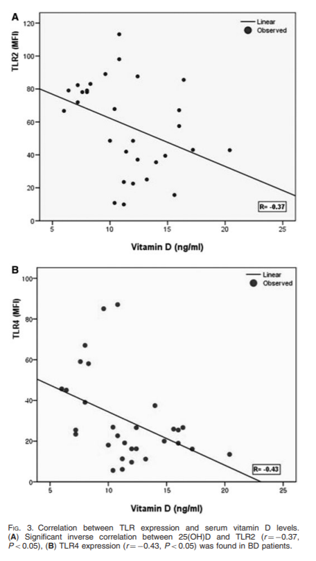
[2008] Effects of vitamin D on expression of Toll-like receptors of monocytes from patients with Behcet's disease
https://pubmed.ncbi.nlm.nih.gov/18411217/
Objectives: Recent studies have shown the immunomodulatory effect of vitamin D(3) through down-regulation of Toll-like receptor (TLR) expression in human monocytes. To understand the implication of innate immunity with the role of vitamin D affecting TLR expression in Behçet's disease (BD), we focused on the association between the TLR expression and the serum vitamin D concentration in BD.

Methods: The expression of TLR2, TLR4 and CD16 on monocytes was detected by flow cytometric analysis and RT-PCR. Serum 25-hydroxyvitamin D [25(OH)D] levels were measured in the patients with BD, psoriasis and healthy controls, and then the expression of TLRs was correlated with the value of serum 25(OH)D levels. To assess the influence of vitamin D(3) on expression and function of TLRs in vitro, human monocytes were treated with increasing concentrations of 1,25(OH)(2)D(3).

Results: We found that the monocytes of active BD patients showed higher expressions of TLR2 and TLR4 than those of controls, and serum 25(OH)D levels tended to be lower in active BD. Furthermore, 25(OH)D levels were inversely correlated with the expressions of TLR2, TLR4 and clinical indicators. In vitro analysis showed that vitamin D(3) was found to dose-dependently suppress the protein and mRNA expressions of TLR2 and TLR4. TNF-alpha synthesis was also decreased upon TLR ligand stimulation in vitamin D(3)-treated monocytes.

Conclusion: These results suggest that the inflammation triggered through TLR2 and TLR4 is important in the pathogenesis of BD. And it seems possible that vitamin D may be used as a therapeutic option by modulating TLR2 and TLR4 expression of monocytes in BD.

LdcLhPU.png


https://pubmed.ncbi.nlm.nih.gov/10549626/
Toll-like receptor (TLR) 2 and TLR4 are implicated in the recognition of various bacterial cell wall components, such as lipopolysaccharide (LPS). To investigate in vivo roles of TLR2, we generated TLR2-deficient mice. In contrast to LPS unresponsiveness in TLR4-deficient mice, TLR2-deficient mice responded to LPS to the same extent as wild-type mice. TLR2-deficient macrophages were hyporesponsive to several Gram-positive bacterial cell walls as well as Staphylococcus aureus peptidoglycan. TLR4-deficient macrophages lacked the response to Gram-positive lipoteichoic acids. These results demonstrate that TLR2 and TLR4 recognize different bacterial cell wall components in vivo and TLR2 plays a major role in Gram-positive bacterial recognition.

https://journals.plos.org/plospathogens/article?id=10.1371/journal.ppat.1007390

What is TLR4 and what are its ligands?

The innate immune system recognizes pathogen-associated molecular patterns (PAMPs) of viral or bacterial intruders via pattern recognition receptors (PRRs). This includes the family of TLRs that consists of related, transmembrane proteins that play a central role in the initiation of inflammatory responses, including the secretion of cytokines and chemokines.

TLR4, which is mainly expressed on cells of the immune system—including monocytes, macrophages and dendritic cells—has long been recognized as a PRR that senses lipopolysaccharide (LPS), a component of the outer membrane of gram-negative bacteria. Activation of TLR4 by LPS, its best studied ligand, is a multistep process. The initial step involves the LPS binding protein (LBP) which extracts LPS from bacterial membranes and LPS-containing vesicles to transfer it to the TLR4 coreceptor cluster of differentiation 14 (CD14). CD14 exists in two forms, soluble and membrane-bound. Both forms are able to interact with LPS-loaded LBP. CD14 breaks down LPS aggregates and transfers monomeric LPS into a hydrophobic pocket on myeloid differentiation factor 2 (MD-2) that is part of the MD-2/TLR4 complex. The high-affinity binding of LPS leads to dimerization and activation of the MD-2/TLR4 complex [6, 7]. Activation of TLR4 results in the recruitment of the intracellular adaptor protein, myeloid differentiation primary response 88 (MyD88), and/or toll/interleukin-1 receptor (TIR)-domain-containing adapter-inducing interferon-β (TRIF), ultimately resulting in the expression and secretion of pro-inflammatory mediators [6, 7].

TLR4 has also been shown to be a sensor for damage-associated molecular patterns (DAMPs). These include a wide variety of molecules released from injured or dying tissues as well as molecules actively released in response to cellular stress from intact cells [6, 8]. In addition to bacterial PAMPs and cellular DAMPs, TLR4 also recognizes PAMPs from other pathogens including fungi, parasites, and viruses [9]. How the TLR4 complex is activated by DAMPs and non-LPS PAMPs, which vary widely in their structure—some with no structural similarities to LPS [8, 10]—remains to be determined. Resolving the structure of these complexes is a critical part toward dissecting their mechanisms of activation.

___

So called "Behcet’s disease" is another good example of basket-case diagnosis of a chronic, body-wide inflammation:

https://www.mayoclinic.org/diseases-conditions/behcets-disease/diagnosis-treatment/drc-20351331
If you have moderate to severe Behcet's disease, your doctor might prescribe: Corticosteroids to control inflammation. Corticosteroids, such as prednisone, are used reduce the inflammation caused by Behcet's disease. Doctors often prescribe them with another medication to suppress the activity of your immune system.

Scientists discover how neuroactive steroids dampen inflammatory signaling in cells
https://www.sciencedaily.com/releases/2019/02/190213142713.htm
For the first time, scientists discovered how neuroactive steroids naturally found in the brain and bloodstream inhibit the activity of a specific kind of protein called Toll-like receptors (TLR4), which have been known to play a role in inflammation in many organs, including the brain.

The effect of systemic corticosteroid therapy on the expression of toll-like receptor 2 and toll-like receptor 4 in the cutaneous lesions of leprosy Type 1 reactions
https://pubmed.ncbi.nlm.nih.gov/22348338/
We have demonstrated that the gene hARP-P0 is a suitable control gene for TLR gene expression studies in this population. The gene and protein expression of TLR2 and TLR4 were both reduced significantly during corticosteroid treatment.

____

Potential Infectious Etiology of Behçet's Disease
https://www.ncbi.nlm.nih.gov/pmc/articles/PMC3255303/
Behçet's disease is a multisystem inflammatory disorder characterized by recurrent oral aphthous ulcers, genital ulcers, uveitis, and skin lesions. The cause of Behçet's disease remains unknown, but epidemiologic findings suggest that an autoimmune process is triggered by an environmental agent in a genetically predisposed individual. An infectious agent could operate through molecular mimicry, and subsequently the disease could be perpetuated by an abnormal immune response to an autoantigen in the absence of ongoing infection. Potential bacteria are Saccharomyces cerevisiae, mycobacteria, Borrelia burgdorferi, Helicobacter pylori, Escherichia coli, Staphylococcus aureus, and Mycoplasma fermentans, but the most commonly investigated microorganism is Streptococcus sanguinis. The relationship between streptococcal infections and Behçet's disease is suggested by clinical observations that an unhygienic oral condition is frequently noted in the oral cavity of Behçet's disease patients. Several viral agents, including herpes simplex virus-1, hepatitis C virus, parvovirus B19, cytomegalovirus, Epstein-Barr virus and varicella zoster virus, may also have some role.
 
Last edited:

Judee

Psalm 46:1-3
Messages
4,461
Location
Great Lakes
This article seems confusing to me. In one place it says:
...lifelong higher 25(OH)D level is associated with an increased risk of Behçet's disease. Special attention should be paid to the potential harm of long-term or high-dose use of vitamin D supplements in clinical practice.

But then further down it says: Conclusion: These results suggest that the inflammation triggered through TLR2 and TLR4 is important in the pathogenesis of BD. And it seems possible that vitamin D may be used as a therapeutic option by modulating TLR2 and TLR4 expression of monocytes in BD.

It also said: We found that the monocytes of active BD patients showed higher expressions of TLR2 and TLR4 than those of controls, and serum 25(OH)D levels tended to be lower in active BD.

Can someone please clarify this for me?

Edit: Generally upping my Vit D makes me feel better as long as I include K2 with it and @lassessen has a good post here regarding how it affects our bodies' production of some of the B vitamins as well as sleep: https://forums.phoenixrising.me/blo...o-b-vit-thus-autoimmune-and-lousy-sleep.2521/
For me it also reverses what a rheumatologist thought was Fibromyalgia. When I get my D levels up, my pain levels go down.
 
Last edited:

uglevod

Senior Member
Messages
220
And it seems possible that vitamin D may be used as a therapeutic option by modulating TLR2 and TLR4 expression of monocytes in BD.

Once one stays on a high level of D25(with chronically suppressed TLR2/4 receptors) long enough(years..decades) he/she could eventually relapse into one of many basket-case inflammatory diseases/diagnoses like(as per above example) Behçet's disease, since the innate immune system was compromised the whole time.

Relapse happens when the overall number of chronic microbiota(bacteria+virus) is reaching the new(much higher) level and the degree of triggering both TLR2 and TLR4 receptors will be enough even for the high D25 to start falling due to the conversion(see below picture) of D25 to the active metabolite(D1.25) through CP27B enzyme, even under the context of semi functional TLR2/4 receptors.

Then the person is offered(as a therapeutic option) a choice between suppressing(modulating) TLRs(and, thus, inflammation) with natural secosteroid(D3 -> D25) or applying synthetic one(like dexamethasone - https://pubmed.ncbi.nlm.nih.gov/28776644/ ) - basically he is returning to square one(again downregulated levels of TLR2/4 with either D25 or steroids) but this time with officially incurable(only manageable with growing doses of steroids) diagnosis and new host of symptoms.
___

P.S:

1598727995753.png 1598730430595.png
from https://www.ncbi.nlm.nih.gov/pmc/articles/PMC2678245/
(Unexpected actions of vitamin D: new perspectives on the regulation of innate and adaptive immunity)
 
Last edited:

Hip

Senior Member
Messages
17,824
lifelong higher 25(OH)D level is associated with an increased risk of Behçet's disease.

Special attention should be paid to the potential harm of long-term or high-dose use of vitamin D supplements in clinical practice.


From reading the Marshall Protocol theory, my understanding is that 25(OH)D, aka calcifediol, is an antagonist of the vitamin D receptor (VDR). It binds to the VDR, but does not activate the VDR.

According to Prof Trevor Marshall, it is activation of the VDR which switches on the intracellular immune response that releases antimicrobial peptides into the cell.

It's only 1,25(OH)2D3, aka calcitriol, the active form of vitamin D, which activates the VDR. But if 25(OH)D is attached to the VDR receptor, it prevents 1,25(OH)2D3 from binding to it.

So higher levels of 25(OH)D can thwart the action of the active form of vitamin D, and in this way high 25(OH)D is immunosuppressive.


Vitamin D Pathway:

If you take a vitamin D3 supplement, this gets converted to 25(OH)D in the liver. Then in turn, the 25(OH)D is converted to 1,25(OH)2D3, the active form of vitamin D, in the kidneys.

In the kidneys 25(OH)D is converted to the active form 1,25(OH)2D3 by the enzyme 1-α hydroxylase.

Parathyroid hormone induces 1-α hydroxylase, so stimulates the production of the active form of vitamin D.
 

uglevod

Senior Member
Messages
220
my understanding is that 25(OH)D, aka calcifediol, is an antagonist of the vitamin D receptor (VDR). It binds to the VDR, but does not activate the VDR.

I think its highly possible partly because TLRs expression increases with a drop of 25(OH)D i.e. with supposed less VDR antagonism and VDR controls the expression level of TLRs too...

In the kidneys 25(OH)D is converted to the active form 1,25(OH)2D3 by the enzyme 1-α hydroxylase.

Its converted in the kidneys but additionally in immune cells too(CP27B aka 1-α hydroxylase exists in monocytes, macrophages maybe in some other immune cells), but since lymphocytes do not express CP27B directly(as far as I know) they are probably depend on the extracellular level of D1.25 for their functionality modulation:

1598730881979.png
 

uglevod

Senior Member
Messages
220
https://www.ncbi.nlm.nih.gov/pmc/articles/PMC6177120/
Upon infection of monocytes with M. leprae, there was no upregulation of CYP27B1[CP27B] nor its enzymatic activity converting the inactive prohormone form of vitamin D (25-hydroxyvitamin D) to the bioactive form (1,25α-dihydroxyvitamin D).
...
Infection of IL-15-derived MΦ, similar to macrophages in lesions from the self-limited form of leprosy, with M. leprae did not inhibit induction of the vitamin D antimicrobial pathway.
...
These results indicate that M. leprae evades the intrinsic capacity of human monocytes/macrophages to activate the vitamin D-mediated antimicrobial pathway via the induction of type I IFN
...
More broadly, our findings suggest that the clinical management of mycobacterial disease using vitamin D supplementation will require simultaneous management of the vitamin D metabolic system to achieve therapeutic benefit.


^^^^^^^^^^^
Some bugs appear to be very dodgy in suppressing VitD pathways, while leaving steroidal effects(on TLRs expression levels) intact.
 

uglevod

Senior Member
Messages
220
Looking from a bright side(mouse model, but still):

http://www.ijmyco.org/article.asp?i...ue=4;spage=349;epage=355;aulast=Wahyunitisari

Supplementation of Vitamin D in the 2nd-line anti-tuberculosis increases the cathelicidin antimicrobial protein:

1598948866153.png

Supplementation of Vitamin D3 in the 2nd-line anti-tuberculosis increases the VDR expression:

1598948943954.png

Supplementation of Vitamin D3 in the 2nd-line anti-tuberculosis decreases the amount of Mycobacterium tuberculosis in the mice's lungs:

1598949035246.png

^^^^^^^^^^^^^^^^^

The viability of intracellular bacteria is influenced by cell death pathways. Apoptosis and autophagy are mycobactericidal, while necrosis precisely causes the bacteria to spread and infects the next cells.
...
M. tuberculosis being trapped in immature phagosome. Vitamin D has mechanisms of controlling this evasion by inducing autophagy. The cathelicidin gene promoter region has 3 Vitamin D response element.[30],[31],[32] This study shows that oral supplementation with Vitamin D effectively increases cathelicidin expression in the lung.
...
We demonstrate that Vitamin D enhances caspase-3 expression in murine model of TB. TNF-alpha promotes caspase activation. It is possible that the interaction of TNF-alpha and calcitriol on mitochondria induces caspase-dependent apoptosis.
...
The results of this study complete the data of other researchers which show that Vitamin D heightens apoptosis by increasing the activity of nitric oxide synthase.
 
Last edited:

uglevod

Senior Member
Messages
220
Vitamin D, serum 25(OH)D, LL-37 and polymorphisms in a Canadian First Nation population with endemic tuberculosis
https://www.ncbi.nlm.nih.gov/pmc/articles/PMC4543449/

Results:
Circulating levels of 25(OH)D were not altered by vitamin D supplementation, but LL-37 levels were significantly decreased. VDBP and VDR SNPs did not correlate with serum concentrations of 25(OH)D, but LL-37 levels significantly decreased in individuals with VDBP D432E T/G and T/T, and with VDR SNP Bsm1 T/T genotypes.

Conclusions:
Our findings suggest that vitamin D supplementation may not be beneficial as an intervention to boost innate immune resistance to M. tuberculosis in the Dene population.
IJCH-74-28952-g004.jpg

Vitamin D supplementation significantly decreased circulating serum LL-37 levels. Serum concentration of LL-37 was monitored by ELISA.
Median and range of serum LL-37 are shown for pre- and post-vitamin D supplementation (a) without seasonal breakdown, or with seasonal analyses in either (b) winter or (c) summer (ns = non-significant).​
 

uglevod

Senior Member
Messages
220
Vitamin D aggravates breast cancer by inducing immunosuppression in the tumor bearing mouse
https://www.ncbi.nlm.nih.gov/pubmed/29852828
The aim of this approach is to test the effects and related mechanism of vitamin D (VD) treatment on the outcomes of breast cancer. BALB/c mice were injected with 4T1 breast cancer cell suspension. The test group was treated with VD reagent. The survival and tumor size of mice were observed. The proliferation of 4T1 in vitro was detected by MTS analysis. The changes of immune parameters and microenvironment in mice were evaluated by flow cytometry and real-time RT-PCR. Our results demonstrate that VD administration caused a decline in survival time and raising the volume of tumor, the decreasing numbers of CD3+CD4+ T, CD3+CD8+ T and CD4+T-bet+IFN-γ+ Th1 cells and transcriptions of T-bet and IFN-γ, an increasing number of myeloid-derived suppressor cells and transcription of TGF-β. Our data suggest that the routine clinical application of any strategies targeting VD status for breast cancer therapy is deserved serious consideration.
...
VD accelerated tumor growth & death of mice in 4T1 tumor-bearing mice Seventeen days after BALB/c mice were inoculated with 4T1 mammary carcinoma cells, the transplanted tumor was already obviously to be observed in the mice, then the tumor-bearing mice were orally treated with vehicle control (soybean oil) or VD for 1 week. Tumor growth was periodically measured and animals were monitored for survival outcome. As shown in Figure 2, around 10 days after 4T1 tumor cell inoculation, we can detect the tumor volume. With the time proceeding, the tumor growth showed a significant promotion in VD treatment mouse group. The tumor size in VD-treated group was bigger than the control group from day 31 postinoculation (Figure 2A) (p < 0.05). The survival rate in the VD-treated group was significantly shortened in comparison with that in the control group (Figure 2B). The result showed that VD could accelerate tumor growth and significantly shorten the survival of 4T1 tumor-bearing mice. On day 8 post VD treatment, we detected the concentrations of VD and calcium in the tumor-bearing mouse sera. The concentration of VD(detected as 25(OH)-Vitamin D)was higher than that of control group (p < 0.05), whereas the concentration of calcium had no difference in these two groups. The results were shown in Figure 3A & B.

VD treatment inhibited the systemic immune response in 4T1 tumor-bearing mice

CD8+ cytotoxic T cells and Th1 cells curb cancer development via mechanisms commonly involving their production of cytotoxins and IFN-γ. To test whether VD treatment could inhibit the immune response mediated by T cells, we tested the numbers of CD3+CD4+, CD3+CD8+ and CD4+T-bet+IFN-γ+ T cells from splenocytes on day 25 and 32 postinoculation. Day 25 postinoculation marks the end of the VD oral administration. Day 32 postinoculation is the day of 1 week after VD oral administration. On day 32 postinoculation, the numbers of CD3+CD4+ (Figure 5A–C) and CD3+CD8+ (Figure 5D–F) T cells in VD treated group dramatically decreased. However, the Th1 cell and CD4+T-bet+IFN-γ+ cells were decreased on day 25 post inoculation (Figure 5G–I). These results indicated that the VD treatment affected the important T cell subsets proliferation, not only help T cell (CD4+) but also cytotoxic T cell (CD8+). Our data also investigated that the decrease of CD8+ T cells might be related to the Th1 cells, which was inhibited on earlier time.

...

VD has been increasingly recognized to have regulatory functions on both innate and adaptive immune [34]. The active form of VD (calcitriol [1,25(OH)2D3]) primarily affects DCs maturation and macrophage differentiation [35,36] and inhibits the production of the cytokines, IL-12 and IL-23. In addition, calcitriol directly acts on T cells to inhibit T-cell proliferation and IFN-γ production [37]. Moreover,calcitriol favors development of Tregs via modulation of DCs [38].Our data demonstrate that VD results in a decrease of the Th1 response in both spleen cells and tumor tissue. Also, suppressive cells of the innate arm of the immune system, such as inflammation-induced MDSCs, are known to be correlated with poor outcome and rapid disease progression [39,40]. MDSCs utilize a variety of mechanisms to suppress T-cell activation and induce other immune-suppressive cell populations [24]. On day 32 post inoculation, the number of MDSCs from spleen cells was dramatically increased. This change is consistent with the change of Th1-related cells. Collectively, these data are in line with earlier descriptions of the suppressive activities of VD with respect to the stimulation of Th1-mediated immunity.

Conclusion & future perspective

In summary, we demonstrate that VD treatment accelerates the growth of breast cancer in mice, which is closely associated with the activation of MDSCs. VD treatment suppressed the Th1 response in both system and tumor microenvironment. These results indicate that VD application might be associated with worse prognosis and seems to promote tumorigenesis. Therefore, these results would sugggest a thoughtful consideration for the routine application of VD replenishment to breast cancer prevention or therapy.
 

uglevod

Senior Member
Messages
220
Immunomonitoring of the different immune subsets in dissociated tumors revealed an increase in tumor infiltrating CD8+ T cells in the vitamin D-treated group.
https://www.ncbi.nlm.nih.gov/pmc/articles/PMC6563618/

...
However, in high-fat diet conditions, we observed an opposite effect of vitamin D on breast cancer tumor growth, associated with a reduction of CD8+ T cell infiltration. Our data show that vitamin D is able to modulate breast cancer tumor growth and inflammation in the tumor microenvironment in vivo. Unexpectedly, this effect is reversed in high-fat diet conditions, revealing the importance of diet on tumor growth. We believe that supplementation with vitamin D can in certain conditions represent a new adjuvant in the treatment of breast cancers.

Some studies have indicated that diet may have an influence in approximately 35% of breast cancer cases (25). Weight gain since early adulthood and obesity have been associated with increased post-menopausal breast cancer risk in several prospective studies (2629). Indeed, obesity favors an increase in circulating estrogens and entails increased risk of hormone-dependent breast cancer (3032). The pathway of chronic inflammation (pro-inflammatory cytokines) induced by adipose tissue also contributes to both tumor transformation and resistance to therapy (33). It is known that the balance between pro-inflammatory and anti-inflammatory T cells is modified during obesity-induced inflammation in visceral adipose tissue, the pool of pro-inflammatory T cells such as CD4+ and CD8+ T cells is increased, and anti-inflammatory T cells such as Tregs are decreased in diet-induced obesity (34). Moreover, macrophages in obese tissue are mostly M1 macrophages (also known as classically activated macrophages), which secrete pro-inflammatory cytokines such as interleukin (IL)-6 and tumor necrosis factor (TNF)-α (3537).

Our work aimed to explore the effects of VD on the breast cancer progression in mice from basal to overweight conditions and its consequences on CD8+ T cell infiltration into the tumor. Our data provide evidence that, depending of diet conditions, the VD supplementation has opposite effects on both in CD8+ T cell infiltration and tumor growth.

In the present study, using female mice injected with EO771 breast cancer cells and supplemented with VD, we report a beneficial role of VD on tumor growth progression and inflammation. Indeed, VD supplementation reduced tumor weight and increased CD8+ T cell infiltration into the tumor. Interestingly, these CD8+ T cells exhibited a more active central/effector memory T cell phenotype, which are considered to have increased anti-tumor properties (43). These data are in agreement with data that highlight TILs as an important predictive and prognostic biomarker in patients with breast cancer (44). Ono et al. observed an association between pathological response and TIL score in patients with triple-negative breast cancer, but not in patients with other tumor types (45). Therefore, a general consensus emerged on the central role played by effector T cells in the anti-tumor response. Similar results were obtained in colorectal cancer in which quantification of immune cell densities revealed the major positive role of cytotoxic and memory T cells for patient survival (46). The present study indeed also reports that CD8+ T cells infiltrating the tumor harbor an activated/cytotoxic phenotype eliciting PD-1/Granzyme B expression. In line with these results, several studies on cancer patients have reported that PD-1 expression is highly expressed on tumor infiltrating CD8+ T cells in various immunogenic tumors (4749).

Surprisingly, in contrast to standard diet condition, VD supplementation of overweight-mice resulted in increased tumor progression without CD8+ T cell infiltration (illustrated in Figure 6). The impact of VD supplementation on inflammation associated with obesity has been largely described. It has been reported a beneficial role of VD supplementation on weight gain limitation in mice (50, 51). Indeed, such supplementation reduced the weight gain induced by high-fat diet and improved insulin sensitivity. The reduced weight gain in VD supplemented mice is primarily due to a limitation of fat mass accumulation (50). We have also shown in a previous study that VD displays immunoregulatory effects and reduces adipocyte inflammation in vitro and in vivo. Indeed, VD supplementation decreases the expression and secretion of a large range of inflammatory markers and macrophage infiltration in the adipose tissue of high-fat diet fed mice (38). In the present study, we report a beneficial role of VD supplementation on inflammation both in basal or overweight conditions. We have shown that VD is able to limit the expression of inflammatory markers in adipocytes (Il6, Ccl5, and Cx3cl1) and their secretion (IL-6 and CCL5) in the plasma. These results are in agreement with the previously reported anti-inflammatory effects of VD on adipocytes (38, 52). Taken together, our results reveal the influence of diet on tumor growth control and CD8+ T cell infiltration by VD supplementation. The difference observed between normal and high-fat diet conditions could be explained by a modification in the VD metabolism in adipocytes, in particular Cyp27a1 in overweight mice, which is involved in the first hydroxylation of cholecalciferol (data not shown). This enzyme is known to be increased in adipose tissue of obese mice, which suggests the possible increase of 25-hydroxylation in adipose tissue, resulting in higher local production of 25(OH)D in adipose tissue (53). Several studies about obesity have demonstrated that 25(OH)D is diluted in a higher volume in obese patients (54). We hypothesized that the increase of adipocytes in overweight-mice reduces 25(OH)D in plasma, which could have an impact on a decreased CD8+ T cell infiltration.

9c0KQhm9oAdzdXKu4DeVatPczfpGkaCTiIg7vqbvCaNDNtyZP8agrNhTjvlWAH_wogfwQifqWpC1QR0GNCj-lkgTafcIw5-zd120mGJM1ndMmrDe6HYaZ4gvPWCra21jSczgOEc1fMln
 

uglevod

Senior Member
Messages
220
Enhanced LL-37 expression following vitamin D supplementation in patients with cirrhosis and spontaneous bacterial peritonitis
https://pubmed.ncbi.nlm.nih.gov/26058412/
Background & aims: The morbidity and mortality of spontaneous bacterial peritonitis (SBP) are high among patients with cirrhosis; however, the mechanisms of SBP pathogenesis are poorly understood. This study aimed to determine the role of the vitamin D-LL-37 pathway in the pathogenesis and treatment in patients with cirrhosis and SBP.
Methods: Serum 25-hydroxyvitamin D concentrations of 119 patients with chronic liver diseases were tested. Vitamin D receptor (VDR) and LL-37 in peritoneal leucocytes of cirrhotic and ascitic patients with SBP were detected and compared with those without SBP. Then the peritoneal macrophages of non-infected patients were cultured and activated by lipopolysaccharide (LPS) to analyse the changes of VDR and LL-37 expressions after incubation with vitamin D.

Results: Vitamin D deficiency or insufficiency was found in all of patients with cirrhosis. LPS inhibited VDR and LL-37 expression in peritoneal macrophages [1.3-fold decrease (P = 0.003) and 20-fold decrease (P = 0.010) respectively]. However, vitamin D could reverse the inhibition of both VDR and LL-37 [1.5-fold increase (P = 0.001) and 2000-fold increase (P < 0.001) respectively]. The effect of the incubation time following vitamin D supplementation was significant for LL-37 expression, with a peak expression found at 36 h (P < 0.001).

Conclusions: When vitamin D levels were low, bacteria inhibited VDR and LL-37 responses in peritoneal macrophages as a mechanism to evade antibacterial defence. Vitamin D supplementation could up-regulate peritoneal macrophage VDR and LL-37 expressions, which resulted in an enhanced immunological defence against SBP in patients with cirrhosis and ascites.

Key points:

• All of patients with cirrhosis were in a state of vitamin D deficiency or insufficiency.

• Under low vitamin D circumstances, bacteria inhibited VDR and LL-37 responses in peritoneal macrophages as a mechanism to evade antibacterial defence.

• Vitamin D supplementation could reverse the inhibition effect of LPS to up-regulate peritoneal macrophage VDR and LL-37 expressions, which resulted in an enhanced immunological defence against SBP in patients with cirrhosis and ascites.

• The results of this study suggest that vitamin D could be an adjunct to antibiotic therapy in the prevention and treatment of SBP.

In our study, we found that in low vitamin D conditions, various concentrations of LPS down-regulated both VDR and LL-37. Pramanik et al. (30) also found that LPS could decrease VDR proteins levels and inhibit VDR functions in the human leukaemic cell line in the absence of 1,25(OH)2D3 stimuli. Furthermore, other researchers have reported that bacterial exotoxins down-regulate LL-37 expression in the intestinal epithelial cells (31) and Neisseria gonorrhoea down-regulated LL-37 expression in cervical epithelial cell line ME180 (32). We speculate that this may be a new bacterial mechanism against antibacterial immunity, which could explain the reasons why cirrhosis and ascites patients with low vitamin D levels are more likely to suffer from bacterial infections, which are often considered serious.

When vitamin D was sufficient, VDR and LL-37 were up-regulated in the peritoneal macrophages, indicating that appropriate levels of vitamin D are necessary for the complete functions of the vitamin D-VDR-LL-37 pathway. This pathway may be a mechanism against bacterial invasion. In particular, LL-37 expression improved with increased vitamin D, indicating that the LL-37 response was vitamin D concentration-dependent. These results are consistent with the results of Adams et al. (33). Furthermore, for the first time, we characterized the time-dependent effect of 1,25(OH)2D3 on LL-37 up-regulation. The maximum LL-37 expression under 1,25(OH)2D3 stimuli was found at 36 h.


In conclusion, vitamin D deficiency as universal in patients with cirrhosis and ascites, and the degree of vitamin D deficiency was more severe with more serious liver diseases. When vitamin D levels are low, bacteria can inhibit the LL-37 response of peritoneal macrophages as a mechanism to evade antibacterial immune defence. However, this inhibition can be corrected by vitamin D supplementation (the higher concentration of vitamin D, the stronger the corrective activity). Therefore, adequate vitamin D is vital for the body to improve antimicrobial immune defence response. The results of this study suggest that vitamin D could be an adjunct to antibiotic therapy in the prevention and treatment of SBP. Our next step is to conduct animal experiments to investigate the effects of vitamin D and LL-37 on the prevention and treatment of SBP in a cirrhosis rat model.

...

Administration of oral vitamin D induces cathelicidin production in atopic individuals
https://www.ncbi.nlm.nih.gov/pmc/articles/PMC2659525/

After supplementation with 4000 IU/d oral vitamin D for 21 days, AD lesional skin showed a statistically significant increase in cathelicidin expression from a median of 3.53 relative copy units (RCU) before supplementation to a median of 23.91 RCU post supplementation (P <.01; Fig 1).

Serum calcium and vitamin D levels were measured in atopic and normal subjects at baseline and after 3 weeks of supplementation with 4000 IU/d of oral vitamin D3. 4000 IU of vitamin D3 has been well documented to effectively increase 25-hydroxyvitamin D levels while maintaining normal serum calcium levels, suggesting that the current recommendation for vitamin D supplementation may be too conservative. Indeed, serum calcium in our normal control subjects did not rise but actually decreased from a median of 9.8 mg/dL to a median of 9.4 mg/dL post supplementation (P > .05). During this time, their serum 25-hydroxyvitamin D levels rose from a median of 24.5 mg/mL to 37 mg/mL (P > .05). Similarly, atopic subjects’ serum calcium levels also decreased from a median of 9.6 mg/dL to a median of 9.4 mg/dL post supplementation (P > .05), and their serum 25-hydroxyvitamin D levels rose from a median of 22.5 mg/mL to 35.5 mg/mL (P > .05). No subject complained of any adverse event during supplementation.
...
Vitamin D deficiency has been linked to increased rates of multiple cancers, autoimmune diseases, infectious diseases, cardiovascular diseases, and hypertension.8 We believe our study has shown that supplementation of oral vitamin D can result in correction of defects in cathelicidins in the innate immune system of atopic subjects.
 

uglevod

Senior Member
Messages
220
Inhibition of IFN-gamma signaling by an Epstein-Barr virus immediate-early protein:
https://pubmed.ncbi.nlm.nih.gov/11728340/

Inhibition of IFN-γ Signaling by Glucocorticoids:
https://www.jimmunol.org/content/170/9/4833

https://pubmed.ncbi.nlm.nih.gov/12707366/
Addition of dexamethasone to PBMC cultures resulted in a dramatic inhibition of IFN-gamma activation of STAT1.

Inhibition of IFN-gamma as a method of treatment of various autoimmune diseases, including skin diseases:
https://pubmed.ncbi.nlm.nih.gov/16329644/
We pioneered anticytokine therapy (ACT) for autoimmune diseases (ADs). In 1974, we proposed that hyperproduced interferon (IFN) can bring AD and anti-IFN can be therapeutic. In 1989, we proposed removing tumor necrosis factor (TNF)-alpha together with certain types of IFN to treat various ADs. We found IFN in patients with different ADs and conducted the first clinical trial of ACT in 1975. Anti-IFN-gamma and anti-TNF-alpha work in similar ways, but the latter brings serious complications in some patients. We obtained good, sometimes striking, therapeutic effects treating many different Th-1-mediated ADs with anti-IFN-gamma, including rheumatoid arthritis, multiple sclerosis (MS), corneal transplant rejection, and various autoimmune skin diseases such as psoriasis, alopecia areata, vitiligo, acne vulgaris, and others. Anti-IFN-gamma was in some ways superior to anti-TNF-alpha, which was ineffective in MS. Anti-IFN-gamma therapy holds great promise for treating many Th-1 ADs, especially skin diseases.

Vitamin D Is Required for IFN-γ–Mediated Antimicrobial Activity of Human Macrophages
https://www.ncbi.nlm.nih.gov/pmc/articles/PMC3269210/

Furthermore, a key role for interferon-γ (IFN-γ) in the immune response to M. tuberculosis infection is corroborated by studies indicating that humans with genetic disorders leading to the decreased production of, or response to, IFN-γ are highly susceptible to tuberculosis and other mycobacterial diseases (3–5). Yet, it has been a matter of faith that IFN-γ can activate human monocytes or macrophages to kill intracellular M. tuberculosis.

Indeed, multiple studies of IFN-γ treatment of human macrophages have consistently failed to demonstrate antimicrobial activity against intracellular M. tuberculosis (6–16). In vitro, IFN-γ is unable to activate human macrophages to restrict or kill virulent M. tuberculosis (17), and human macrophages infected with M. tuberculosis are desensitized to this cytokine (17). However, the discovery that the innate immune response, activated by Toll-like receptors (TLRs) on human monocytes and macrophages, triggers an antimicrobial response against M. tuberculosis that is vitamin D–dependent (18, 19) provided a new opportunity to investigate this phenomenon. We found that the acquired T cell response could activate antimicrobial activity in human cells of the monocyte/macrophage lineage.
...
Next, we investigated the functional roles of CYP27b1 and the VDR in this system. IFN-γ treatment of human monocytes resulted in enhanced conversion of 25D to 1,25D, indicating increased CYP27b1 enzymatic activity (Fig. 2C, P < 0.05). To determine whether CYP27B1 and VDR induction was required for IFN-γ–mediated expression of antimicrobial peptides, we performed small interfering RNA (siRNA) knockdown of each gene on primary monocytes. Both siVDR and siCYP27B1 knocked down the IFN-γ–induced expression of their respective targets by about 50% as measured by qPCR. Transfection of siRNA oligos targeting CYP27B1 and VDR, but not the non-specific control oligo, significantly reduced IFN-γ–induced expression of both cathelicidin and DEFB4 (Fig. 2D, P < 0.05). In contrast, knockdown of CYP27B1 and VDR did not inhibit IFN-γ–induced CD64, but instead may have increased CD64 expression, consistent with the ability of 1,25D to inhibit CD64 expression (29). Furthermore, the VDR antagonist VAZ, shown to inhibit 1,25D induction of cathelicidin (30), blocked induction of antimicrobial peptides (Fig. 2E, P < 0.05), but not CD64, which was again slightly increased (Fig. 2E). Finally, IFN-γ also induced the VDR downstream gene CYP24, providing evidence for activation of the VDR (fig. S2). These data indicate that IFN-γ induction of cathelicidin and DEFB4 mRNAs is dependent on both CYP27b1 and the VDR.
...
On the basis of these studies, we hypothesized that previous studies failed to detect an antimycobacterial activity by IFN-γ–treated human macrophages because of the different amounts of 25D and/or its availability in the sera used for the in vitro killing experiments. Because IFN-γ was able to up-regulate antimicrobial peptide mRNA expression, we compared the types of sera used in the previous reports in which IFN-γ treatment did not induce an antimycobacterial activity in human macrophages (6, 7, 9–12, 14, 15). We found that IFN-γ–induced antimicrobial peptide expression was optimal in cultures containing 10% or greater vitamin D–sufficient human serum (25D = 98 nM), but could not be induced in 2 or 10% heat-inactivated human serum or 10% vitamin D–deficient fetal calf serum (FCS) (25D = 16 nM) (fig. S5, A to C, P < 0.05), conditions used in one or more of the previously mentioned studies. Markedly, 10% vitamin D–deficient human serum (25D = 45 nM) also failed to support antimicrobial peptide expression in monocytes (Fig. 5B, P < 0.05), but this could be restored by supplementation of vitamin D–deficient serum to sufficient concentrations by the addition of 25D in vitro (Fig. 5C, P < 0.05).
...
Because macrophages are the natural hosts of mycobacteria during the course of infection (46), we tested whether IFN-γ treatment would induce the vitamin D antimicrobial pathway in monocyte-derived macrophages (MDMs). As observed in human monocytes, IFN-γ induced cathelicidin and DEFB4 gene expression in MDMs in 10% vitamin D–sufficient serum (Fig. 5D, P < 0.05), but not in 10% vitamin D–deficient serum. Likewise, the induction of cathelicidin and DEFB4 gene expression in MDMs could be restored by supplementation of vitamin D–deficient serum by addition of 25D in vitro (Fig. 5D, P < 0.05). Moreover, vitamin D–deficient serum did not support autophagolysosomal fusion and phagosome maturation in M. tuberculosis–infected macrophages, but this could be rescued by in vitro supplementation with 25D (fig. S6).
...
Next, we infected MDMs with virulent M. tuberculosis and stimulated the infected cells with IFN-γ in 10% vitamin D–sufficient human serum or 10% vitamin D–deficient human serum in the same experiments. IFN-γ treatment of M. tuberculosis–infected MDMs using vitamin D–sufficient human serum resulted in a 90% reduction of M. tuberculosis growth at 3 days, relative to untreated cells (Fig. 5F, P < 0.01). Markedly, IFN-γ treatment resulted in an 85% reduction in M. tuberculosis compared to the initial inoculum (Fig. 5F, P < 0.001), formally demonstrating that IFN-γ induced killing of the bacteria.

In contrast, IFN-γ treatment of MDM using vitamin D–deficient serum had little effect on the bacterial viability (Fig. 5F); however, antimicrobial activity could be restored by in vitro supplementation of vitamin D–deficient serum with 25D3 (fig. S7). These data reveal a marked difference in IFN-γ–induced antimicrobial activity against intracellular M. tuberculosis depending on the serum 25D concentration. Furthermore, these in vitro data are consistent with abundant clinical evidence that links low amounts of serum 25D to both tuberculosis disease susceptibility and progression (47–50).

nihms351009f6.jpg


IFN-γ induces an antimicrobial pathway in human monocytes/macrophages. This model shows that STAT1-dependent induction of IL-15 by IFN-γ leads to the up-regulation of VDR and CYP27b1. CYP27b1 hydroxylates the inactive form of vitamin D (25D) into the active form (1,25D), which mediates the up-regulation of antimicrobial peptides cathelicidin and DEFB4. The intracrine-produced 1,25D also triggers autophagy, which overcomes the M. tuberculosis–induced phagosome maturation block, leading to autophagolysosomal fusion and antimicrobial activity against M. tuberculosis
...
The present results provide at least one explanation for a fundamental difference in antimicrobial mechanisms seen between mice and humans. The IFN-γ–induced antimicrobial pathway described here is vitamin D–dependent in humans but not in the mouse. The human promoter of cathelicidin contains three vitamin D response elements (VDREs), whereas the promoter of the murine homolog does not contain a single VDRE (57). In addition, the human promoter of DEFB4 contains one VDRE (19), whereas there is no mouse homolog for this gene. Therefore, the biological significance of the IFN-γ–induced, vitamin D–dependent antimicrobial pathway must be investigated in human cells and simply cannot be studied in a mouse model. The evolution of distinct antimicrobial mechanisms makes sense teleologically as well because mice are nocturnal animals and humans are not, and the amount of vitamin D increases with sun exposure.

^^^


Zinc deficiency impairs interferon-γ production on post-transcriptional level:
https://www.sciencedirect.com/science/article/abs/pii/S0946672X20301632
Zinc is a trace element and is thus commonly known to be indispensable for regular cellular function. Until today, zinc deficiency is a widespread health problem, affecting approximately one sixth of the world's population. Especially the immune system has proven to be highly dependent on zinc. Interferon-γ (IFN-γ) is a key element in the defense against intracellular pathogens. A lack of this cytokine results in immunological impairment, whereas an excess can lead to autoimmunity, highlighting the importance of a well-regulated IFN-γ expression. In a state of zinc deficiency, the production of this cytokine has long been shown to be reduced.
 
Last edited: