• Welcome to Phoenix Rising!

    Created in 2008, Phoenix Rising is the largest and oldest forum dedicated to furthering the understanding of, and finding treatments for, complex chronic illnesses such as chronic fatigue syndrome (ME/CFS), fibromyalgia, long COVID, postural orthostatic tachycardia syndrome (POTS), mast cell activation syndrome (MCAS), and allied diseases.

    To become a member, simply click the Register button at the top right.

Is Apheresis an effective treatment for Long Covid and ME?

GlassCannonLife

Senior Member
Messages
819
More details on microclot detection via light microscopy. Claim that only a blood smear is needed. I sort of doubt that as it would be hard to see the microclots among the RBCs, platelets, & other cells. Pertorius used Platelet Free Plasma (PFP), which is plasma that's been centrifuged twice. A smear of PFP would be clearer, I'd think.

View attachment 45973


One Long Covid patient, who is a doctor, discovered microclots in here blood using a light microscope. She is a dermatologist, so you'd think she'd definitely have a microscope in her office.
View attachment 45975


She describes her procedure on how she detected them here. At only 40x (I assume x10 w/ eye piece, so 400x total), a child's microscope or cheapest microscope on amazon for $150 should be able to see that. Maybe even those digital ones for $50.
View attachment 45978

Images of her results, and description of procedure:

View attachment 45977

That's interesting! Shame I can't work anymore, we had all of these things in the lab.

A 4x objective is pretty common so I don't know if it's necessarily 400x vs actually 10x eye piece and 4x objective. We had some small bench microscopes that would have 2.5, 4, 10, and 25 IIRC.
 

godlovesatrier

Senior Member
Messages
2,555
Location
United Kingdom
I find it interesting how he's postulating a starkly different immune abnormality between the LC and ME patients. Makes me wonder if someone with ME can even get LC if they are two totally different diseases (if you go off what he's said) and what it would look like if they could.
 
Messages
67
More details on microclot detection via light microscopy. Claim that only a blood smear is needed. I sort of doubt that as it would be hard to see the microclots among the RBCs, platelets, & other cells. Pertorius used Platelet Free Plasma (PFP), which is plasma that's been centrifuged twice. A smear of PFP would be clearer, I'd think.

She describes her procedure on how she detected them here. At only 40x (I assume x10 w/ eye piece, so 400x total), a child's microscope or cheapest microscope on amazon for $150 should be able to see that. Maybe even those digital ones for $50.
View attachment 45978

She talks about finding a derm who does PRP (Platelet Rich Plasma) because then they will know how to obtain PPP (Platelet Poor Plasma). PPP is what's needed to see the microclots.

I'm trying to find a microscope for doing this, and in my research I found that for obtaining PPP for a lab test, normally you'll need to do a two steps process, centrifuge for 15 min at 3000rpm, then extract the PPP into another tube, then centrifuge again at 3000rpm for another 15min. This way you eliminate the risk of clotting or something like it?
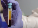
I think here's the difference with Pretorious method, she does only one spin at 3000rpm for 15 min, you can find her process in her papers. The result should be something like this pic I think, where the clearer part is the PPP.

Captura de pantalla 2021-12-10 a las 14.34.44.png

Then you draw a small blood from the clearer part and create the smear like Angela Bowers said.
In USA you have the AmScope microscopes, pretty cheap microscopes made in china, probably enough to see the microclots.
 

junkcrap50

Senior Member
Messages
1,335
A 4x objective is pretty common so I don't know if it's necessarily 400x vs actually 10x eye piece and 4x objective. We had some small bench microscopes that would have 2.5, 4, 10, and 25 IIRC.
Yes, but I don't think that 40x power would be able to see microclots. I have looked into this to see how powerful a microscope one would need and if they're available off amazon. I think 400x would be the minimum and 1000x or higher might even be better, assuming microclots are ~2-5 micrometers based on Pertorius's paer. I reached those magnification numbers based on this diagram:
1639169166148.png

And also based it by googling images and youtube videos of "RBCs at 400x/1000x" or "Platelets at 400x/1000x/etc."


She talks about finding a derm who does PRP (Platelet Rich Plasma) because then they will know how to obtain PPP (Platelet Poor Plasma). PPP is what's needed to see the microclots.

I'm trying to find a microscope for doing this, and in my research I found that for obtaining PPP for a lab test, normally you'll need to do a two steps process, centrifuge for 15 min at 3000rpm, then extract the PPP into another tube, then centrifuge again at 3000rpm for another 15min. This way you eliminate the risk of clotting or something like it?
Obtaining PPP (Platelet Poor Plasma) aka Platelet Free Plasma (PFP) is very easy and can be done by any place that draws blood.

I think the reason why PPP is preferred is that you want a very clean plasma solution free of any particles except for the microclots. After looking at lots of microscopy images of blood online, I'm surprised that there's a lot of "junk" that can be in there. By junk I mean very small particles compared to RBCs. Most of the time they are platelets, which can be various sizes including very small. So one might mistake a platelet or broken parts of a platelets as the microclots.

I'm not sure why Dr. Bowers did just one centrifuge (maybe she made a mistake). But if only 1 centrifuge is needed for detection, then that would simplify the process for patients and doctors in getting this done (more straight forward, less explaining, less time, etc.). Most probably have never heard of Plaletet Poor Plasma.
 

SNT Gatchaman

Senior Member
Messages
302
Location
New Zealand
This is an interesting discussion on Long Covid and the micro clots are mentioned. Apparently, the UK is already treating them.

Thank you for posting. The host was excellent. I understood that the patient (Hannah) who had evidence of micro-clots in her right lung (by V/Q scan) was on a single — albeit quite powerful — direct oral anticoagulant (apixaban). The other two (Gez and Shaun) sounded like they were on triple platelet blockade, but I wouldn't infer that that is anywhere near the standard of practice in the UK — quite the opposite.

Most LC clinics there, if they have been set up, are based around CBT and exercise rehabilitation. They are of course ineffective.
 

keepontruckin

Senior Member
Messages
214
At one point he was asked if they had plans to write long covid into the health policy and he answered they don't feel comfortable talking about that. They want them to go to work and Christmas parties. Such a denial of reality!
People in the health system sacrificed themselves without proper equipment etc.. There is a huge mound of bodies unfit to work and care for their families and the government does not feel comfortable writing a policy about long covid? They should be saying these people who put their lives on the line deserve the best cutting edge research our science can provide. It is our responsibility in appreciation for their service to the country.
 
Last edited:

GlassCannonLife

Senior Member
Messages
819
@junkcrap50 yeah that makes sense, I didn't look into it beyond my assumption that she didn't share the wrong magnification - many people forget that the eyepiece is 10x though so that must have been it!

From the images, however, I'd be hesitant to diagnose ourselves by looking at blood with just light microscopy. It's so easy to have some type of contaminant appear in the field and misinterpret the result.. It would be best to stain the clots in some way (and ideally use fluorescence microscopy for specificity and clarity).

I wonder if we'd be able to use a basic histological stain and still use light microscopy though? I haven't checked what we'd be wanting to stain (sorry very crashed atm still), but for example something basic like H&E could maybe be an option? I briefly googled and found this article about the analysis of a thrombus.

Thrombus material was histologically analyzed using hematoxylin and eosin, Martius Scarlet Blue stain (red blood cells and fibrin), Feulgen stain (DNA), von Kossa stain (calcifications) and immunohistochemical analysis of von Willebrand factor, platelets, leukocytes and neutrophil extracellular traps.
 

junkcrap50

Senior Member
Messages
1,335
From the images, however, I'd be hesitant to diagnose ourselves by looking at blood with just light microscopy. It's so easy to have some type of contaminant appear in the field and misinterpret the result.. It would be best to stain the clots in some way (and ideally use fluorescence microscopy for specificity and clarity).

I wonder if we'd be able to use a basic histological stain and still use light microscopy though? I haven't checked what we'd be wanting to stain (sorry very crashed atm still), but for example something basic like H&E could maybe be an option? I briefly googled and found this article about the analysis of a thrombus.
Yes. I agree that using only light microscopy could lead to some misdiagnoses. Why centrifuging twice and getting PPP would be important. But I think it would be a pretty good, initial, basic screening test that's easily available or relatively inexpensive. However, if light microscopy found microclots in me, I would want to confirm with flourescent microscopy before spending $1500 (in Germany) or $4500 (in US) per HELP apheresis treatment.

Great idea about looking to see if there is a histology stain unique and selective for fibrin/thrombi! I'm sure basic stains could also stain fibrin as well as other things. And that would make it much easier to get tested by sending PPP to a pathology lab, as that would be very familiar to them.

Doing some quick googling, I was able to confirm your finding that there are stains to identify fibrin.
https://stainsfile.info/stain/fibrin/fibrin.htm

According to the microclot paper, the fluorescent dye they used, Thioflavin T, binds to "amyloid fibrils." They also found within the microclot that might be stain targets (I don't know) "α(2)-antiplasmin (α2AP), various fibrinogen chains, [and] Serum Amyloid A."
 
Last edited:
Messages
67
Yes. I agree that using only light microscopy could lead to some misdiagnoses. Why centrifuging twice and getting PPP would be important. But I think it would be a pretty good, initial, basic screening test that's easily available or relatively inexpensive. However, if light microscopy found microclots in me, I would want to confirm with flourescent microscopy before spending $1500 (in Germany) or $4500 (in US) per HELP apheresis treatment.

I completely agree with this, light microscopy would lead to some misinterpretations, but I don't want to go for HELP without at least having some proof leading to microclots. I'm still trying to find someone with access to fluorescent microscopy.

Pretorius used a Plan-Apochromat 63x DIC objective (from Zeiss). With a 10x eyepiece, her captured images would be taken at 630x. Since Dr. Bowers's capture clearly is from afar, I would say 400x seems right.

the fluorescent dye they used, Thioflavin T, binds to "amyloid fibrils." They also found within the microclot that might be stain targets (I don't know) "α(2)-antiplasmin (α2AP), various fibrinogen chains, [and] Serum Amyloid A.

This dye is also used to light amyloid plaque cells in Alzheimer's samples. Pretorius demonstrated in COVID-19 healthy fibrinogen changes to an amyloid form, that's the reason it stains.
It would be interesting to see both images in us (fluorescence and bright-field microscopy). We can then see if we have some kind of microclots and if it's fibrin inside or other molecules with amyloid changes (don't know if this has sense or every microclot is formed inherently with fibrin molecules).
 

junkcrap50

Senior Member
Messages
1,335
This dye is also used to light amyloid plaque cells in Alzheimer's samples. Pretorius demonstrated in COVID-19 healthy fibrinogen changes to an amyloid form, that's the reason it stains.
Hmm.. That makes me think that a Pathology Lab might likely be able to test and check for this. But would have to call and see if they have a fluorescent microscope.

EDIT: Actually, I think nearly all pathology labs or pathology doctor groups would have a fluorescent microscope. Looking at a few websites, they list services/test that use fluorescent detection, some even directly say "direct immunofluorescent testing" as a service. But other tests/services that use fluorescent microscopy would be: Fluorescent In Situ Hybridization (FISH), Cytogenetics, Flow Cytometry, and Immunohistochemical Staining (some are fluorescent stains). So, I'd think a doctor's order explaining Pretorius's finding, and a request to do the same, and with a sample of PPP, then any pathology group could do this. They could also look for it using traditional stains too, unique to fibrin.
 
Last edited:
Messages
55
Hmm.. That makes me think that a Pathology Lab might likely be able to test and check for this. But would have to call and see if they have a fluorescent microscope.

EDIT: Actually, I think nearly all pathology labs or pathology doctor groups would have a fluorescent microscope. Looking at a few websites, they list services/test that use fluorescent detection, some even directly say "direct immunofluorescent testing" as a service. But other tests/services that use fluorescent microscopy would be: Fluorescent In Situ Hybridization (FISH), Cytogenetics, Flow Cytometry, and Immunohistochemical Staining (some are fluorescent stains). So, I'd think a doctor's order explaining Pretorius's finding, and a request to do the same, and with a sample of PPP, then any pathology group could do this. They could also look for it using traditional stains too, unique to fibrin.
I tried to start the “microclots” discussion with my PCP and messaged her, sending her an article about evidence of inflammatory microclots discovered by Resia Pretorius and she said “the issue is there is no way to test for this as this is experimental. While that may be happening, there is not a test we can do here to see if that is the case and there is no treatment for it.“
I wanted to say - THERE IS A WAY to test for it if you will think outside of the box.
It’s almost futile to argue though.
Edit- please forgive the rant 😊. I pretty much expected this response from PCP but wanted to educate her on progress being made in the world for treatment of post viral disease! And that it might apply to me!
 
Last edited:

Shanti1

Administrator
Messages
3,220

GlassCannonLife

Senior Member
Messages
819
I tried to start the “microclots” discussion with my PCP and messaged her, sending her an article about evidence of inflammatory microclots discovered by Resia Pretorius and she said “the issue is there is no way to test for this as this is experimental. While that may be happening, there is not a test we can do here to see if that is the case and there is no treatment for it.“
I wanted to say - THERE IS A WAY to test for it if you will think outside of the box.
It’s almost futile to argue though.

I think what they mean is that there is no standardised and validated test that is available - experimental "just have a quick look on the microscope" testing is not acceptable for the medical community for obvious reasons. You would probably have to use someone that is willing to either do it off the books in a path lab or have a researcher do it for you.
 

junkcrap50

Senior Member
Messages
1,335
I'm wondering if they aren't larger than 2-5 μmeters. I didn't see a size in the text of Pretorius's main paper, but the scale on the pictures shows they are at least 10μm, but generally larger. It seems they would be quite visible at 400x.
https://www.ncbi.nlm.nih.gov/pmc/articles/PMC8381139/
View attachment 46002
You're correct. I meant "as small as 2-5 micometers" and that was just my estimation.

I think what they mean is that there is no standardised and validated test that is available - experimental "just have a quick look on the microscope" testing is not acceptable for the medical community for obvious reasons. You would probably have to use someone that is willing to either do it off the books in a path lab or have a researcher do it for you.
Yes. It's not standardized and validated. But you wouldn't be asking the pathology lab for an interpretation (because they'd be liable), but rather ask for microscopy images to be sent back, after the stains or fluorescent markers. Paying for procedure rather than for conclusion.
 

GlassCannonLife

Senior Member
Messages
819
You're correct. I meant "as small as 2-5 micometers" and that was just my estimation.


Yes. It's not standardized and validated. But you wouldn't be asking the pathology lab for an interpretation (because they'd be liable), but rather ask for microscopy images to be sent back, after the stains or fluorescent markers. Paying for procedure rather than for conclusion.

Yeah sure, I was just explaining why the PCP wouldn't seem interested.

I'm also not convinced we'd be able to get a pathology lab to do it unless we got very lucky. I worked in medical device development, and it is highly likely that they follow protocols that are strictly governed by a QMS. Doing something that isn't a standardised and approved process would just be unacceptable. They'd possibly have to do calibration, cleaning, revalidation, etc depending on what their QMS documentation requires and what equipment they used for the test. This is to ensure that uncontrolled practices don't affect their highly impactful work (diagnosing potentially serious illnesses), and this is standard practice in pharmaceuticals/medical device work. It's all about traceability and control of various risks.

I've never worked in pathology so I'm not sure what they specifically do, but I am just sceptical that we'd be able to convince validated, high throughput labs to just do a few off-the-books samples for us.. I could be wrong though, could contact some and see.

I'd think we'd be much more likely to get a research group to run samples/imaging for us. They in the vast majority of cases don't work in a quality system space at all (unless they do a specific GLP study or something) and can do random tests of whatever they want.