

Okay, so I've got the full set of flexion, extension and neutral MRIs.
It would be really cool if @jeff_w, @JenB and @Hip could give their opinion.")
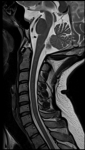
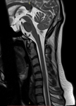
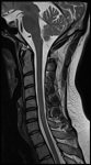
From my amateurish analysis, a lot of the measurements seem borderline or pathological:
CXA in neutral is around 139 degrees, which is borderline.
Grabb-Oakes line is around 7mm, which is borderline.
Translational BAI seems definitely equal to or above 2mm, which is pathological. BAI in flexion is around 9.5mm and BAI in extension is around 7mm.
I'm also seeing an empty sella, which could indicate intercranial hypertension.
There also seems to be spinal cord compression at around C5-C6 vertebrae when in the flexion position.
Are my observations anywhere near the mark?
My primary symptoms are fatigue and brain fog, and I don't have many of the ancillary symptoms that many others with CFS have.
However, the fatigue seems to get progressively worse every year. And recently I've developed mild facial pain as a symptom when fatigue is increased. My whole face feels like it's filled with lead.
There's also the constant feeling of a heavy head and a mild bobblehead feeling, and some degree of neck stiffness.
It would be really cool if @jeff_w, @JenB and @Hip could give their opinion.
From my amateurish analysis, a lot of the measurements seem borderline or pathological:
CXA in neutral is around 139 degrees, which is borderline.
Grabb-Oakes line is around 7mm, which is borderline.
Translational BAI seems definitely equal to or above 2mm, which is pathological. BAI in flexion is around 9.5mm and BAI in extension is around 7mm.
I'm also seeing an empty sella, which could indicate intercranial hypertension.
There also seems to be spinal cord compression at around C5-C6 vertebrae when in the flexion position.
Are my observations anywhere near the mark?
My primary symptoms are fatigue and brain fog, and I don't have many of the ancillary symptoms that many others with CFS have.
However, the fatigue seems to get progressively worse every year. And recently I've developed mild facial pain as a symptom when fatigue is increased. My whole face feels like it's filled with lead.
There's also the constant feeling of a heavy head and a mild bobblehead feeling, and some degree of neck stiffness.
Attachments
Last edited: