• Welcome to Phoenix Rising!

    Created in 2008, Phoenix Rising is the largest and oldest forum dedicated to furthering the understanding of and finding treatments for complex chronic illnesses such as chronic fatigue syndrome (ME/CFS), fibromyalgia (FM), long COVID, postural orthostatic tachycardia syndrome (POTS), mast cell activation syndrome (MCAS), and allied diseases.

    To become a member, simply click the Register button at the top right.

MRI: White Matter Lesions?!

Prefect

Senior Member
Messages
307
Location
Canada
My MRI shows 10 white matter lesions in the periventricular regions of my brain, each about 4mm in size. Funny thing is the neurologist who ordered it told me my MRI is normal except for compacted debris in my right maxillary sinus, and made no mention of the lesions. I found out later by obtaining my hospital records. Is this because he found the lesions clinically insignificant?

I'm 50 (not old enough to be getting brain lesions).

Does anyone else know enough about this subject to comment on the significance of this? Has anyone else had white matter lesions show up in MRI?
 

Zebra

Senior Member
Messages
851
Location
Northern California
Hi, @Prefect.

I have not had white matter lesions present on my brain MRIs, but I wanted to respond today because I am concerned for you.

I am under the care of an excellent neurologist, and I know that if a radiology report indicated that I had *10* white matter lesions that 1) she would tell me, 2) she would answer any questions I had about them, and 3) she would find them clinically significant, given the severity of my neurological symptoms/conditions.

I am appalled that you did not receive the same level of care from your neurologist. Ack!

Do you feel like you can talk to your present neuro about these findings?

If not, I would encourage you to seek a second opinion.

I usually do not write such strongly worded posts, but as mentioned above, I am concerned for you.

I hope you will keep us posted!

Best regards,
Z
 

andyguitar

Moderator
Messages
6,595
Location
South east England
Does anyone else know enough about this subject to comment on the significance of this? Has anyone else had white matter lesions show up in MRI?
A friend of mine had this show up in an MRI scan. They were told that it was probably associated with the migraines that had been having for years. So in their case NOT an indication of MS, Dementia or any of the other nasty things! Results of scans on their own are not a very good way of coming up with a diagnosis. Other things need to be considered as 'Abnormal' scans are not always clinically significant. Sounds like your neurologist needs to improve their bedside manner and at least tell you the lesions are not clinically significant.
 

PatJ

Forum Support Assistant
Messages
5,288
Location
Canada
I've had them show up on multiple MRIs. None of the neurologists I've seen thought that the lesions had any significance. One neuro said they can happen from rough sports, and that they're common in people with a history of migraine.

Here is a search of Phoenix Rising that lists a lot of past posts about white matter lesions.
 

Pyrrhus

Senior Member
Messages
4,172
Location
U.S., Earth
Periventricular T2 hyperintensities on MRI (sometimes referred to as "white matter abnormalities") are commonly found in the elderly, but are less common in younger people.

Nonetheless, neurologists routinely ignore these lesions as "normal" or "clinically insignificant", even though they clearly indicate some disease or aging process, & have been linked to an increased risk of cognitive problems.[1][2][3]

These T2 hyperintensities are also more common in ME patients:
https://www.me-pedia.org/wiki/Brain#T2_Hyperintensities_in_MRI
  • 1993: A comparison of brain MRI scans from 52 CFS patients and 52 controls found that 27% of CFS patients had findings considered abnormal, while only 2% of controls had findings considered abnormal. Abnormalities included T2 hyperintensities and ventricular enlargement.[4]
  • 1999: A comparison of brain MRI scans from 39 CFS patients and 19 controls found that the 21 CFS patients who did not have a psychiatric diagnosis had significantly more T2 hyperintensities, compared to either controls or the 18 CFS patients with a psychiatric diagnosis.[5]

Hope this helps.

References:
[1] https://dnd.or.kr/DOIx.php?id=10.12779/dnd.2015.14.3.114
[2] https://jnnp.bmj.com/content/76/3/362
[3] https://n.neurology.org/content/early/2021/01/27/WNL.0000000000011537
[4] https://doi.org/10.1016/0022-510x(93)90276-5
[5] https://doi.org/10.1016/s0022-510x(99)00243-9
 
Last edited:
Messages
8
20 years ago at the age of 40 my MRI reflected white matter lesions in the pons and midbrain. A neurologist and my primary said that it appeared to be the beginnings of Multiple Sclerosis. Other neurologists have not agreed. Subsequent MRI's indicated that there is no change of note.
 

Pyrrhus

Senior Member
Messages
4,172
Location
U.S., Earth
Dr. Michael Van Elzakker discusses enlarged perivascular spaces (those "T2 Hyperintensities" also known as dilated Virchow-Robin spaces) in this presentation from the CMRC2020 conference:

 
Messages
15
Ditto what everyone else is saying, though I've been told same as OP before and I'm 33.

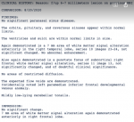
Here's my last MRI report if anyone's curious.
Screen Shot 2021-02-19 at 7.26.05 PM.png
 

Pyrrhus

Senior Member
Messages
4,172
Location
U.S., Earth
These T2 hyperintensities are also more common in ME patients:
https://www.me-pedia.org/wiki/Brain#T2_Hyperintensities_in_MRI

Oh, I forgot to mention that these T2 hyperintensities often correspond to "dilated perivascular spaces".

Now what on earth is a "dilated perivascular space"?

I try to explain it here:
Yes, the exact structure of the blood-brain-barrier (BBB) is very complicated to describe, even in a diagram.
It took me quite a while to understand it myself, and it's an active area of research so we're still learning.

The first thing to explain is that the blood-brain-barrier actually consists of multiple layers or barriers:
  • The outermost layers ("dura mater" and "arachnoid membrane/mater") enclose the brain on the outside, and do not penetrate into the interior of the brain.
  • Below the arachnoid membrane is a space filled with cerebrospinal fluid (CSF). This space is called the sub-arachnoid space.
  • Blood vessels that supply the brain first run through these outer layers and then penetrate into the interior of the brain.
  • The inner layers of the blood-brain-barrier ("pia mater" and the "astrocyte end-feet" AKA "glia limitans") surround and follow the penetrating blood vessels into the interior of the brain. So, the further into the interior of the brain, the fewer layers in the blood-brain-barrier.
  • However, between the penetrating blood vessels and the inner layers there is a space called the perivascular space. Toward the outside of the brain, this space is connected to the sub-arachnoid space and receives some cerebrospinal fluid from there.
  • But as you go further into the interior of the brain, the composition of the fluid in the perivascular space changes. This inner perivascular space may be filled with immune cells and too many immune cells clumped in one spot can result in a "dilated perivascular space".
  • Eventually, the fluid in this perivascular space drains out of the brain along the cranial nerves, emptying into lymph nodes. There, the fluid is processed before returning to the bloodstream.
Here are some rough diagrams that might help:

1621036538822-png.42887


1621035637318-png.42886


1621036608975-png.42888
 

heapsreal

iherb 10% discount code OPA989,
Messages
10,089
Location
australia (brisbane)
For what its worth. December 2020 had an unsual headache that was constant until about march but varying in intensity. Often wake at 2am or some crazy time from a headache and sometimes vomitting from them. Abit weird waking up suddenly like that.

Saw a neuro who eventually ordered the mri. A soon as i walked in the door and not even sitting down she ranted about seeing guys like me. I just have chronic migraines. She ranted on for 20mins. I couldnt hardly get a word out. Then i left with a form for an mri. I didnt return to the neuro, just a B___h and thats coming from my wife who was also in disbelief. Got mri done and had results also sent to gp.

Empty sellar syndrome, which i knew from ct scan
Periventricular T2/Flair hyperintensities (white matter lesions)
Chronic microangiopathic changes.

The neuro said the empty sellar syndrome that was on ct scan is nothing. But when you read into these things, yes it can be normal as in no symptoms or symptoms like headaches and different hormonal defiencies and their symptoms. I never got a chance to even talk about it and never looked at my recent hormone test where i had low dhea. Normally on dhea but stopped it the week before the test.

White matter lesions from my research can be from many things but they arent normal. MS comes up alot as do infections, migraines and many other condition.

Chronic microangiopathic changes is damage to small blood vessels. My guess probably inflammation from cfsme.

So my gp was shocked at the neuro and i said i wont be seeing her again. All the other neuro's have long waiting lists. I said to my gp that not much theyare going to do other than control bp and treat symptoms anyway. He agreed but also said we need to get ypur cholesterol lower. My bp ive had normal BPs 120s/70s during this period as well as high bp 180/110. But extra meds have helped the bp.

The weird headache i had finally stopped in march/april now just get my normal headaches woohoo. I actually think a big part of all this has been the intermittent supply of famvir. One of those herpes viruses, probably varicella has minced my brain.

12 months ago i tested high in homocysteine levels but have been on a b12/b6/folate formula. Maybe a sign of neuroinflammation.

The only thing i got out of it was when i saw the white matter lesions and instantly thought of dr cheney finding them in cfsme pts. Might help confirm cfsme for you or have you more confused and concerned. I think one needs an experienced neurologist in cfsme. Chronic migraines is their go too when they dont know.
 

vision blue

Senior Member
Messages
1,877
For My bp ive had normal BPs 120s/70s during this period as well as high bp 180/110.

The weird headache i had finally stopped ...I actually think a big part of all this has been the intermittent supply of famvir. One of those herpes viruses, probably varicella has minced my brain.

12 .

Can you say more about the herpes virus? My weird headaches and related (as well as lbp spikes) started with one as well. (The high bp can also cause white matter hyper intensities hough usually that takes longer )
 
Messages
97
Have had them show on MRI’s. A decent sized one in the periventricular region. My MRI report stated to follow up for suspicion of MS. The neurologist I was seeing mentioned getting spinal tap, but I declined it. just watching and waiting at this time.
 

heapsreal

iherb 10% discount code OPA989,
Messages
10,089
Location
australia (brisbane)
Can you say more about the herpes virus? My weird headaches and related (as well as lbp spikes) started with one as well. (The high bp can also cause white matter hyper intensities hough usually that takes longer )

About 20yrs ago my cfs started with 3 different herpes viruses within 6 months period, cmv, ebv and chickenpox age 31. I had chickenpox as a kid also.

2015 had shingles on left side of my head and have had many reactivations all were quite minor compared to initial infection, other than early 2016 when i stopped antivirals and had another severe episode. All on left side of my head. Prior to any shingles i was on antivirals mostly for cmv and i did stop them in early 2015 also when i got the initial shingles outbreak.

So for many years i thought my viral issues were probably cmv then ebv but as things have unfolded id say its varicella, although im not ruling out the other viruses. Different type of headaches and more severe headache episodes since shingles.

I cant give viral titre numbers because they just give igg or igm positive here in Australia. So drs dont believe in chronic viral infections unless its hiv or hepatitis or on immune suppressants for organ transplant. General blood work usually indicates im fighting some bug.

There is a condition called mollarettes meningitis. It involves herpes 2 or varicella and it causes benign meningitis. People usually get severe headaches lethargy, basically sounds like a cfs crash. Only way to diagnose it is with a spinal tap when in the middle of an attack but even then it can be negative. I have heard of some people having 3 or more episodes where they recieved spinal taps before they picked up these viruses in the spinal fluid that surrounds the brain. It wouldnt suprise me if other herpes viruses do the same thing or that those with cfsme who respond well to antivirals, probably have mollarettes. The treatment is IV antivirals or oral antivirals and pain killers. Many have ongoing cfsme type issues.
 

vision blue

Senior Member
Messages
1,877
Thanks much for the elqboration. Interesting history. I think i posted my history on my frequently recurrent/chronic herpetic infection. In my case i cant tolerate the antivirals and no doc has been willing to work with me to find a solution Ive read some of tge papers on hard to diagnose encephalitis which i believe i have from it and it (or autoantibodies) are progressing