mitoMAN
Senior Member
- Messages
- 629
- Location
- Germany/Austria
Discriminatory cytokine profiles predict muscle function, fatigue and cognitive function in patients with Myalgic Encephalomyelitis/Chronic Fatigue Syndrome (ME/CFS)
Abstract:
https://www.medrxiv.org/content/10.1101/2020.08.17.20164715v1
Full paper:
https://www.medrxiv.org/content/10.1101/2020.08.17.20164715v1.full.pdf
Abstract
Personal selection of outtakes:
The association of plasma cytokines with diagnosis and symptoms of ME: .... diagnosis of ME/CFS is significantly associated with increased plasma content of IL-10, MIP-1β, and RANTES and lower PDGF, IL-6, eotaxin, MIP-1α and VEGF. ...... higher plasma values of IL-1β, IL-8, IL-10, IP-10, and RANTES were significantly associated with higher Chalder Fatigue scores i.e. greater perceived fatigue"
Dysfunctional mitochondria have been reported in non-muscle cells from patients with ME/CFS (eg (28)) and so although the current study suggests that there are no major mitochondrial abnormalities in skeletal muscle mitochondria, it does not rule out the potential role of dysfunctional non-muscle mitochondria.
The source of plasma cytokines in patients with ME/CFS are unclear, but a number of cytokines can be produced by muscle (75-78). Studies have shown that the acute release of cytokines by muscle during exercise, a process which is quickly resolved, is likely to contribute to the positive effects of activity (78). In contrast, a chronic release of pro-inflammatory cytokines will undoubtedly have a detrimental effect on both muscle and non-muscle organs including the brain, and such cross-tissue interactions have been relatively well described during ageing (77). The specific increased production of IP-10 and MCP-1 by muscle is likely to have an effect on local tissue such as the peripheral nerve. This is supported by studies in inflammatory demyelinating neuropathies which imply a pathogenic role for IP-10 in the genesis of these neuropathies (79).
Myself:
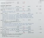
I am moderate-severe (can still leave the flat for around 20mins per day and go for a very slow walk) and have very high IL-1B, IL-8, TGF-B and borderline maximum IL-10. IP-10 is in range. Never checked RANTES tho.
My IL-6 is low as suggested by the study.
Their Cytokine profile seems to be matching my personal CFS profile pretty well.
Sadly some of the markers are not possible to be checked privately in Germany:
MIP-1B - MIP-1a, eotaxin, PDGF, G-CSF.
Abstract:
https://www.medrxiv.org/content/10.1101/2020.08.17.20164715v1
Full paper:
https://www.medrxiv.org/content/10.1101/2020.08.17.20164715v1.full.pdf
Abstract
Myalgic Encephalomyelitis (ME) /Chronic Fatigue Syndrome (CFS) is a severely debilitating and complex illness of uncertain aetiology, affecting the lives of millions and characterised by prolonged fatigue. The initiating factors and mechanisms leading to chronic debilitating muscle fatigue in ME/CFS are unknown and are complicated by the time required for diagnosis. Both mitochondrial dysfunction and inflammation have been proposed to be central to the pathogenesis of ME/CFS. This original and extensive study demonstrated that although there was little dysfunction evident in the muscle mitochondria of patients with ME/CFS, particular blood plasma and skeletal muscle cytokines, when adjusted for age, gender and cytokine interactions could predict both diagnosis and a number of measures common to patients with ME/CFS. These included MVC and perceived fatigue as well as cognitive indices such as pattern and verbal reaction times. We employed advanced multivariate analyses to cytokine profiles that leverages covariation and intrinsic redundancy to identify patterns of immune signaling that can be evaluated for their predictions of disease phenotype. The current study identified discriminatory cytokine profiles that can be sufficiently used to distinguish HCs from patients with ME/CFS and provides compelling evidence that a limited number of cytokines are associated with diagnosis and fatigue. Moreover, this study demonstrates significant potential of using multiplex cytokine profiles and bioinformatics as diagnostic tools for ME/CFS, potentiating the possibility of not only diagnosis, but also being able to individually personalise therapies.
Personal selection of outtakes:
The association of plasma cytokines with diagnosis and symptoms of ME: .... diagnosis of ME/CFS is significantly associated with increased plasma content of IL-10, MIP-1β, and RANTES and lower PDGF, IL-6, eotaxin, MIP-1α and VEGF. ...... higher plasma values of IL-1β, IL-8, IL-10, IP-10, and RANTES were significantly associated with higher Chalder Fatigue scores i.e. greater perceived fatigue"
Dysfunctional mitochondria have been reported in non-muscle cells from patients with ME/CFS (eg (28)) and so although the current study suggests that there are no major mitochondrial abnormalities in skeletal muscle mitochondria, it does not rule out the potential role of dysfunctional non-muscle mitochondria.
The source of plasma cytokines in patients with ME/CFS are unclear, but a number of cytokines can be produced by muscle (75-78). Studies have shown that the acute release of cytokines by muscle during exercise, a process which is quickly resolved, is likely to contribute to the positive effects of activity (78). In contrast, a chronic release of pro-inflammatory cytokines will undoubtedly have a detrimental effect on both muscle and non-muscle organs including the brain, and such cross-tissue interactions have been relatively well described during ageing (77). The specific increased production of IP-10 and MCP-1 by muscle is likely to have an effect on local tissue such as the peripheral nerve. This is supported by studies in inflammatory demyelinating neuropathies which imply a pathogenic role for IP-10 in the genesis of these neuropathies (79).
Myself:
I am moderate-severe (can still leave the flat for around 20mins per day and go for a very slow walk) and have very high IL-1B, IL-8, TGF-B and borderline maximum IL-10. IP-10 is in range. Never checked RANTES tho.
My IL-6 is low as suggested by the study.
Their Cytokine profile seems to be matching my personal CFS profile pretty well.
Sadly some of the markers are not possible to be checked privately in Germany:
MIP-1B - MIP-1a, eotaxin, PDGF, G-CSF.
Last edited: