• Welcome to Phoenix Rising!

    Created in 2008, Phoenix Rising is the largest and oldest forum dedicated to furthering the understanding of and finding treatments for complex chronic illnesses such as chronic fatigue syndrome (ME/CFS), fibromyalgia (FM), long COVID, postural orthostatic tachycardia syndrome (POTS), mast cell activation syndrome (MCAS), and allied diseases.

    To become a member, simply click the Register button at the top right.

Association between Chronic Pain and Alterations in the Mesolimbic Dopaminergic System

pattismith

Senior Member
Messages
3,932
Association between Chronic Pain and Alterations in the Mesolimbic Dopaminergic System


oct 2020


Seoyon Yang
1, Mathieu Boudier-Revéret 2, Yoo Jin Choo 3, Min Cheol Chang 4

Free PMC article
Abstract

Chronic pain (pain lasting for >3 months) decreases patient quality of life and even occupational abilities. It can be controlled by treatment, but often persists even after management.

To properly control pain, its underlying mechanisms must be determined.

This review outlines the role of the mesolimbic dopaminergic system in chronic pain.

The mesolimbic system, a neural circuit, delivers dopamine from the ventral tegmental area to neural structures such as the nucleus accumbens, prefrontal cortex, anterior cingulate cortex, and amygdala. It controls executive, affective, and motivational functions.

Chronic pain patients suffer from low dopamine production and delivery in this system.

The volumes of structures constituting the mesolimbic system are known to be decreased in such patients.

Studies on administration of dopaminergic drugs to control chronic pain, with a focus on increasing low dopamine levels in the mesolimbic system, show that it is effective in patients with Parkinson's disease, restless legs syndrome, fibromyalgia, dry mouth syndrome, lumbar radicular pain, and chronic back pain.

However, very few studies have confirmed these effects, and dopaminergic drugs are not commonly used to treat the various diseases causing chronic pain.

Thus, further studies are required to determine the effectiveness of such treatment for chronic pain.

1608208131408.png
 

xebex

Senior Member
Messages
840
Yes I tried modafinil too and it was helpful for motivations but not pain and not as helpful as Ritalin - I couldn’t get out of bed the next day I think it did something to my acetylcholine receptors because I get the same day after hangover/extreme sleepiness from mestinon and Benadryl. Ritalin was the lesser of the 3 in side effects/hangover/ rebound.
 

pattismith

Senior Member
Messages
3,932
This 2018 study found the same link between hyperalgesia and an altered mesolimbic dopamine circuit.


Activation of ventral tegmental area dopaminergic neurons reverses pathological allodynia resulting from nerve injury or bone cancer
Moe Watanabe 1, Michiko Narita 1, Yusuke Hamada 1, Akira Yamashita 1, Hideki Tamura 2, Daigo Ikegami 1 3, Takashige Kondo 1, Tatsuto Shinzato 1, Takatsune Shimizu 4, Yumi Fukuchi 4, Akihiro Muto 4, Hideyuki Okano 2 5, Akihiro Yamanaka 6, Vivianne L Tawfik 7, Naoko Kuzumaki 1, Edita Navratilova 3, Frank Porreca 3, Minoru Narita 1 2
Affiliations expand
Free PMC article

Abstract

Chronic pain induced by nerve damage due to trauma or invasion of cancer to the bone elicits severe ongoing pain as well as hyperalgesia and allodynia likely reflecting adaptive changes within central circuits that amplify nociceptive signals.

The present study explored the possible contribution of the mesolimbic dopaminergic circuit in promoting allodynia related to neuropathic and cancer pain.

Mice with ligation of the sciatic nerve or treated with intrafemoral osteosarcoma cells showed allodynia to a thermal stimulus applied to the paw on the injured side.

Patch clamp electrophysiology revealed that the intrinsic neuronal excitability of ventral tegmental area (VTA) dopamine neurons projecting to the nucleus accumbens (N.Acc.) was significantly reduced in those mice.

We used tyrosine hydroxylase (TH)-cre mice that were microinjected with adeno-associated virus (AAV) to express channelrhodopsin-2 (ChR2) to allow optogenetic stimulation of VTA dopaminergic neurons in the VTA or in their N.Acc. terminals.

Optogenetic activation of these cells produced a significant but transient anti-allodynic effect in nerve injured or tumor-bearing mice without increasing response thresholds to thermal stimulation in sham-operated animals.

Suppressed activity of mesolimbic dopaminergic neurons is likely to contribute to decreased inhibition of N.Acc. output neurons and to neuropathic or cancer pain-induced allodynia suggesting strategies for modulation of pathological pain states.
 

pattismith

Senior Member
Messages
3,932
@Hip

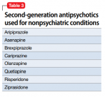
this 2020 paper is about antipsychotic drugs for non psychiatric illnesses. Some alternatives to Aripiprazole may be possible...
For each drug, there is a short paragraph about effect on brain Dopamine...
1615789146501.png

View attachment 42000


Most antipsychotics will work on dopamine, as the dopamine theory of schizophrenia posits that an over-active dopamine D2 receptor drives the positive symptoms of schizophrenia. Whereas the negative symptoms of schizophrenia are thought due to D1 under-activation.

Traditional antipsychotics work by blocking D2 in a blunt way, but these third generation antipsychotics use the more subtle dopamine system stabilization mechanism, and don't just uniformly block D2, but rather block the strong dopamine signals on D2, but let the weaker dopamine signals through.


If this dopamine system stabilization is the mechanism by which Abilify helps ME/CFS, then other dopamine system stabilizer drugs may work equally well for ME/CFS. Dopamine system stabilizer drugs include:

Amisulpride
Aripiprazole
Brexpiprazole
Cariprazine
Pridopidine
OSU61612

The first four are all third generation antipsychotics. The last two are not available as licensed drugs.
 

Attachments

  • 13075_2014_Article_480.pdf
    432.9 KB · Views: 15
  • 1615789138175.png
    1615789138175.png
    36.1 KB · Views: 15
Last edited:

pattismith

Senior Member
Messages
3,932
Disruption of brainstem monoaminergic fibre tracts in multiple sclerosis as a putative mechanism for cognitive fatigue: a fixel-based analysis
Tiziana Carandini
2021
https://doi.org/10.1016/j.nicl.2021.102587

Abstract
..... In this study, we aimed to compare the integrity of the monoaminergic white matter fibre tracts projecting from brainstem nuclei in a group of patients with MS (n = 68) and healthy controls (n = 34), and to investigate its association with fatigue.

Fibre tracts integrity was assessed with the novel fixel-based analysis that simultaneously estimates axonal density, by means of ‘fibre density’, and white matter atrophy, by means of fibre ‘cross section’.

We focused on ventral tegmental area, locus coeruleus, and raphe nuclei as the main source of dopaminergic, noradrenergic, and serotoninergic fibres within the brainstem, respectively.

Fourteen tracts of interest projecting from these brainstem nuclei were reconstructed using diffusion tractography, and compared by means of the product of fibre-density and cross-section (FDC).

Finally, correlations of monoaminergic axonal damage with the modified fatigue impact scale scores were evaluated in MS.

Fixel-based analysis revealed significant axonal damage – as measured by FDC reduction – within selective monoaminergic fibre-tracts projecting from brainstem nuclei in MS patients, in comparison to healthy controls; particularly within the dopaminergic-mesolimbic pathway, the noradrenergic-projections to prefrontal cortex, and serotoninergic-projections to cerebellum.

Moreover, we observed significant correlations between severity of cognitive fatigue and axonal damage within the mesocorticolimbic tracts projecting from ventral tegmental area, as well as within the locus coeruleus projections to prefrontal cortex, suggesting a potential contribution of dopaminergic and noradrenergic pathways to central fatigue in MS.

Our findings support the hypothesis that axonal damage along monoaminergic pathways contributes to the reduction/dysfunction of monoamines in MS and add new information on the mechanisms by which monoaminergic systems contribute to MS pathogenesis and fatigue.

This supports the need for further research into monoamines as therapeutic targets aiming to combat and alleviate fatigue in MS.
 

pattismith

Senior Member
Messages
3,932
The Role of Mesostriatal Dopamine System and Corticostriatal Glutamatergic Transmission in Chronic Pain
by
Barbara Ziółkowska


Department of Molecular Neuropharmacology, Maj Institute of Pharmacology, Polish Academy of Sciences, 12 Smętna Str., 31-343 Kraków, Poland

Brain Sci. 2021, 11(10), 1311; https://doi.org/10.3390/brainsci11101311

Published: 2 October 2021

Abstract

There is increasing recognition of the involvement of the nigrostriatal and mesolimbic dopamine systems in the modulation of chronic pain.

The first part of the present article reviews the evidence indicating that dopamine exerts analgesic effects during persistent pain by stimulating the D2 receptors in the dorsal striatum and nucleus accumbens (NAc).

Thereby, dopamine inhibits striatal output via the D2 receptor-expressing medium spiny neurons (D2-MSN).

Dopaminergic neurotransmission in the mesostriatal pathways is hampered in chronic pain states and this alteration maintains and exacerbates pain.

The second part of this article focuses on the glutamatergic inputs from the medial prefrontal cortex to the NAc, their activity changes in chronic pain, and their role in pain modulation.

Finally, interactions between dopaminergic and glutamatergic inputs to the D2-MSN are considered in the context of persistent pain.

Studies using novel techniques indicate that pain is regulated oppositely by two independent dopaminergic circuits linking separate parts of the ventral tegmental area and of the NAc, which also interact with distinct regions of the medial prefrontal cortex.
 

pattismith

Senior Member
Messages
3,932
1635143589834.png

Figure 1.
Interactions between the Ventral Mesostriatal Dopaminergic Pathway and Corticostriatal Glutamatergic Afferents in Chronic Pain. The scheme presents only those neuronal connections, whose participation in pain processing and involvement in chronic pain have been documented directly.

Glutamatergic, dopaminergic, and GABAergic neurons are depicted as orange, blue, and green ovals, respectively, and their projections are depicted as arrows. “+” and “−“ signs denote excitatory and inhibitory effects on the postsynaptic cell, respectively.

Left panel. “A” and “H” placed along projections denote effects of activation of the particular projection on pain responses: analgesia and hyperalgesia/pain, respectively. These signs reflect effects on both sensory and affective aspects of pain, or either (see text for details), and show general tendencies, some of which become explicit only under tonic or chronic pain.

Right panel. Changes in the activity of particular neuronal populations and their projections under chronic pain have been reflected by the “cell body” size and the arrow width. ↑ and ↓ arrows denote the final effect on pain (increase and decrease, respectively) resulting from both the function of a particular projection in pain modulation (refer to “A” and “H” on the left panel) and changes in the activity of this projection in chronic pain.

?*–In contrast to some other studies, Ren et al. [67] provide evidence of an increase in dopaminergic neurotransmission in the medial VTA–medial NAc shell projection in chronic pain. This scheme has been based on the studies by Schwartz et al. [89], Lee et al. [81], Ren et al. [63,67], and Gao et al. [85]. The nigrostriatal dopamine pathway has not been included in the figure due to the lack of corresponding studies on the dorsal striatum.

ACC—anterior cingulate cortex;
BLA—basolateral amygdala;
D1-MSN—D1 receptor-expressing medium spiny neuron;
D2-MSN—D2 receptor-expressing medium spiny neuron;
DA—dopamine;
IL—infralimbic cortex;
LTD—long-term depression;
LTP—long-term potentiation;
mNAc-sh—medial nucleus accumbens shell;
NAc-c—nucleus accumbens core;
PL—prelimbic cortex;
VTA—ventral tegmental area.
 
Last edited:

pattismith

Senior Member
Messages
3,932
Lipopolysaccharide (LPS) and Amyloid Beta (Aβ) activate astrocytes and microglial cells via toll-like 4 (TLR4) receptors leading to neuroinflammation.

Reactive (activated) astrocytes mainly comprising of A1 astrocytes (A1s) are involved in neuroinflammation, while A2 astrocytes (A2s) possess neuroprotective activity.

Studies link low dopamine (DA) levels during the early stages of neurodegenerative disorders with its anti-inflammatory and immuoregulatory properties.

DA mediates neuroprotection via inhibition of the A1 astrocytic pathway through blockade of NF-kB and nucleotide-binding oligomerization domain-like receptor pyrin domain-containing 3 (NLRP3); and promotion of A2 astrocytic pathways leading to the formation of neurotrophic factors like BDNF and GDNF

Dopaminergic Signaling as a Plaus... Neuroinflammation and Cognition: Ingenta Connect Fast Track Article
2022
 

Wishful

Senior Member
Messages
5,684
Location
Alberta
It's really amazing how complicated all those interactions are, forming an organism that works fairly reliably. If cars or computers had that level of complex interaction, they would be breaking down constantly.

It's not good for finding the root cause of ME though. Which one of those interactions is slightly abnormal possibly in a limited region of the brain? How to find it? That's why I think we're more likely to discover a factor that reliably affects PWME--positive or negative--by accident, which can then lead us to the true cause. So, everyone should keep trying new things and reporting anything that has a reliable effect.