Role of dysfunctional glycolysis...?

Messages
10
TL;DR - is it possible that instead of insufficient ATP, we're wasting it via different mechanisms than mitochondrial ATP, such as too much glycolysis?

As an (on-hold) national-level athlete, the worst symptom for me is myopathy. Most of my research centers around mechanisms to improve myopathy and fatigue, primarily skeletal muscle-related as well as general. In addition to all my own health-related research, I do a lot of tutoring in AP/college level bio, among other things, so I'm well-versed in the cellular mechanisms and energetics. I came across this today:

https://www.ncbi.nlm.nih.gov/pmc/articles/PMC5065105/

https://en.wikipedia.org/wiki/Inborn_errors_of_carbohydrate_metabolism#Galactose

Which surprised me, since everything else I've read to date seems to think that we have errors in cellular/mitochondrial energetics, sometimes specified as Complex IV/V energy transport issues. To be clear, our errors are less likely to be inborn, but we still may have issues with how our bodies process carbohydrates, which could also explain why some people do better on low carb/keto diets. As an ethical vegetarian, I did go low carb/keto for a few months, but I didn't gain much benefit, but my body did seem to appreciate going back to carbs once I discovered that ketosis is bad for muscle-fiber type switching (I need to stay fast-twitch dominant).

What really resonated with me was the one that referred to potential crossovers with severe asthma, which I also have. This makes it sound like our little hamsters are spinning in their wheels; as opposed to being unable to produce sufficient ATP, they're wasting it through too much glycolysis, which is known to speed up aging and aging-related issues.

Anyone have any insight? To the best of my knowledge, this hasn't been discussed much in these communities.
 

Zebra

Senior Member
Messages
1,000
Location
Northern California
Hi, @shadowraith

Have you already investigated Glycogen Storage Diseases as an alternative diagnosis to ME/CFS?

Many years ago I had a very smart and committed neurologist who tested me for Pompes's Disease, and we were both surprised when it came back negative.

It would have been nice to have done a wider genetic panel for all of the Glycogen Storage Diseases.

Best wishes to you regarding your health!
 

Murph

:)
Messages
1,803
One theory I have on ATP is cells might be expelling it as a signal.

They call this purinergic signalling and it is a danger signal. https://en.wikipedia.org/wiki/Purinergic_signalling.

Cells use it to signal to other cells that something is wrong.

One potentially very relevant example is red blood cells. They use ATP to signal to blood vessels that the vessel is too narrow and they need to get through. The cloud of ATP tells the blood vessel to relax and open up.

Of course in people with autonomic or endothelial issues that ATP expulsion may not achieve anything and the red blood cells might just run out of ATP. Which makes them lose their shape.

When ATP is expelled from a cell it doesn't last long before it is broken down to other things. We can't measure it directly.

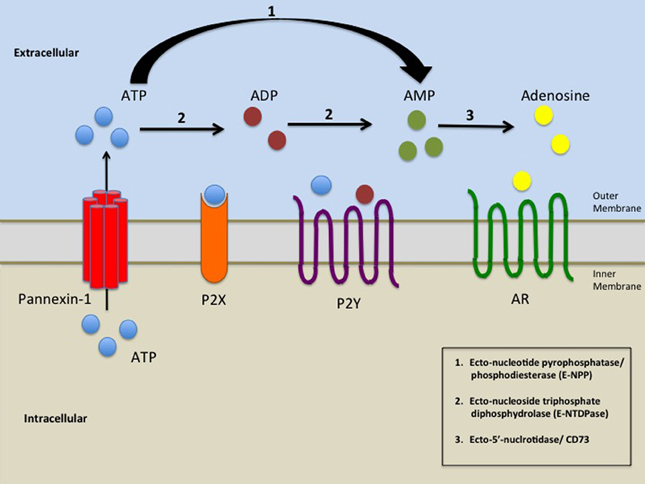
There are some signs that we have issues with the breakdown of ATP. Here's the pathway. It goes from ATP to ADP then AMP then adenosine.

Purinergic_signalling.jpg


MECFS patients have high amounts of AMP but low levels of adenosine, according to measurements from Hanson, which you can see here on my webapp: (https://jasemurphy.shinyapps.io/Germainetal2022/ use the dropdown menu at top left to choose amp and then adenosine.)

it's possible something's wrong with our cd73 enzyme that is supposed to be converting AMP to the immune-suppressing adenosine.

Then high amp is likely to trigger the Ampk sensor, telling the cell to go into catabolic mode to try to make atp, Rather than muscle-building mode. the body perceives it is starving and mtor is inhibited (mtor can be turned on by glucose and amino acids). although importantly this would cause less glycolysis not more! the body uses glycolysis more during anabolism, when it is using lipids and amino acids to build stuff. it uses glycolysis less and relies on lipids for energy more during catabolism

Just one theory of one possible set of connections in the body!

Link explaining ampk-mtor a little bit: https://www.sciencedirect.com/science/article/abs/pii/S1874604710280034
 
Last edited:

linusbert

Senior Member
Messages
1,466
just watched that video and think its somewhat related to post here,


basically excessive sugar or oxidative stress can shut down energy production to stop excessive radicals...
 
Messages
10
i need carbs, fast easy carbs. every 2 hours.
what improved me was pioglitazon. just started a thread about this https://forums.phoenixrising.me/thr...zon-actos-which-works-on-same-pathways.91261/
it also shifts energy production towards fats.

methylene blue can help with complex 4/5 issues. b2 with complex 1-2
pioglitazone (and other glitazones) can cause water retention and weight gain, which is why I haven't tried them. methylene blue can turn you blue, lol and it can also worsen muscle weakness. I used to do b2, but it never made a difference. Glad pioglitazone worked for you though! A lot of things that agonize ppar-(various Greek letters) cause muscle fiber type switching as well, so that's a red flag also
 
Messages
10
Hi, @shadowraith

Have you already investigated Glycogen Storage Diseases as an alternative diagnosis to ME/CFS?

Many years ago I had a very smart and committed neurologist who tested me for Pompes's Disease, and we were both surprised when it came back negative.

It would have been nice to have done a wider genetic panel for all of the Glycogen Storage Diseases.

Best wishes to you regarding your health!
I just came across them in more detail the other day, so I need to do more digging. Thanks, best wishes to you as well!
 
Messages
10
One theory I have on ATP is cells might be expelling it as a signal.

They call this purinergic signalling and it is a danger signal. https://en.wikipedia.org/wiki/Purinergic_signalling.

Cells use it to signal to other cells that something is wrong.

One potentially very relevant example is red blood cells. They use ATP to signal to blood vessels that the vessel is too narrow and they need to get through. The cloud of ATP tells the blood vessel to relax and open up.

Of course in people with autonomic or endothelial issues that ATP expulsion may not achieve anything and the red blood cells might just run out of ATP. Which makes them lose their shape.

When ATP is expelled from a cell it doesn't last long before it is broken down to other things. We can't measure it directly.

There are some signs that we have issues with the breakdown of ATP. Here's the pathway. It goes from ATP to ADP then AMP then adenosine.

Purinergic_signalling.jpg


MECFS patients have high amounts of AMP but low levels of adenosine, according to measurements from Hanson, which you can see here on my webapp: (https://jasemurphy.shinyapps.io/Germainetal2022/ use the dropdown menu at top left to choose amp and then adenosine.)

it's possible something's wrong with our cd73 enzyme that is supposed to be converting AMP to the immune-suppressing adenosine.

Then high amp is likely to trigger the Ampk sensor, telling the cell to go into catabolic mode to try to make atp, Rather than muscle-building mode. the body perceives it is starving and mtor is inhibited (mtor can be turned on by glucose and amino acids). although importantly this would cause less glycolysis not more! the body uses glycolysis more during anabolism, when it is using lipids and amino acids to build stuff. it uses glycolysis less and relies on lipids for energy more during catabolism

Just one theory of one possible set of connections in the body!

Link explaining ampk-mtor a little bit: https://www.sciencedirect.com/science/article/abs/pii/S1874604710280034
^ which is why I stay away from things that are bad for MTOR, exactly. We do know we have endothelial dysfunction, but ISRIB didn't help for me.
 
Messages
10
just watched that video and think its somewhat related to post here,


basically excessive sugar or oxidative stress can shut down energy production to stop excessive radicals...
Well we do know we have insane amounts of oxidative stress, but no amount of any antioxidant has ever made a difference for me, so it doesn't seem like targeting that directly has been productive for me at least. Oxidative stress can do a bunch of nasty things
 

datadragon

Senior Member
Messages
429
Location
USA
There are some signs that we have issues with the breakdown of ATP. Here's the pathway. It goes from ATP to ADP then AMP then adenosine. MECFS patients have high amounts of AMP but low levels of adenosine, according to measurements from Hanson
Hmm, this was interesting...

Zinc deficiency potently decreases the activities of extracellular adenine-nucleotide-hydrolyzing ectoenzymes, delaying both extracellular ATP clearance and adenosine generation. Our findings indicate that the activity of ENPP1, ENPP3, NT5E/CD73, and TNAP, which are involved in the regulation of purinergic signaling, decreased under zinc-deficient conditions.. Thus, the zinc status can alter extracellular adenine-nucleotide metabolism. Zinc can promote non-REM sleep, whereas defective non-REM sleep responses to sleep deprivation are found in Nt5e/Cd73-KO mice. Zinc can prevent diarrhea, whereas allergen-induced diarrhea (inflammatory diarrhea) is found in Enpp3-KO mice. Both ATP/ADP and adenosine are widely recognized to mediate opposite physiological effects, such as pain signaling by ATP and ADP vs. pain relief by adenosine, and pro-inflammatory effects of ATP vs. anti-inflammatory effects of adenosine; zinc alleviates pain, whereas zinc deficiency is associated with pain. Zinc is also a potent anti-inflammatory nutrient, and its deficiency leads to a pro-inflammatory state. Moreover, zinc supplementation has been shown to ameliorate some defects associated with the loss of zinc-requiring ectoenzymes involved in extracellular adenine-nucleotide metabolism.

Zinc deficiency causes delayed ATP clearance and adenosine generation in rats and cell culture models. Considering that several ectoenzymes involved in purinergic signaling through extracellular adenine-nucleotide hydrolysis possess zinc ions in their active sites, and disorders in purinergic signaling result in diverse diseases that are frequently similar to those caused by zinc deficiency, herein we examine whether zinc deficiency affects extracellular adenine-nucleotide metabolism. Zinc deficiency severely impairs the activities of major ectoenzymes (ENPP1, ENPP3, NT5E/CD73, and TNAP), and also strongly suppresses adenine-nucleotide hydrolysis in cell-membrane preparations or rat plasma, thereby increasing ATP and ADP levels and decreasing adenosine levels. Thus, zinc deficiency delays both extracellular ATP clearance and adenosine generation, and zinc modulates extracellular adenine-nucleotide metabolism. Since the finely tuned balance between extracellular adenine nucleotides and adenosine is critical for purinergic signaling, these findings provide a novel insight into why zinc deficiency results in diverse symptoms. Furthermore, the regulatory roles of zinc in insulin metabolism can suggest its association with dysregulation of glucose metabolism under zinc deficiency https://www.nature.com/articles/s42003-018-0118-3

Extracellular ATP can cause P2X receptors to activate the NOD-like receptor 3 (NLRP3) inflammasome and cause IL-1β and IL-18 maturation and release. https://pubmed.ncbi.nlm.nih.gov/23434541/

NLRP3 inflammasome activation leads to endoplasmic reticulum stress which causes high WASF3 and disrupts Mitochondrial function, while blocking ER stress lowered WASF3 levels and restored mitochondrial function. Zinc deficiency evokes the endoplasmic reticulum (ER)-stress response https://pubmed.ncbi.nlm.nih.gov/23748779/ SOD1 as a molecular switch for initiating the homeostatic ER stress response under zinc deficiency https://pubmed.ncbi.nlm.nih.gov/24076220/ Butyrate inhibits ER stress as mentioned but also zinc https://pubmed.ncbi.nlm.nih.gov/32549180/ (and low butyrate is a downstream effect of the unavailability of zinc during inflammation/infection.

Acute immune activation suppresses oxidative phosphorylation-mediated ATP production and favors Warburg glycolysis, a state that is coupled with increased succinate levels. Notably, both of these changes facilitate NLRP3 inflammasome activation. high levels of the metabolite succinate can support IL-1β expression by stabilizing hypoxia-inducible factor 1-alpha (HIF-1α) for IL-1 β transcript expression to occur. This process has recently shown to be inhibited by the anti-inflammatory metabolite itaconate, a TCA cycle off-shoot metabolite that is produced by decarboxylation of cis-aconitate of the TCA cycle by the enzyme immune responsive gene 1. Itaconate inhibits the activity of succinate dehydrogenase and mitochondrial respiration in LPS-activated macrophages, concurrently, decreasing LPS-induced mtROS. Itaconate can also activate the anti-inflammatory cellular programming of nuclear factor erythroid 2-related factor 2 (Nrf2) via alkylation of cysteine residues on the protein KEAP1. Additionally, itaconate can inhibit LPS-induced IL-1β secretion by impairing glycolytic flux via targeting glycolytic enzymes GAPDH or fructose-bisphosphate aldolase A https://www.ncbi.nlm.nih.gov/pmc/articles/PMC7463618/

Succinate accumulation, along with the induction of the glycolytic enzyme hexokinase-1, increases the activity of the respiratory chain complex II, promotes the production of mROS, stabilizes HIF-1α, regulates the transcription of pro-IL-1β, and activates the NOD-like receptor protein 3 (NLRP3) inflammasome, increasing the production of IL-1β (which increases c-reactive protein). https://forums.phoenixrising.me/threads/an-acod1-genomics-question.90574/#post-2441131

NLRP3 lowers levels of shank3, which is also linked to zinc uptake in the gut. During inflammation or infection, zinc uptake levels are lowered by also lowering levels of Shank3. we have reported expression of SHANK3 in human enterocytes, where SHANK3 was functionally linked to zinc (Zn) transporter levels mediating Zinc absorption. We detected decreased expression of Zn uptake transporters ZIP2 and ZIP4 on mRNA and protein level correlating with SHANK3 expression levels, and found reduced levels of ZIP4 protein co-localizing with SHANK3 at the plasma membrane. We demonstrated that especially ZIP4 exists in a complex with SHANK3. Zip2 and Zip4 proteins act as key players of zinc absorption in enterocytes. Low enterocytic SHANK3 levels result in diminished Zn transporter levels that could eventually explain reduced Zn concentrations in tissues and organs https://www.ncbi.nlm.nih.gov/pubmed/28345660/
 
Last edited:
Messages
10
And WASF3 is the evil me/cfs from that one article where that lady went into remission from an experimental drug that would be very difficult/expensive to obtain…
 
Back