• Welcome to Phoenix Rising!

    Created in 2008, Phoenix Rising is the largest and oldest forum dedicated to furthering the understanding of, and finding treatments for, complex chronic illnesses such as chronic fatigue syndrome (ME/CFS), fibromyalgia, long COVID, postural orthostatic tachycardia syndrome (POTS), mast cell activation syndrome (MCAS), and allied diseases.

    To become a member, simply click the Register button at the top right.

Possible Brain Biomarker for ME/CFS Found

CantThink

Senior Member
Messages
800
Location
England, UK
When I first came down with Infectious Mono as a teenager I went from being a straight A student to one who couldn't read a single sentence and make any sense of it in any of the languages that I spoke and still speak.

I struggled and suffered without understanding what was going on with me, through many many ups and downs I managed to finish high school, but had to repeat junior high school year. Back then there was no home schooling support in Italy and I wonder if nowadays doctors and teachers are more aware of what is the best treatment for young people with IM and/or ME: rest, rest, rest and more rest!

I managed to go to law school and graduate, it was also a brutal and grueling experience, as many of you unfortunately know and can imagine I had to follow the waxing and waning of all the physical and neurological symptoms. It was like living through pure hell!

Same here! I went from being a grade A student who was in the top 5 students in my class to being unable to do things I'd learnt. This especially hit me in maths - I suddenly couldn't do long division, fractions, multiplication and anything mental arithmetic was a no go as I couldnt hold information in my head, do another part of the sum and then retrieve the first part's answer to combine with the second answer.

I struggled through school and years later at university I went through that gruelling, brutal living hell.. It was like torturing myself! I had to quit one class and I spent 4 years doing double the work of my peers to compensate for my mental 'deficiencies'/disabilities. It was like mentally wading through mud.
 

Marco

Grrrrrrr!
Messages
2,386
Location
Near Cognac, France
Specificity is MUCH more important for us than sensitivity. Its critical. Sensitivity would be expected to be lower because even under a tight definition it would still be a somewhat heterogeneous group. If 100% specificity could be shown in a larger replication, we might have a biomarker, provided only that other disorders don't have a similar finding.

Specificity is high c/w controls but I wonder how it would stack up against ASD for example?

White matter connectivity in children with autism spectrum disorders: a tract-based spatial statistics study

Results

The TBSS analysis revealed widespread increase of fractional anisotropy (FA) in major WM pathways. The tractographic approach showed an increased fiber length and FA in the cingulum and in the corpus callosum and an increased mean diffusivity in the indirect segments of the right arcuate and the left cingulum. Mean diffusivity was also correlated with expressive language functioning in the left indirect segments of the arcuate fasciculus.

http://www.biomedcentral.com/1471-2377/12/148


alex3619

Senior Member
Messages
13,810
Location
Logan, Queensland, Australia
Specificity is high c/w controls but I wonder how it would stack up against ASD for example?

Exactly. Follow-up studies will have to be more carefully designed if they want to look at a possible biomarker, and the selection of control groups will be important. However this study was more exploratory ... the first of its kind?
 

Nielk

Senior Member
Messages
6,970
Exactly. Follow-up studies will have to be more carefully designed if they want to look at a possible biomarker, and the selection of control groups will be important. However this study was more exploratory ... the first of its kind?

We seem to have many of these small studies that seem promising but no one replicates them with a larger study. Is it all because of the lack of funding? This is beyond frustrating. It's like being constantly teased and no follow up.
 

NK17

Senior Member
Messages
592
We seem to have many of these small studies that seem promising but no one replicates them with a larger study. Is it all because of the lack of funding? This is beyond frustrating. It's like being constantly teased and no follow up.
Maybe and I hope to be right this group of researchers from Stanford will continue to do and publish studies.
 

MeSci

ME/CFS since 1995; activity level 6?
Messages
8,231
Location
Cornwall, UK
I have trouble speaking when my symptoms are worse. I can picture the words in my head, but they are just out of reach. I try to talk and the words don't come. When I get migraines I can't speak. Twice I have ended up in an ambulance as people think I have had a stroke.

I had quite serious aphasia two days in a row, with 'ME normality' in between, a few weeks ago. I kept writing and saying the wrong words, and couldn't make sense of the radio listings, being unable to recognise the one-word title of a programme I listened to regularly. I also had a headache, and a chill one day and fever the next, and had nausea and loss of appetite. My GP thought I must have had a TIA, although I could speak, and smile, I think, and move normally, and had no facial paralysis at the time. Had an MRI and neck scan, and neuro decided it was migraine - not something I am known to have a history of.
 

Sidereal

Senior Member
Messages
4,856
It's good to see a study like this using diffusion tensor imaging which is a more sensitive technique for picking up subtle white matter abnormalities (as opposed to gaping holes) than normal T2 weighted MRI.

We have of course had the perennial problem of one patient after another being sent for an MRI and then having their white matter hyperintensities (also known as unidentified bright objects or high signal intensity areas) dismissed as "nothing" or "nonspecific" or "don't worry about it" since the earliest days of this technology when, back in the mid-1980s, Cheney and Peterson began referring their Incline Village ME victims for MRI scans. Of course even today no one is sure of the clinical significance of these. Some neurologists seem to think that once they've ruled out MS, they should to refer us to psychiatry to get "help".

@NK17, I am not a lawyer but apart from that detail you might as well be describing my life. Graduate school was complete hell with waxing and waning but nonetheless ever-worsening encephalopathy which was painfully embarrassing & incomprehensible to me at the time. This went on for several years until I collapsed completely with severe ME. All this came after a period of near remission in my late teens/early twenties which enabled me to get through college and first year of grad school relatively unscathed, although I still had significant SNS excess (in hindsight, I was running on norepinephrine fumes during those years) and exertion-related FM type pain and other symptoms, including suboptimal energy levels. Back then I foolishly thought I had left behind those nightmarish high school years of my first housebound spell due to what I now know to be POTS but what was at the time diagnosis-free hell regarded by others as being nothing/stress/perfectionism/trying to avoid school/etc.

It's interesting that you should bring up Byron Hyde because when I saw this press release I was immediately reminded of Sheila Bastien's chapter in Hyde's 1992 textbook on myalgic encephalomyelitis. Bastien is/was a clinical neuropsychologist who did detailed neuropsych testing on the Peterson/Cheney Incline Village cohort that formed the sample for the "famous" 1992 Annals of Internal Medicine paper. You don't need expensive neuroimaging to show these deficits. In actuality, pen and paper tests and somebody with the right training who knows how to interpret them can tell you what the issues are. She studied 81 patients who met the 1988 CDC (Holmes) criteria for CFS. I don't know if this was ever published in the peer-reviewed literature. Here's a quote (page 452):

The patient sample had the following neuropsychological impairments: word-finding problems; subtle problems with receptive and expressive aphasia, including intermittent dysnomias; decreased concentration; distractability, problems in recall, verbal more than visual, including a remote memory disturbance; dyscalculia (for example, over 50 percent of these patients could not accurately subtract $6.50 from $17.00); both gross and fine motor problems; spatial-perceptual dysfunction, including losing their way while driving; some abstract reasoning disturbance, primarily non-verbal; decreased visual discrimination; and problems in sequencing. In addition, the patients had trouble making decisions and planning. Many of the patients had a personality change from a previously even-tempered individual, to someone easily frustrated, irritable, impulsive, angry, and sometimes verbally out of control.

This describes my pattern of cognitive impairment very well. These days I struggle to subtract one integer from another in my head despite having a masters in statistics. The word finding difficulty has been a major problem for me for many years (and was an issue even during my years of partial remission when I was able to work full time and even go to the gym sometimes). It waxes and wanes depending on my current energy levels.

I think when all of this is said and done the pattern of cognitive dysfunction in ME will be regarded as a type of subcortical dementia. In addition to this study showing white matter abnormalities, we already have that fMRI study from the CDC / Miller et al earlier this year showing dysfunction of the basal ganglia. Plus, given the prominent nature of autonomic dysfunction in this illness, brain stem involvement is a given.
 

daisybell

Senior Member
Messages
1,613
Location
New Zealand
@MeSci
Aphasia is I think much more common in migraine than the medical info would have us believe. I have seen quite a few people with aphasia and also apraxia of speech due to migraine in my work as a speech therapist. It can present just like a TIA or mild stroke.
I hope you are feeling better now...

My first sign of migraine is yawning! If I take aspirin immediately, I can sometimes stop it developing.

@Sidereal
That pattern of cognitive impairment also fits very nicely with symptoms you get after a head injury, or with forms of encephalitis I think.
 

Simon

Senior Member
Messages
3,789
Location
Monmouth, UK
here's the official paper:

Radiology: Right Arcuate Fasciculus Abnormality in Chronic Fatigue Syndrome


Abstract

Purpose
To identify whether patients with chronic fatigue syndrome (CFS) have differences in gross brain structure, microscopic structure, or brain perfusion that may explain their symptoms.

Materials and Methods
Fifteen patients with CFS were identified by means of retrospective review with an institutional review board–approved waiver of consent and waiver of authorization. Fourteen age- and sex-matched control subjects provided informed consent in accordance with the institutional review board and HIPAA.

All subjects underwent 3.0-T volumetric T1-weighted magnetic resonance (MR) imaging, with two diffusion-tensor imaging (DTI) acquisitions and arterial spin labeling (ASL).

Open source software was used to segment supratentorial gray and white matter and cerebrospinal fluid to compare gray and white matter volumes and cortical thickness. DTI data were processed with automated fiber quantification, which was used to compare piecewise fractional anisotropy (FA) along 20 tracks. For the volumetric analysis, a regression was performed to account for differences in age, handedness, and total intracranial volume, and for the DTI, FA was compared piecewise along tracks by using an unpaired t test. The open source software segmentation was used to compare cerebral blood flow as measured with ASL.

Results
In the CFS population, FA was increased in the right arcuate fasciculus (P = .0015), and in right-handers, FA was also increased in the right inferior longitudinal fasciculus (ILF) (P = .0008). In patients with CFS, right anterior arcuate FA increased with disease severity (r = 0.649, P = .026). Bilateral white matter volumes were reduced in CFS (mean ± standard deviation, 467 581 mm3 ± 47 610 for patients vs 504 864 mm3 ± 68 126 for control subjects, P = .0026), and cortical thickness increased in both right arcuate end points, the middle temporal (T = 4.25) and precentral (T = 6.47) gyri, and one right ILF end point, the occipital lobe (T = 5.36). ASL showed no significant differences.

Conclusion
Bilateral white matter atrophy is present in CFS. No differences in perfusion were noted. Right hemispheric increased FA may reflect degeneration of crossing fibers or strengthening of short-range fibers. Right anterior arcuate FA may serve as a biomarker for CFS.

=====================

I've not read the full text yet but thought it might help to explain a bit about the techniques the researchers used. Basically it's MRI imaging with a couple of extras:
  • Arterial spin labelling looks at blood flow but as they found no difference between patients and controls we can ignore that.
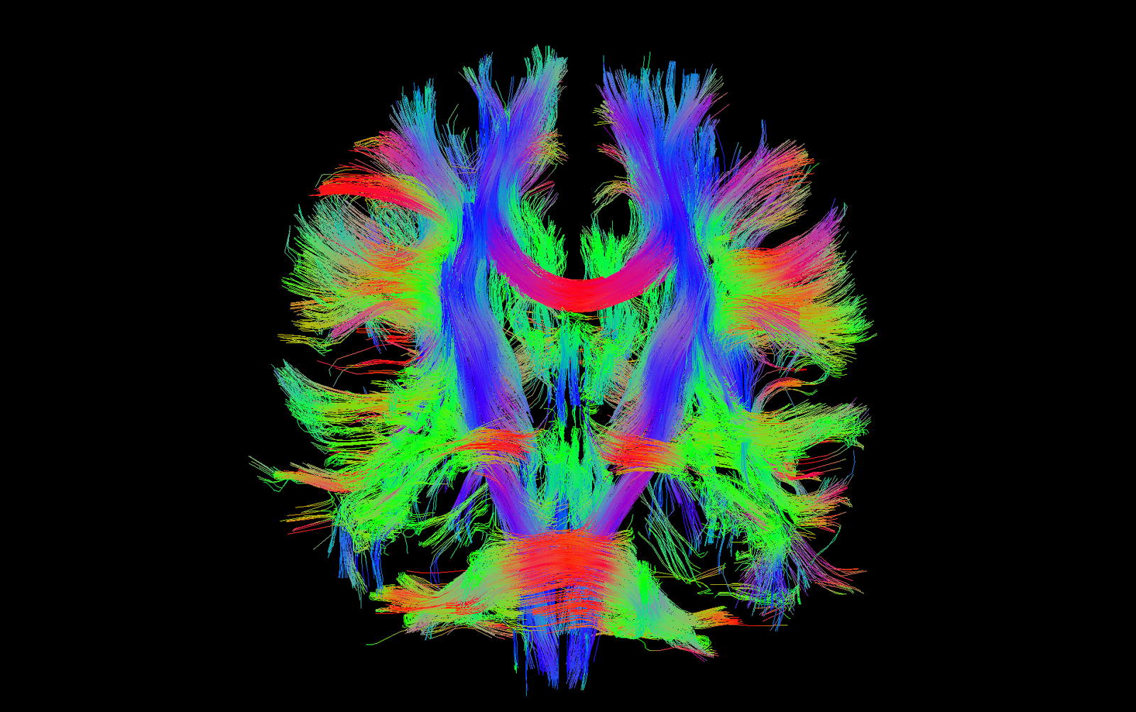
  • 'Diffusion Tensor Imaging', DTI, is where the action is, and it's used for mapping white matter, the bundles of nerve fibres that connect 'grey matter', which are the brain's main processing centres.

Despite the ugly name, DTI produces some spell-bindingly beautiful images, like this one, a colour-coded map of nerve tracts in the brain:

DTI+white+matter+tracks.png


From Wikipedia:
Diffusion Tensor Imaging's main clinical application has been in the study and treatment of neurological disorders, especially for the management of patients with acute stroke. Because it can reveal abnormalities in white matter fiber structure and provide models of brain connectivity, it is rapidly becoming a standard for white matter disorders.[6]

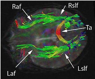
This new study from Stanford found that the nerve tract called the Arcuate fasciculus showed increased anisotropy in patients compared with controls. What does this mean? Dunno, but the conclusion says: "Right hemispheric increased FA [anisotropy] may reflect degeneration of crossing fibers or strengthening of short-range fibers." Sounds like the authors aren't too sure either.

Here's a pretty picture of the Arcuate fasciculus (R is the right side, L the left side) using the same DTI technology used in this study. Whatever else, this technology is good looking.

310px-DTI_Brain_Tractographic_Image_A_panal.jpg
 
Last edited:

Forbin

Senior Member
Messages
966
A memory problem I've had since coming down with ME is a reduced ability to store information that is spoken to me. For instance, if someone gives me driving directions it's almost as if my mind turns off the input the first time I hear it. I have to ask for the directions to be repeated and then I really have to focus on them. This is also true when I'm given other serial lists of instructions, or try to remember names when I'm introduced to more than one person. Everyone may have this to a degree, I suppose, but this got a lot worse when I got ME.

[Not to be flip, but it's actually kind of reminiscent of a recurring gag used on the old TV show "Get Smart." The Chief turns to Max and says, "Now this is what I want you to do." He then gives Max a lengthy list of instructions and says, "Now, have you got that?" and Max responds, "I think so Chief. There's just one part I don't get." "What part don't you get?" "The part after 'Now this is what I want you to do.'"]
 
Last edited:

Hip

Senior Member
Messages
17,865
I remember, especially in my early years of ME/CFS, I had some intriguing language difficulty phenomena: in addition to forgetting words, losing vocabulary, and having sudden difficulty in spelling words, perhaps the oddest language symptom I experienced was the frequent selecting of incorrect words while talking, often in a bizarre way, where I would select a word which was incorrect, but the incorrect word was from the same category as the right word – for example, I might incorrectly say “pun” when I meant to say “irony” – both are in the same category of literary devices.

Has anyone else had this, where you say the wrong word, but that wrong word is from the same general category as the right word that you should of said?
 

Hip

Senior Member
Messages
17,865
The intriguing thing about this new finding of abnormalities in the right arcuate fasciculus in the brains of ME/CFS patients is that it occurs asymmetrically, on just one side of the brain.

Thinking in terms of a viral etiology, what would confine the effects of a virus to just one particular side of the brain? I cannot think of any anatomical structures that are asymmetric in the brain, that could explain why this abnormality was present on just the right side of the brain.
 

snowathlete

Senior Member
Messages
5,374
Location
UK
We have of course had the perennial problem of one patient after another being sent for an MRI and then having their white matter hyperintensities (also known as unidentified bright objects or high signal intensity areas) dismissed as "nothing" or "nonspecific" or "don't worry about it" since the earliest days of this technology when, back in the mid-1980s, Cheney and Peterson began referring their Incline Village ME victims for MRI scans. Of course even today no one is sure of the clinical significance of these. Some neurologists seem to think that once they've ruled out MS, they should to refer us to psychiatry to get "help".

Indeed, my scan this year for example: "Two tiny areas of altered signal are seen in the peripheral white matter, on the right lying in the centrum semiovale posteriorly and on the left in the anterior frontal lobe. Thee are not thought to be of clinical significance."
Of course, that equates to 'normal' and it always will until research proves otherwise. This new study is at least a potential step toward that.
 

WillowJ

คภภเє ɠรค๓թєl
Messages
4,940
Location
WA, USA
My father who had crippling migraines used to have severe speech problems with them as well as other symptoms similar to ME/CFS which has always made me wonder if we both had similar problems, presenting as two different diseases.

I have both, and I think so. I think some of the pathology overlaps (blood flow interruptions in the brain, subtle inflammation in the brain).

http://www.aphasia.org/content/aphasia-definitions

I don't fit neatly into any of those categories on any consistent basis, but I have experienced most of them transiently with both migraine and ME. Normally I'd say I fit the last:

Other varieties of aphasia
In addition to the foregoing syndromes that are seen repeatedly by speech clinicians, there are many other possible combinations of deficits that do not exactly fit into these categories.

But rather than being fixed as in resulting from a single exact one-time injury, it's fluid as if it results from changing bloodflow or metabolic conditions in the brain, or something dynamic like that.

Except that I can never reach the level of thought I was capable prior to being ill. Not remotely close.
 
Last edited:

WillowJ

คภภเє ɠรค๓թєl
Messages
4,940
Location
WA, USA
We seem to have many of these small studies that seem promising but no one replicates them with a larger study. Is it all because of the lack of funding? This is beyond frustrating. It's like being constantly teased and no follow up.

This time, Montoya has funding.

http://stanmed.stanford.edu/2014fall/immune-system-disruption.html
Opportunity knocked in 2008 when a wealthy donor met with Montoya to talk about the ME/CFS problem. He asked if a $5 million donation for research could make a difference.

Montoya could hardly believe the sum, replying, “Yes, give me five years.”

With the freedom of private funding, Montoya was able to take a multifaceted and rigorous approach to analyzing ME/CFS.

Montoya’s game plan was to use a big-picture, big-data strategy to find out what was wrong with patients like Erin. His first step in launching the Stanford Initiative on Infection-Associated Chronic Diseases was to convince a dozen or so academic investigators to venture out of their comfort zones to research a wildly unpopular disease using technologies yet to be developed.

Montoya convinced experts in immunology, rheumatology, genetics, bioengineering, anesthesiology, neuroradiology, cardiology, psychiatry, infectious diseases and bioinformatics to all work together. The team members would be searching blood samples for infectious microbes, inflammation-related molecules and genetic flaws. They’d do brain scans and physical exams. They’d survey study subjects for fatigue levels and medical histories. Then they’d compare all this data with that of healthy people to see what was different. Next, he launched a Bay Area recruitment campaign for 200 patients who met the Centers for Disease Control’s definition for chronic fatigue syndrome, including Erin, and 400 age- and sex-matched healthy volunteers, all of whom agreed to donate eight tubes of blood and be poked, scanned and surveyed over the next decade.
 

daisybell

Senior Member
Messages
1,613
Location
New Zealand
I remember, especially in my early years of ME/CFS, I had some intriguing language difficulty phenomena: in addition to forgetting words, losing vocabulary, and having sudden difficulty in spelling words, perhaps the oddest language symptom I experienced was the frequent selecting of incorrect words while talking, often in a bizarre way, where I would select a word which was incorrect, but the incorrect word was from the same category as the right word – for example, I might incorrectly say “pun” when I meant to say “irony” – both are in the same category of literary devices.

Has anyone else had this, where you say the wrong word, but that wrong word is from the same general category as the right word that you should of said?

Semantic paraphasias are quite common in aphasia. The other thing that can occur but is less common, is a difficulty in selecting the right word, which can result in using esoteric words rather than common ones. I think that issue is one of the brain having difficulty selecting between options, so the result is that it selects words that are uncommon and therefore don't have a good alternative... This is mostly seen with problems in the fronto-temporal region of the left hemisphere.....
 

Hip

Senior Member
Messages
17,865
@daisybell
Very interesting. I thought that there might be a name for this phenomenon, but could not find the term for it. Now I know what it is: semantic paraphasias. Many thanks.

I found a list of various semantic paraphasia types in this Wikipedia article.
 

zzz

Senior Member
Messages
675
Location
Oregon
Indeed, my scan this year for example: "Two tiny areas of altered signal are seen in the peripheral white matter, on the right lying in the centrum semiovale posteriorly and on the left in the anterior frontal lobe. Thee are not thought to be of clinical significance."
Of course, that equates to 'normal' and it always will until research proves otherwise. This new study is at least a potential step toward that.

After reading the main article here as well as some of the posts such as @snowathlete's, I dug out my "normal" MRI from last year. A few interesting points from the report:
The sella turcica is partially empty.

That's certainly not normal; see the thread Empty Sella Syndrome and CFS: A patient study. It appears that whatever is not understood is dismissed as "normal".
Hyperintense T2 and FLAIR signal foci are small in size and few in number. These are nonspecific but statistically most likely due to mild microvascular ischemic disease. Sequelae of migraine headaches could also have this appearance.
Except that from my history, there's no reason to suspect mild microvascular ischemic disease, nor have I ever had migraines. My primary diagnosis was CFS, but radiologists don't know any more about this than the man in the street.
Mild cerebral volume loss is likely age-related.
I looked at the pictures when I got them, and compared to an MRI taken before my final relapse, the volume loss did not look mild, nor did it look like a typical volume loss for that amount of time. Instead, the pictures looked much closer to those of my mother, who was diagnosed with Alzheimer's at the time hers were taken, than to my previous ones. That much volume loss shook me up a bit.


And of course, no one did diffusion-tensor imaging on me. That would have been interesting.

I would suspect that there are plenty of abnormalities on "normal" MRIs of PWME that correlate with known abnormalities found in ME/CFS. There's a lot of data out there in the MRIs that a lot of us have had taken that would be very interesting to analyze. Of course, this would best be done by professionals who were up to date on the latest ME/CFS research. In the mean time, people with recent "normal" MRIs might find it useful to go over their reports. It may also be useful to bring to the attention of your doctor those abnormalities that were missed but that are consistent with what is found in ME/CFS. This assumes, of course, that your doctor is at least somewhat open-minded.
 
Last edited:

WillowJ

คภภเє ɠรค๓թєl
Messages
4,940
Location
WA, USA
I would suspect that there are plenty of abnormalities on "normal" MRIs that correlate with known abnormalities found in ME/CFS. There's a lot of data out there in the MRIs that a lot of us have had taken that would be very interesting to analyze. Of course, this would best be done by professionals who were up to date on the latest ME/CFS research. In the mean time, people with recent "normal" MRIs might find it useful to go over their reports. It may also be useful to bring to the attention of your doctor those abnormalities that were missed but that are consistent with what is found in ME/CFS. This assumes, of course, that your doctor is at least somewhat open-minded.

It also assumes the radiologist documented findings that didn't strike them as odd/pathological. Many of them classify brain scans as MS/not MS... brain tumor/not brain tumor... and so on through whatever main condition(s) they were looking for.

Yours seems to have been unusually thorough.