• Welcome to Phoenix Rising!

    Created in 2008, Phoenix Rising is the largest and oldest forum dedicated to furthering the understanding of and finding treatments for complex chronic illnesses such as chronic fatigue syndrome (ME/CFS), fibromyalgia (FM), long COVID, postural orthostatic tachycardia syndrome (POTS), mast cell activation syndrome (MCAS), and allied diseases.

    To become a member, simply click the Register button at the top right.

Microglia and immune function in brain affects circulation

Learner1

Senior Member
Messages
6,305
Location
Pacific Northwest
Though this news came out about Alzheimer's, I'm wondering if it affects patients with POTS and ME/CFS:

"Discovery of New Role for the Brain’s Immune Cells Could Have Alzheimer’s Implications - Neuroscience News" https://neurosciencenews.com/microglia-blood-brain-19485/

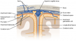
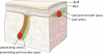
“Precise blood vessel function is critical to accommodate the extreme energy demands of the brain for normal brain function,” said UVA’s Ukpong B. Eyo, PhD, of UVA’s Department of Neuroscience, the UVA Brain Institute and UVA’s Center for Brain Immunology and Glia (BIG). “These findings suggest previously unknown roles for these brain cells in the proper maintenance of blood delivery to the brain and provide novel opportunities to intervene in contexts where blood perfusion to the brain is impaired.”
 

Pyrrhus

Senior Member
Messages
4,172
Location
U.S., Earth
Briefly, the paper describes how some of the tissue-resident macrophages of the brain (referred to in the article as "parenchymal microglia") are shown to cozy up to blood vessels (these are then referred to in the article as "capillary-associated microglia").

It is important to note that these "capillary-associated microglia" stay on the brain side of the blood-brain-barrier, and do not enter the perivascular space, so they are indeed tissue-resident macrophages (microglia), not blood-borne macrophages (monocytes).

These "capillary-associated microglia" use P2RY12 receptors to respond to purine signals from the blood vessels, released through pannexin 1 channels in the blood vessels, that help to regulate the constriction or dilation of the blood vessels. This purine signal could be as simple as extracellular ATP molecules.

Although it is well known that tissue-resident macrophages in the brain can release cytokines that affect the dilation or constriction of nearby blood vessels, this is the first evidence that I have seen that blood vessels can release ATP that is sensed by the nearby tissue-resident macrophages in the brain.

Abstract:
Microglia are brain-resident immune cells with a repertoire of functions in the brain. However, the extent of their interactions with the vasculature and potential regulation of vascular physiology has been insufficiently explored.

Here, we document interactions between ramified CX3CR1 + myeloid cell somata and brain capillaries. We confirm that these cells are bona fide microglia by molecular, morphological and ultrastructural approaches.

Then, we give a detailed spatio-temporal characterization of these capillary-associated microglia (CAMs) comparing them with parenchymal microglia (PCMs) in their morphological activities including during microglial depletion and repopulation.

Molecularly, we identify P2RY12 receptors as a regulator of CAM interactions under the control of released purines from pannexin 1 (PANX1) channels. Furthermore, microglial elimination triggered capillary dilation, blood flow increase, and impaired vasodilation that were recapitulated in P2RY12−/− and PANX1−/− mice suggesting purines released through PANX1 channels play important roles in activating microglial P2RY12 receptors to regulate neurovascular structure and function.
 
Last edited:

Pyrrhus

Senior Member
Messages
4,172
Location
U.S., Earth
I am also including the following, as the structure of the blood-brain barrier surrounding blood vessels can be very hard to visualize and understand:

Yes, the exact structure of the blood-brain-barrier (BBB) is very complicated to describe, even in a diagram.
It took me quite a while to understand it myself, and it's an active area of research so we're still learning.

The first thing to explain is that the blood-brain-barrier actually consists of multiple layers or barriers:
  • The outermost layers (dura mater and arachnoid membrane) enclose the brain on the outside, and do not penetrate into the interior of the brain.
  • Below the arachnoid membrane is a space filled with cerebrospinal fluid (CSF). This space is called the sub-arachnoid space.
  • Blood vessels that supply the brain first run through these outer layers and then penetrate into the interior of the brain.
  • The inner layers of the blood-brain-barrier (pia mater and glia limitans) surround and follow the penetrating blood vessels into the interior of the brain. So, the further into the interior of the brain, the fewer layers in the blood-brain-barrier.
  • However, between the penetrating blood vessels and the inner layers there is a space called the perivascular space. Toward the outside of the brain, this space is connected to the sub-arachnoid space and receives some cerebrospinal fluid from there.
  • But as you go further into the interior of the brain, the composition of the fluid in the perivascular space changes. This inner perivascular space may be filled with immune cells and too many immune cells clumped in one spot can result in a "dilated perivascular space".
  • Eventually, the fluid in this perivascular space drains out of the brain along the cranial nerves, emptying into lymph nodes. There, the fluid is processed before returning to the bloodstream.
Here are some rough diagrams that might help: