• Welcome to Phoenix Rising!

    Created in 2008, Phoenix Rising is the largest and oldest forum dedicated to furthering the understanding of and finding treatments for complex chronic illnesses such as chronic fatigue syndrome (ME/CFS), fibromyalgia (FM), long COVID, postural orthostatic tachycardia syndrome (POTS), mast cell activation syndrome (MCAS), and allied diseases.

    To become a member, simply click the Register button at the top right.

Comparison between four studies on energy metabolism

paolo

Senior Member
Messages
198
Location
Italy
There is a European study which has checked for the expression of 1007 mitochondrial proteins in platelets from 2 twins, one with ME/CFS and the other one healthy (Ciregia F et al 2016). Of these proteins, 194 were significantly modified in the sick twin, in comparison with the healthy one. I have checked for differencies in pyruvate dehydrogenase complex, ADP/ATP translocase subunits and pyruvate dehydrogenase kinases. This is what I have found in these two twins:

1) Pyruvate dehydrogenase E1 subunits alpha (PDHA) and beta are both inceased in the sick twin, which is in partial accordance with the increse in PDHA found by Fluge and Mella (Fluge et al. 2016);

2) ADP/ATP translocase, sub unit 2 and 3, are low in the sick twin, compared with the healthy one, which could be in accordance with the study by Myhill and colleagues (Myhill S et al. 2009), (Booth, N et al 2012), if only we assume that the problem with this enzyme found in group HiBlk is not due to blockage from a molecule or an autoantibody, but is instead due to under expression of the enzyme itself (@Hip);

3) Pyruvate dehydrogenase kinases 1 and 3 are over expressed, which again is in partial accordance with what FLuge and Mella have found in their recent paper, where PDK 1, 2, 4 are over expressed in ME/CFS patients.

In conclusion, the sick twin does not seem to have any blockage of ADP/ATP translocase, because if that was true he would have an over expression of the enzyme, while the enzyme is under expressed. On the other hand he does seem to have a problem with his pyruvate dehydrogenase, in fact there is inhibition by over expressed PDK 1 and 3 and - at the same time - he is expressing more PDHA than his heathy twin.
 
Last edited:

paolo

Senior Member
Messages
198
Location
Italy
The same European study (Ciregia F et al 2016) then selected three enzymes from the 194 significantly modified in the sick twin: ACON, ATPB e MDHM. They then evaluated the expression of these enzymes in a cohort of 45 Italian patients with ME/CFS and in 45 matched controls. In this case they considered mitochondria from saliva. They found that both ACON and ATPB are over expressed in patients.

ACON stands for aconitase, which is an enzyme of the TCA cycle, which catlyzes the step from citrate to cis-aconitate. Thus its over-expression in this cohort of patients is in agreement with the depletion of these two metabolites, found in a recent japanes study (Yamano E, et al. 2016) and with the study by Fluge and Mella: in fact, if we assume that the TCA cycle is deficiently supplied by glycolysis, it would over express one or more enzymes in order to increase the energy production from the substrate available. Thus, this finding seems in agreement with both the norvegian and the japanese study, and seems to complete the picture.

ATPB is subunit beta of ATP synthase, and is involved in the last step of mitochondrial metabolism, the conversion of ADP into ATP. Again, an over expression of this enzyme seems to be in agreement with poor energy supply.
 

Snow Leopard

Hibernating
Messages
5,902
Location
South Australia
This is interesting! I never got around to looking at the supplementary data of this study.

Some other notes after a quick glance:

Carnitine O-palmitoyltransferase 1 lower in CFS twins P~0.001, whereas Carnitine O-palmitoyltransferase 2 is increased, P~0.001

Acetyl-CoA acetyltransferase is increased, P~0.00003
 
Last edited:

paolo

Senior Member
Messages
198
Location
Italy
In this picture I have put together the levels of the TCA cycle metabolites from the japanese study with the representation of amino acids catabolism used by Fluge and Mella in their own work. I have indicated also the position of aconitase in the TCA cycle.

upload_2017-1-14_12-58-9.png
 

Hip

Senior Member
Messages
17,824
Great work paolo!

I take it you looked at the mitochondrial protein levels in ME/CFS patients and healthy control provided in Supplementary Table S1 of the Ciregia study to arrive at your conclusions.


2) ADP/ATP translocase, sub unit 2 and 3, are low in the sick twin, compared with the healthy one, which could be in accordance with the study by Myhill and colleagues (Myhill S et al. 2009), (Booth, N et al 2012), if only we assume that the problem with this enzyme found in group HiBlk is not due to blockage from a molecule or an autoantibody, but is instead due to under expression of the enzyme itself (@Hip);

That is very interesting paolo: so the Ciregia study found that the amount of ADP/ATP translocase protein in the mitochondria was lower in ME/CFS patients compared to healthy controls.

I see what you are saying about explaining the low levels of ADP/ATP translocase in terms of reduced gene expression of the ADP/ATP translocase.

However, although I don't know the details of how proteomic analysis works, if we assume in ME/CFS patients there is an autoantibody that targets and attaches to the ADP/ATP translocase protein, then possibly the combined molecular structure of ADP/ATP translocase protein + attached autoantibody protein might be seen as a different protein structure in proteomic analysis, and therefore not detected as ADP/ATP translocase. Could that be the case?

If this is the case, then the low levels of ADP/ATP translocase found in this study might be explained in terms of an autoantibody bound onto the ADP/ATP translocase protein.

In the Myhill, Booth and McLaren-Howard studies, they talk about xenobiotic substances blocking translocator protein. Myhill et al use the term "translocator protein" to refer to the adenine nucleotide translocator (ANT).

But it seems to me that autoantibodies could be responsible for the translocator blockage, because it is known that in coxsackievirus B myocarditis, the virus appears to trigger an autoantibody that targets ADP/ATP translocase and then blocks mitochondrial function — see this thread. So from this myocarditis study we know that ADP/ATP translocase autoantibodies exist, and that they can inhibit mitochondrial function.
 
Last edited:

paolo

Senior Member
Messages
198
Location
Italy
Great work paolo!

Thank you @Hip !

I take it you looked at the mitochondrial protein levels in ME/CFS patients and healthy control provided in Supplementary Table S1 of the Ciregia study to arrive at your conclusions.

Yes, I have searched for pyruvate dehydrogenase, pyruvate kinases, and ADP/ATP translcase both in Table 2 and in Table S1 of supplementary material. I have forgotten to mention that.

That is very interesting paolo: so the Ciregia study found that the amount of ADP/ATP translocase protein in the mitochondria was lower in ME/CFS patients compared to healthy controls.

Yes, exactly.

However, although I don't know the details of how proteomic analysis works, if we assume in ME/CFS patients there is an autoantibody that targets and attaches to the ADP/ATP translocase protein, then possibly the combined molecular structure of ADP/ATP translocase protein + attached autoantibody protein might be seen as a different protein structure in proteomic analysis, and therefore not detected as ADP/ATP translocase. Could that be the case?

If this is the case, then the low levels of ADP/ATP translocase found in this study might be explained in terms of an autoantibody bound onto the ADP/ATP translocase protein.

Wow, I really don't know. I think that the bond between an autoantibody and its specific epitope is not stronger than the bond between an enzyme and its natural substrate. But I don't know if that bond can interfere with a proteomic analysis. This is a good question!

But it seems to me that autoantibodies could be responsible for the ADP/ATP translocase blockage, because it is known that in coxsackievirus B myocarditis, the virus appears to trigger an autoantibody that targets ADP/ATP translocase and then blocks mitochondrial function — see this study. (Note that ADP/ATP translocase is also called the adenine nucleotide translocator). So from this myocarditis study we know that ADP/ATP translocase autoantibodies exist, and that they can inhibit mitochondrial function.

It is very interesting, but in order to demonstrate this theory you have to explain how an autoantibody can reach the mitochondrial inner membrane. It sems that in the study that you have indicated (Schultheiss HP et al. 1996), they didn't demonstrate the internalization of the autoantibody, but only the effect on the energy metabolism of guinea pigs immunized with myocardial ANT protein and in mice infected with the Coxsackie B3 virus.
 
Last edited:

alex3619

Senior Member
Messages
13,810
Location
Logan, Queensland, Australia
ACON stands for aconitase, which is an enzyme of the TCA cycle, which catlyzes the step from citrate to cis-aconitate. Thus its over-expression in this cohort of patients is in agreement with the depletion of these two metabolites
One confounding factor is this could be partially or fully due to oxidative stress, as aconitase is imported into the mitochondria and needs proper folding, which means a strict need for reduced glutathione (reduced, not decreased, its the opposite of oxidated).

I should add that I would then expect to see lots of aconitase, but little properly folded aconitase.
 

alicec

Senior Member
Messages
1,572
Location
Australia
Thank you @paolo - excellent work.

However, although I don't know the details of how proteomic analysis works, if we assume in ME/CFS patients there is an autoantibody that targets and attaches to the ADP/ATP translocase protein, then possibly the combined molecular structure of ADP/ATP translocase protein + attached autoantibody protein might be seen as a different protein structure in proteomic analysis, and therefore not detected as ADP/ATP translocase. Could that be the case?

No.

Several different techniques are used to separate proteins prior to analysis. These are designed to break all non-covalent bonds. An antibody-antigen complex wouldn't survive the treatment.
 

Hip

Senior Member
Messages
17,824
It is very interesting, but in order to demonstrate this theory you have to explain how an autoantibody can reach the mitochondrial inner membrane. It sems that in the study that you have indicated (Schultheiss HP et al. 1996), they didn't demonstrate the internalization of the autoantibody, but only the effect on the energy metabolism of guinea pigs immunized with myocardial ANT protein and in mice infected with the Coxsackie B3 virus.

That's a good point. Though it seems it may be more complicated than that, as I just found this Schultheiss 1990 viral myocarditis / cardiomyopathy study (full paper here) which says:
In conclusion, the biochemical and functional data clearly suggest the hypothesis that antibodies against the ADP-ATP carrier [ADP/ATP translocase] cause a dysfunction of the heart by an antibody-mediated disturbance of cellular energy metabolism.

The antibody-mediated alteration of the carrier function can be realized by several ways.

First, the antibodies might inhibit carrier function as a direct result of antibody binding to the carrier protein.

Second, as the carrier is synthesized in the cytosol and imported post-translationally into the mitochondria, the antibodies might react with the primary translation product, which has the same apparent molecular weight as the mature protein, hindering the complete and functional active incorporation of the carrier protein into the mitochondrial inner membrane.

Third, the anti-carrier antibodies might cause antigenic modulation of the protein, increasing carrier degradation.

Fourth, the antibody binding to the cell surface might influence carrier function indirectly by activating a messenger system.

It's interesting that ADP/ATP translocase is synthesized in the cytosol of the cell, and then later transported into the mitochondria. So the anti-ADP/ATP translocase autoantibodies could bind to the ADP/ATP translocase while this protein is still in the cytosol.

Interesting also that in the study they say the autoantibody could modify the protein structure of ADP/ATP translocase; could this perhaps could account for the lower levels of ADP/ATP translocase subunits 2 and 3 found in the Ciregia 2016 study?


Though a further complexity about ADP/ATP translocase protein expression is that in the Schultheiss 1996 viral myocarditis / cardiomyopathy study (full paper here), they found a shift in the relative expression of the three ADP/ATP translocase isoforms:
We found a markedly lowered transport capacity of the translocator accompanied by an elevation in total ANT protein content. The alteration in ANT protein amount is caused by an ANT isoform shift characterized by an increase in ANT 1 isoform protein associated with a decrease in ANT 2 isoform and an unchanged ANT 3 content.

This increase in ANT1 expression, incidentally, may be detrimental to the host in enterovirus (EV) heart infections, as this Schultheiss et al 2014 study (full paper here) found that:
reduced ANT1 expression is linked to spontaneous EV elimination in human and murine EV-infected hearts. In contrast, elevated ANT1 expression supports EV infection and is associated with EV persistence
The authors say that ANT1 overexpression influences the expression of other genes that may affect viral replication.


Interestingly, this study found reducing IL-17 inhibits the production of anti-ADP/ATP translocase autoantibodies (in CVB3-induced acute viral myocarditis).
 
Last edited:

eljefe19

Senior Member
Messages
483
@Hip Reading the self hacked article on th17 and I see the picture gets even more muddled after reading this;

mTOR increases glycolysis, which is what allow Th17 cells to proliferate. This works through HIF1α. Blocking glycolysis inhibited Th17 development while promoting Treg cellgeneration. (R)

Increased mTOR promotes Th1 and Th17 immunity, leading to increased intestinal inflammation (R), among other issues.
 

paolo

Senior Member
Messages
198
Location
Italy
That's a good point. Though it seems it may be more complicated than that, as I just found this Schultheiss 1990 viral myocarditis / cardiomyopathy study (full paper here) which says:


It's interesting that ADP/ATP translocase is synthesized in the cytosol of the cell, and then later transported into the mitochondria. So the anti-ADP/ATP translocase autoantibodies could bind to the ADP/ATP translocase while this protein is still in the cytosol.

Interesting. But you have still to explain how can the anti-ANT antibody enter the cytosol.
 

paolo

Senior Member
Messages
198
Location
Italy
Thank you @paolo - excellent work.


No.

Several different techniques are used to separate proteins prior to analysis. These are designed to break all non-covalent bonds. An antibody-antigen complex wouldn't survive the treatment.

Thank you @alicec for this information!

It is worth noting that the comparison we are talking about is on only two persons, two twins, one of whom has ME/CFS. Moreover, the level of these mitochondrial proteins has been calculated on mitochondria from platelets, which may not be representative for other tissues.
 
Last edited:

Hip

Senior Member
Messages
17,824
Interesting. But you have still to explain how can the anti-ANT antibody enter the cytosol.

I read that there are a number of different mechanisms by which certain antibodies (but not all) can cross the cellular membrane and enter the cytosol, but these processes are not well understood.

In the Schultheiss 1996 myocarditis study (full paper here), they found antibody immunoglobulin deposits in the mitochondrial membranes of the heart muscle cells:
Using immunofluorescence techniques and peroxidase-antiperoxidase staining, we found immunoglobulin deposits in mitochondrial membranes in cryosections of the myocardium of immunized animals [34]. In isolated cardiac myocytes, the formation and cytosolic internalization of immunoglobulin-containing membrane-coated vesicles could be shown.

The results might support the hypothesis of a direct binding of the cytosolically internalised antibodies to ANT.

There are actually a number of anti-mitochondrial autoantibodies known: Anti-mitochondrial antibody - Wikipedia
 

nandixon

Senior Member
Messages
1,092
The same European study (Ciregia F et al 2016) then selected three enzymes from the 194 significantly modified in the sick twin: ACON, ATPB e MDHM. They then evaluated the expression of these enzymes in a cohort of 45 Italian patients with ME/CFS and in 45 matched controls. In this case they considered mitochondria from saliva. They found that both ACON and ATPB are over expressed in patients.
…..

ATPB is subunit beta of ATP synthase, and is involved in the last step of mitochondrial metabolism, the conversion of ADP into ATP. Again, an over expression of this enzyme seems to be in agreement with poor energy supply.
The findings of the 2016 Ciregia study (full text here:
https://www.ncbi.nlm.nih.gov/pmc/articles/PMC5048217/) have now been essentially validated to a fairly good extent with respect to ATPB, which is also known as ATP5B, a subunit of ATP synthase (aka Complex V).

In Paul Fisher's recent study of this thread:

An Isolated Complex V Inefficiency and Dysregulated Mitochondrial Function in Immortalized Lymphocytes from ME/CFS Patients
https://forums.phoenixrising.me/thr...lized-lymphocytes-from-me-cfs-patients.77577/

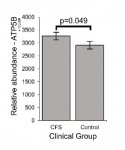
It was also found that the level of ATP5B protein was increased in immortalized lymphocytes from ME/CFS patients:

20191003_050221.jpg

Complex V subunits ATP5A1, ATP5B, and ATP5H were significantly upregulated (iBAQ) in whole cell mass spectrometry proteomics experiments (independent t-test).


The p-value was not impressive at 0.049, but related findings in Fisher's study have excellent p-values and strongly suggest that ATP5B is indeed upregulated as Ciregia found.

I have a hypothesis that the "something in the blood" that Ron Davis and several other groups have noted may be targeting ATP5B and I may have found an interesting specific candidate for what that something is. I'll be posting more about this on the Fisher thread fairly soon. (I'm still working on trying to disprove it.)