• Welcome to Phoenix Rising!

    Created in 2008, Phoenix Rising is the largest and oldest forum dedicated to furthering the understanding of, and finding treatments for, complex chronic illnesses such as chronic fatigue syndrome (ME/CFS), fibromyalgia, long COVID, postural orthostatic tachycardia syndrome (POTS), mast cell activation syndrome (MCAS), and allied diseases.

    To become a member, simply click the Register button at the top right.

Am I “Allergic to Life”?

View the Post on the Blog


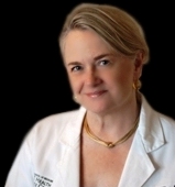
Claudia S. Miller, M.D., M.S., is an allergist/immunologist and tenured Professor at the University of Texas School of Medicine at San Antonio.

Janis Bell (JanisB) reviews her personal journey of chronic illness after being inspired by a new article written by Jill Neimark and appearing online today in Discover magazine. It highlights the work of Dr Claudia Miller (pictured) and her theories relating to extreme chemical sensitivity and toxicants, a condition she terms, Toxicant-induced Loss of Tolerance or TILT.


By the time I finished Jill Neimark’s “Extreme Chemical Sensitivity Makes Sufferers Allergic to Life,” (November 2013 issue of Discover Magazine - available free and online today), I was reconsidering my own story, the story I tell myself about the illness that changed my life.

Jill explains the groundbreaking theory of Dr Claudia Miller (pictured), a physician and environmental health expert, and writes:

“Miller studies a phenomenon she calls Toxicant-induced Loss of Tolerance (TILT). The word toxicant refers to a man-made poison, such as Dursban, whereas a toxin is a naturally occurring poison produced by living cells or organisms, such as spider venom.”

TILT happens when a susceptible individual gets sick after toxic exposures and then, instead of recovering, stays sick.

Maybe that sounds like your story, but instead of chemical exposure it’s a tick bite, an unknown virus, a moldy house or workplace, or a traumatic event.

I certainly felt it applied to my own descent into chronic illness, and as I read the article I thought carefully about my journey in a very new light - even though Miller's research and Jill's article, deal mainly with chemical sensitivity.

TILT is believed to play a role in neuroendocrine diseases such as Gulf War Illness, and I couldn't help but wonder what kind of role it might play in ME/CFS.

Reappraising my life...


For years my story appeared straightforward - a sudden onset virus, that hit on the first Saturday of May 1987, led to chronic symptoms and no recovery. It changed me from an upwardly mobile professional to a downward-spiraling searcher of anything that would restore me to health.

Over the next two decades, the name of my condition changed as did the clueless physicians I travelled to see, and the all-too-confident alternative practitioners whose fees ate into my meager income.

While mainstream physicians waited for the virus to be identified by researchers and offered drugs for symptomatic relief - most of which I couldn’t tolerate - I immersed myself into research on nutritional, herbal, and energy therapies.

Was I too toxic? Was I allergic? Perhaps deficient in some vitamin or antioxidant? Maybe full of parasites or infected with candida?

Over the years, symptoms disappeared and new ones emerged. I experienced hope and disappointment. I was lucky to have three glorious periods of almost total recovery, until they ended within a few months in crushing relapses, each one pushing me lower on the Bell Disability Scale and giving me new challenges to deal with.

I tried to understand what caused each relapse but my story didn’t change significantly until I read Martin Pall’s “Explaining Unexplained Illnesses” and learned how all of my extra diagnoses were connected by a vicious cycle of oxidative stress.

Pall explains how nitric oxide and peroxynitrite, abbreviated NO and ONOO (like Oh! no!) damage cell membranes. The membrane is the “brain” of every cell as it decides what nutrient and messengers come in and what toxins and metabolites get out. He himself had CFS for many years.

Through extensive reading as a biochemist at the University of Washington, Pall discovered that the NO/ONOO cycle, which is elevated in acute infections, becomes locked in that elevated state, interfering with cellular functions.

I ordered the anti-oxidant supplements he recommends from Allergy Research Group/Nutricology, and I bought home test kits (Oxidata test kit from Apex Energetics) to monitor a urinary marker of oxidative stress known as MDA (Malondialdehyde), so that I could see if the antioxidants were doing anything for me.

I’ve learned over the years that most of the things that happen to me don’t show up in medical tests, but my scientific curiosity won over me. Would MDA go up when I felt worse and down when I felt better? There did appear to be a correlation: I felt a little better when I moved MDA into the low range.

Yet Dysautonomia/POTS, my most disabling symptom, together with PEM (Post-exertional Malaise), persisted with the tenacity of a pit bull. I was far from recovered, and far from the highpoints of remission I’d experienced in the past.

Another influence on my own story came after reading Ritchie Shoemaker’s, “Surviving Mold”, while on a family trip to Costa Rica, lying in bed with a view of the Arenal volcano. After I laughed about having “first year medical student syndrome,” we uncovered a coating of the fuzzy black stuff on my bed frame and the underside of my mattress.

We hurriedly checked out and moved into a breezy room at the beach. Within a few days, I could participate in conversations again. By the end of our stay I was walking to nearby restaurants.

When I returned to the States, Dr Vinitsky (Enlightened Medicine) told me I had biotoxin illness as well as a few other opportunistic and causative pathogens no other physician had found, including Lyme disease.

Thoughts about mold pushed the onset of my illness back to 1982, at a time when I took a teaching job at Kenyon College and was assigned an office in the modern Biology building.

It was, I felt, the combination of windows that didn’t open, research animals in the basement, central air, and damp weather, that combined to spread mold spores throughout the building and into the air I breathed all day.

I believed I had the classic respiratory onset, then endocrine and immune issues, all preparing the way for that 1987 virus to knock me down.

When the ERMI (Environmental Relative Moldiness Index) test for mold in my current house came back with a high score (ERMI analyzes dust for fungal DNA and gives a score relative to other US households), we found mold in the basement under the crawlspace batts.

Survivors online warned me that remediation rarely worked for people like me. Still my partner valiantly tried to save the life we knew, replacing and cleaning and sealing off the affected areas around the house.

I would make a little progress but then crash again, reacting more and more violently to dust scattered in the air from his latest cleaning endeavor.

In desperation, we contacted the remediator in “Surviving Mold”, the book that had become my bible, and took his recommendation to fog the space with Aerosolver. Even though it was custom made for my sensitive body, with ingredients I used in tinctures and eyewash (glycine and borax), it did me in and less than two months later, I was in the car heading out west, my loving partner left to deal with the wreck of our lives.

My attempt at escape...


Although many people online suggested I go camping, I never seriously considered it until the emergence of this crisis.

My partner had been an avid backpacker in his twenties and thirties, yet whenever he suggested a camping trip Christine Lavin’s lyrics to her song, “Camping,” would run through my head:

“I’ve got stars on my ceiling that glow in the dark,

I’ll open up the windows we can hear the birds in the park,

I’ll put Wild Kingdom on my TV…

This might not be camping but it’s close enough for me.”

Nevertheless, camping is what I ended up doing when other accommodation I tried to rent affected my health even more.


My home under the stars

The camping lifestyle is difficult, yet I never regretted my choice. As symptom after symptom lessened or entirely disappeared, I put up with the hard ground, the cold, the heat, wind, the endless packing and moving, the whiffs of a neighbor’s cigar or cigarettes, the lousy WiFi, the inadequate cell phone service, the dirt, the noise, the diesel fumes of a one ton pickups and large motor-homes.

And I complained whenever the cold wind ripped through my jacket or the rain left me huddled inside dripping nylon walls.

When my most disabling symptoms, PEM and POTS, began to clear in that low mold environment, I often thought, “Maybe all I have is mold illness, not ME/CFS.

Yet my ME/CFS diagnosis was sound, and approved by specialists. I’d met the Canadian Consensus Criteria and had been a subject for research studies.

I felt there was an association between symptom onset and exposure. And it was interesting, if painful, to witness how certain exposures could suddenly bring on POTS and PEM again, but also satisfying when I experienced the relief as they went quickly away.

Reading about Dr Miller’s theory of TILT, I found myself re-thinking the crisis that sent me in exile from my house and the community where I’d lived most of my adult life:

What made me react to buildings?

Why had my sense of smell become dog-like in its acuteness, picking up fabric softener on a woman across the room and food smells long after a meal had been cooked?

Did everyone who left a moldy house have to go through this misery, and if so, why didn’t Shoemaker mention it?

It perplexed me that, while camping in the desert, I now seemed to be more reactive than I ever was back in Ohio. I had traded a disabling illness for one that left me as isolated and in my worst periods of relapse. I was spending too much time on survival and not enough on productive, joyful activity that could help me heal.

I wanted a unit like the one Jill described in her article and that Dr Theron Randolph had created for his own patients. These specially constructed units with filtered air, cotton bedding, and purified water, allowed patients to improve enough that he could test them to find out what was making them sick.

Willliam Rea of the Environmental Health Center in Dallas, houses his patients in similar rooms, but I was deterred by the expense, as well as the fact that my exploratory trip to Dallas had been a disaster (an experience I describe in this blogpost from October 2011 ).

Camping was the closest I could get to an Environmental Control Unit.

“Why am I more sensitive?” I asked Dr. Rea on my initial visit. He pointed to a diagram of a barrel filled with toxins until not one single drop could be added without spilling over the edge. “But I had more toxins in my house in Ohio!” I protested and left feeling very misunderstood.

The toxic load theory of chronic illness was not unfamiliar to me. I had read the book from Dr Sherry Rogers, a former patient of Rea's, who had said, “Detox or Die”. But what did any of this have to do with ME/CFS?

I and my friends were reaching the end of our patience. We had tried every classic detox protocol - juice fasting, liver cleanses, raw foods, sauna, and heavy metal chelation - with little or no noticeable benefits.

Worse, my experience with juice fasting at Dr Gabriel Cousens’ Tree of Life Rejuvenation Center had brought on my second relapse.

Yet there appeared to be some connection. After living in a tent for just over a month, I could tolerate CSM (Cholestyramine), the second step of Shoemaker’s mold illness protocol, although I had yet to benefit from its binding of fat soluble toxins.

When I met with Dr Scott McMahon eighteen months later, my Visual Contrast Sensitivity had greatly improved from the reduction of systemic mycotoxins and other toxins that CSM, charcoal, and bentonite pulled out. Maybe Scott knew why I was hypersensitive?

“We don’t know why some people develop MCS [Multiple Chemical Sensitivity],” he told me honestly, “And we don’t really know what to do about it.”

I braced myself on the edge of my seat as I waited for his answer to the next question, only relaxing my grip as I heard his encouraging words,

“Some get well when treated for mold illness.”

Phew! Maybe I was on the right track.

People with MCS, according to Dr Miller, are TILT'ed


We are more sensitive to minute amounts of things that others tolerate with little or no problem. Sure, all these toxicants and toxins eventually catch up to some people, but they can stand at a bus stop in Manhattan or LA without getting congested, losing their balance or becoming suicidal.

Dr Miller has proven that our sensitivity is real, validating the physical changes through publications in peer-reviewed journals. So why am I finding myself beset by gloomy thoughts as I get to the end of Jill’s article?

A few sentences are running through my head on repeat:

“...instead of recovering, the neurological and immune systems remain damaged...”

“Once genes are switched on […] and once you are sensitized, you essentially have a reprogrammed cell...”

No, no, NO! I’ve been clinging to the hope that someday I’ll live in the world again, go to stores, go to parties. This camping thing has got to end!

I try not to dwell on the thought that I might always be living on the edge, vulnerable to the next exposure, a semi-hermit in our toxic world. But gloom settles into my bones.

However, I remember that Lisa Petrison (Paradigm Change), an ME/CFS survivor who recovered her life through mold avoidance, once told me that nearly everyone is hypersensitive at first, but if they stick with a program of avoidance, they eventually improve.

I mentally compared the intensity of my reactions in the past and my reactions now and noticed a slight improvement.

I contacted Jill to clear things up. Yes, she says, Dr Miller argues that we’ll always be more vulnerable than a undamaged person, but we can recover with proper avoidance.

Dr Miller herself was inspired by Randolph’s work because she saw people getting well in those non-toxic chambers as they unmasked and eventually were treated for their specific sensitivities.

She believes isolation from toxicants is key to healing and, in her blog, Dr Miller writes about how to help veterans with Gulf War Illness:

“The single most important task is to sort out and “unmask” the causes or triggers for their symptoms. This requires an environmental medical unit, or EMU. Congress once endorsed EMU research for the Gulf War veterans but never funded it. Only a few EMUs exist in the world. They are environmentally controlled in-patient hospital units designed to isolate patients from exposures that set them off.”

This encourages me in my plans to find land and build a non-toxic, healthy home where I can avoid mold and other toxicants.

Since Dr Miller isn’t a clinician, the article did not go into therapeutic approaches beyond avoidance of toxicants and toxins. Yet Jill’s summary on the path of toxicants - through the olfactory nerve in the nose and directly into the limbic brain - got me thinking about therapies for the limbic brain that have allegedly benefited patients with MCS and ME/CFS such as Gupta’s Amygdala Retraining and Hopper’s Dynamic Neural Retraining. Individuals swear these approaches made a profound difference to their health.

My own preference for “mind-control” is yoga. Since Jill's article mentioned one patient who recovered from electo-shock therapy, a treatment used widely in the past to change the brain chemistry of depression, I hold to the belief that we can mobilize and direct the electric patterns of our brains through our thoughts. For millennia, yogis in India have defied the tenets of modern science by walking on hot coals, warming a circle of snow with their body heat, and stopping their hearts - as have Christian mystics.

Then I find myself wondering if toxins (made by nature as compared to man-made toxicants), shift a body into TILT in the same way as toxicants.

Can fungal, bacterial, and viral toxins provoke neurological and immune system damage that persists even after the offending organisms have left?

Could these infections set up an inflammatory response which gets locked into place and becomes chronic, due, in part, to a vicious cycle of elevated NO/ONOO?

Bodies damaged by toxicant exposure (Gulf War Veterans, Gulf oil spill workers) get opportunistic infections just like those of us with ME/CFS as well as those infected by chronic Lyme Disease.

Are you at risk of Toxicant-induced Loss of Tolerance?


If you think that as you can live in a house and can tolerate a few medications, you are not at risk of TILT, think again. Maybe you remember some doctors who judged you as malingering when you reacted to one medication after another. Maybe family members or former friends think you are crazy and overreacting because you seemed fine one day and complaining of misery the next.

I recommend you give them a photocopy of Jill's article, “Extreme Chemical Sensitivity Makes Sufferers Allergic to Life,” so they can see that there is scientific validity for TILT. You might just be too masked to see what you are reacting to.

According to Dr Lisa Nagy, a survivor of mold illness and TILT, and now an advocate for the environmentally injured, 60% of CFS patients have chemical sensitivity. If you react to supplements or to standard doses of medications, chances are you have more sensitivity than you realize.

If I’d understood the value of Dr Claudia Miller’s QEESI questionnaire, I would have known before using Aerosolver that I was among the 12% deemed vulnerable enough to go into full TILT. My doctor could have advised me to avoid the risk because it was just too great, and I could be healing from mold now in a nice house or condo in the desert city of my choice.

My friends and blog followers, if you have ME/CFS, be careful to avoid going into severe TILT.

Click HERE for the QEESI questionnaire to see where you score. Print it out (it takes a while to answer and calculate everything). Then come back to share your score in the comments section below.


You can follow Janis's journey on her blog: “Search for the Cure: My Healing Journey”.


Phoenix Rising is a registered 501 c.(3) non profit. We support ME/CFS and NEID patients through rigorous reporting, reliable information, effective advocacy and the provision of online services which empower patients and help them to cope with their isolation.

There are many ways you can help Phoenix Rising to continue its work. If you feel able to offer your time and talent, we could really use some more authors, proof-readers, fundraisers, technicians etc. and we'd love to expand our Board of Directors. So, if you think you can help then please contact Mark through the Forum.

And don't forget: you can always support our efforts at no cost to yourself as you shop online! To find out more, visit Phoenix Rising’s Donate page by clicking the button below.


View the Post on the Blog
 
Last edited by a moderator:
Not everyone is helped by restoring glutathione and methylation. I've been doing this since 2005, but got nowhere living in an environment with my primary trigger, mold. I still take glutathione in various ways daily as well as methylation supplements. Be aware that there are other major detoxification pathways in the body, such as acetylation, which is the one used for pesticides and other toxic chemicals, and this requires nutrients such as niacinamide and B1.

Right, you have to do both - avoid the toxin and do methylation supps, as they both affect methylation. This is the thing that a lot of people don't seem to get - you will likely have to do a total program addressing all aspects that affect methylation, not just taking methylation supps and thinking it will fix everything.

You might get lucky and one aspect will be extremely helpful, but that doesn't occur for most people. So you have to do an overall assessment and treat all aspects:

1. any toxins still going in - get rid of the source of those
2. any ongoing infections - get rid of those
3. gut problems - address those
4. poorly operating methylation cycle - address that
5. still have metals despite killing gut bugs and addressing methylation (both of which will detox metals) - do further metal detox
6. you have a great deal of ongoing stress or past trauma still causing issues - address that
7. nutritional deficiencies - test for and supplement for those

I'm probably missing something, but you get the idea. All of these things affect methylation, therefore you have to do all of them to completely address methylation and get back in balance.
 
Hi JanisB, and Jill Neimark,

Thanks SO much for these excellent articles. I normally don't read much at a time, but felt "fairly captivated" as I read both of your articles back to back. Your articles reminded me of an experience I had several years ago, and which I posted about on the ProHealth board. Thought I'd paste my original post here, as it seems relevant to the discussion.

Thanks again; double thumbs up! :thumbsup::thumbsup: --- Best, Wayne

Originally Posted This on 5/31/07
I’ve had extreme MCS/EI in the past but I’ve improved somewhat over time. I’ve wondered a lot as to how a person could be so sensitive to such small amounts of exposure. I had an experience about a year ago that gave me some insight (for myself).

I was in the waiting room of the chiropractor/naturopath I go to who also does cranial sacral therapy. Before I knew it, his wife was spraying some “spot remover” on the carpet in the hallway. At the same time she was musing “Hmmm, I just remembered that some people can be sensitive to things like this”.

Well, about the time I started to reel from it, the doctor came out and escorted me into his office. I told him I could still smell the chemicals and I was getting sicker by the moment and getting so dizzy I absolutely had to leave.

He persuaded me to go with him into a back room that was still clear. I was very reluctant to do so because I did not want to waste a cranial/sacral session at a time when I was feeling extremely ill. I’m glad I did however, because within seconds of him putting his hands on my head, my symptoms began to wane almost immediately. Most symptoms were gone within a minute. I could hardly believe it as an exposure such as that would normally take me at least hours to start recovering from and sometimes days to get back my equilibrium.

What I learned is that the exposures I experience seem to primarily affect me neurologically. And I’ve come to believe that so many of symptoms we with CFS/FM deal with are neurological symptoms. Dr. Jay Goldstein’s drug therapy seems to have been specifically developed to alter/modulate neurological responses.

More natural therapies such as the cranial/sacral I mentioned and some of the NAET experiences others have shared seem to also modulate neurological responses. It appears in most cases that these all seem to be palliative measures and have to be continued indefinitely to sustain any improvement we may experience. My goal is to try to correct what is making us have such extreme neurological responses in the first place. To that end, I am slowly starting the methylation cycle block therapy.

Regards, Wayne

P.S. I had an experience a couple of months ago where some cooking done in the house was bothering me on a particularly sensitive day for me. Hours and hours later when I no longer was smelling it at all in the house, I kept waking up in the night smelling it over and over again. Felt like another indication to me that this is primarily neurological, with "imprints" being able to linger around long after one removes themselves from the exposure.
 
Last edited:
It's not at all that simple, Caledonia. 'Nuff said

@jenbooks I didn't say that application of the glutathione depletion hypothesis to real life treatment was simple, if that's what you mean. It's pretty tricky for most people, including myself. There is still much to learn.

Or are you saying that the glutathione depletion hypothesis is incorrect in regards to MCS and/or mold sensitivity?

My point was, assuming glutathione depletion hypothesis is correct, that there is already one all encompassing hypothesis that explains all the things in the article, which the article never mentioned.

So assuming it's correct, it gives you a framework of why things are the way they are, and how to proceed forward, instead of only understanding pieces but not the big picture, or wandering around randomly trying out things hoping they work. This is huge.
 
Not everyone is helped by restoring glutathione and methylation. I've been doing this since 2005, but got nowhere living in an environment with my primary trigger, mold. I still take glutathione in various ways daily as well as methylation supplements. Be aware that there are other major detoxification pathways in the body, such as acetylation, which is the one used for pesticides and other toxic chemicals, and this requires nutrients such as niacinamide and B1.

Ben Lynch has two new videos where he talks about glutathione, which is relevent to this thread.

Folate and Methylation Defects and Metabolism in 2013: Clinical Breakthroughs (Part 1 - Presentation)

7 minute mark: Too much glutathione supplementation causes feedback inhibition
48 minute mark: explains how NAD (niacin) helps convert oxidized glutathione to reduced glutathione and why this is important

MTHFR and Methylation Advancements: Q and A with Dr. Lynch (Part 2 - Question and Answer Session)

29 minute mark: don't supplement with glutatione if you're deficient in NAD as it will just make oxidized glutathione. If you're deficient in NAD, you may take a glutathione test and have the results come out ok, when you're actually deficient in glutathione. This is because the low NAD masks the deficiency.
 
Caledonia, I'm saying....

1) Glutathione depletion is only one cause of inability to detoxify (or inadequate detox)
2) Methylation is such a basic function that you cannot necessarily predict from the few polymorphisms being studied, whether a person needs the basic methylation supplements that are so popular today, or whether they will react adversely. Methylating genes and viruses is both good and bad. For instance, you don't want to overmethylate your tumor suppressor genes
3) So much is yet to be learned, and when a doctor sells his own supplements I raise an eyebrow as to his objectivity
4) Lots of other pathways can be involved in sensitivity to chemicals or poor detox. Pon1 for pesticides, SOD inside the mitochondria, various liver pathways and many more.
In addition, chronic infections may be playing a role and once treated, detoxification might resume
 
I'm not sure about something: based on this article, Dr. Miller didn't seem to have any recommendation as to what can be done about MCS (granted, it's only one article, which cannot replace a doctor's visit, but I would think there might have been some hints in her wording as to whether she has any answers to the problem, if she does have any answers). If she is just trying to define the existance of mcs, ok, I guess that's something for the future of getting the disorder recognized by mainstream. I just wish the article had been clearer as to whether she does anything for it.

I have low grade mcs -- enough to have extreme empathy for those who have it worse than I do - and if I get frustrated at the lack of doctor interest in solving the problem, then I'm sure those who have it worse than I do must get frustrated as well. I went to an alternative MD once who discussed his awareness of the existence of the problem, but again, had no answers at all as to what to do about it, and I left his office completely frustrated.
 
I went to an alternative MD once who discussed his awareness of the existence of the problem, but again, had no answers at all as to what to do about it, and I left his office completely frustrated.

Hi South,

I just ran across this 2 1/2 min. video a couple days ago as I was researching deep liver and gallbladder cleansing. This woman claims to have completely recovered from MCS, and now runs a business (Optimal Health Network) trying to help others do the same. I haven't had a chance to persue her website yet, but thought I'd at least post a link here while it's on my mind (never know how long that will last). :rolleyes:

How to Heal from Multiple Chemical Sensitivities
 
I'm not sure about something: based on this article, Dr. Miller didn't seem to have any recommendation as to what can be done about MCS (granted, it's only one article, which cannot replace a doctor's visit, but I would think there might have been some hints in her wording as to whether she has any answers to the problem, if she does have any answers). If she is just trying to define the existance of mcs, ok, I guess that's something for the future of getting the disorder recognized by mainstream. I just wish the article had been clearer as to whether she does anything for it.

I have low grade mcs -- enough to have extreme empathy for those who have it worse than I do - and if I get frustrated at the lack of doctor interest in solving the problem, then I'm sure those who have it worse than I do must get frustrated as well. I went to an alternative MD once who discussed his awareness of the existence of the problem, but again, had no answers at all as to what to do about it, and I left his office completely frustrated.
She's not acting in the capacity of a physician, but a researcher trying to establish the validity of the condition. Were she to start offering solutions it would be very problematic--since she isn't treating patients. She just can't do that. If you want to see good research by an MD in a university setting, google Stephen Genuis (he's Canadian) and look at his treatments.
 
Thanks for the great blog Janis!

We have a lot in common. I'm going to PM you with some of the things you mentioned that really resonated with me. It's important for us to post when approaches that are not widely accepted by our community, but have such relevance for so many of us, are incredibly helpful and reveal so much about the disease. The fact that extreme mold avoidance has helped most people that have tried it with the cardinal symptom of PEM deserves a whole lot more attention than it has received.
 
NOt read all but allergies are very common in EDS - Ehlers- Danlos Syndrome
for unknown reasons

EDS has many symptoms similar to ME and so far many that I know of - over 100 - have been re- diagnosed with EDS after and ME/cfs diagnosis more info here


http://forum.notcrazy.net/index.php?topic=9571.0

cheers
 
https://www.liebertpub.com/doi/full/10.1089/eco.2017.0003
wanted to put it some place
A Comparison of Multiple Chemical Sensitivity with Other Hypersensitivity Illnesses Suggests Evidence and a Path to Answers
Laurie Dennison Busby
Published Online:1 Jun 2017https://doi.org/10.1089/eco.2017.0003


Abstract


Throughout history, when doctors and scientists have encountered illnesses that were not clearly visible and didn't have an obvious explanation, those illnesses have often been dismissed as either not real or as psychological. Asthma was among the illnesses once thought to be psychological. In order for doctors and researchers to reach similar conclusions about multiple chemical sensitivity, they have had to overlook a great deal of valuable evidence including the signs and symptoms of patients as well as the commonalities this illness shares with other now well-recognized hypersensitivity illnesses. Researching the relationship between multiple chemical sensitivity and other hypersensitivity illnesses may help narrow down the potential mechanisms in this illness and bring answers to patients, which might ease the burden of having an illness that is once again unrecognized by the people patients are most dependent on for help.
Key Words: Multiple chemical sensitivity—Multiple drug hypersensitivity—Nonallergic asthma.
Introduction
Multiple chemical sensitivity (MCS) and other hypersensitivity illnesses share signs and symptoms, similar triggers, a female predominance, and some test results. It has been known for a decade or more that some patients with MCS were found to have nasal pathology abnormalities (defects in tight junctions and “desquamation” of the respiratory epithelium); increased airway hyperresponsiveness to capsaicin, a TRPV1 agonist; increased plasma neuropeptides, which increased further after provocation; and xenobiotic-metabolizing enzyme (XME) polymorphisms (Kimata, 2004; Meggs, 1997; Millqvist, 2000; Schnakenberg et al., 2007). In the years since then, while vast strides have been made in uncovering some of the other mechanisms in well-recognized hypersensitivity illnesses such as asthma and chronic urticaria (CU), that same pace of advancement has not been made in MCS research. This is in part due to a lack of illness recognition despite the overlap between MCS and other hypersensitivity illnesses. Researching those relationships might bring answers and legitimacy to MCS. This review highlights some of the relevant past findings in MCS and other hypersensitivity conditions, their contribution thus far to the proof of MCS, and the need to focus on the findings in well-researched hypersensitivity illnesses in order to advance MCS research.
MCS Signs and Symptoms
In MCS, chronic fatigue syndrome (CFS) is often a comorbidity. CFS was found in 88.5% and 68.1% of non-occupational and occupational cases of MCS, respectively (Nogué Xarau et al., 2010). Patients with MCS or sometimes CFS may present with headaches; airway symptoms, especially upper airway symptoms; sometimes cutaneous symptoms; and/or adverse drug reactions to multiple medications (Baraniuk & Zheng, 2010; Ferre Ybarz et al., 2005; Nijs et al., 2003; Ross, 1997; Ziem & McTamney, 1997).
In patients with MCS and MCS-related disorders, “Preliminary data indicate the nasal pathology of these disorders is characterized by defects in tight junctions between cells, desquamation of the respiratory epithelium, glandular hyperplasia, lymphocytic infiltrates, and peripheral nerve fiber proliferation” (Meggs, 1997, p. 473). In CFS, there was a “high prevalence” of idiopathic nonallergic rhinopathy, and a subgroup of patients had bronchial hyperresponsiveness (Baraniuk & Zheng, 2010; Nijs et al., 2003).
Patients with MCS and CFS have reported drug reactions or drug “allergies” (Ferre Ybarz et al., 2005; Ross, 1997) Some of the reported drug “allergies” may actually have been drug reactions. IgE-mediated drug allergies are “rare,” especially to multiple medicines, and researchers noted total IgE was not highly elevated in most patients with MCS or CFS (Hasegawa et al., 2005; Kowal et al., 2002; Thalayasingam et al., 2013).
Findings in Common with Other Hypersensitivity Illnesses
Similar triggers
Airborne chemicals or irritants, even in natural fragrances, encountered in everyday life have been reported as triggers of symptoms in other hypersensitivity illnesses including asthma and migraine. “Air pollution is linked to increased emergency room visits for headache, and migraine patients frequently cite chemicals or odors as headache triggers” (Kunkler et al., 2015, p. 1192). “Odorants, … especially perfume, may trigger migraine attacks after a few minutes of exposure” (Silva-Neto et al., 2014, p. 14). In asthmatics, after exposure to cologne, pulmonary function declined 18–58% (Shim & Williams, 1986). This study concluded, “Odors are an important cause of worsening asthma” (Shim & Williams, 1986, p. 18).
Gender prevalence
In adults, there is a female predominance in MCS, CFS, and many of the hypersensitivity conditions including severe asthma, CU, multiple drug hypersensitivity (MDH), some types of angioedema (including acquired angioedema type II), and idiopathic anaphylaxis (Colombo et al., 2009; Moneret-Vautrin & Gay, 1991; Nogué et al., 2007; Vohra et al., 2011; Zein et al., 2016).
The immune system
Based on what is known about other hypersensitivity conditions, it is likely that there are subgroups of and multiple mechanisms behind MCS. Some patients with MCS or CFS have been found to have IgE-mediated allergies; elevated inflammatory immune cells including C-reactive protein; or an immune deficiency, which could contribute to respiratory infections and reactions (Hasegawa et al., 2005; Hilgers & Frank, 1994; Kowal et al., 2002; Rea, 1979; Ziem & McTamney, 1997). These were found in 10%, 10%, and 15% of patients with MCS, respectively (Rea, 1979). What about the rest of the patients?
In MCS and CFS, there is a female predominance (88% in one MCS cohort), an increased frequency of Hashimoto's thyroiditis (HT)-related autoantibodies or positive anti-nuclear antibodies (ANA), and/or total IgE and eosinophils, which are not highly elevated in most patients (eosinophils were “depressed” in 90% of patients with MCS) (Galland, 1987; Hilgers & Frank, 1994; Nogué et al., 2007; Rea, 1979; Ziem & McTamney, 1997). In addition, female gender and thyroid involvement have been associated with the propensity for having a reaction in MCS and CFS, respectively. The patients with MCS, who had trouble taking medicine, were more likely to be female (Suzuki et al., 2004). The subgroup of patients with CFS and bronchial hyperresponsiveness were more likely to have thyroid inflammation (Nijs et al., 2003). These findings suggest allergies are less likely and autoimmune mechanisms may be more likely to play at least a partial role in the hypersensitivity reactions in a subgroup of patients with MCS.
Within many of the hypersensitivity conditions, there are subgroups thought to have an autoimmune basis to their illness. These include many of the patients with nonallergic asthma, aka intrinsic asthma, and MDH, and patients with chronic autoimmune urticaria. Patients within these autoimmune subgroups, despite having different conditions, may be more likely than their counterparts with the same hypersensitivity condition to share similarities, including a female predominance; an increased frequency of HT-related autoantibodies or positive ANA; a propensity toward other autoantibodies, that may contribute to their hypersensitivity reactions; a positive autologous serum skin test (ASST), a marker of self-reactivity to a patient's own serum; total IgE and eosinophils that are normal or even low; and/or more severe reactions than most patients with allergies.
The female predominance in some hypersensitivity illnesses may in part be due to an increased female prevalence within the autoimmune subgroups. In a cohort of nonallergic asthmatics, 65% were female. When patients were further subgrouped based on test results thought to reflect potential autoimmunity, the female prevalence was even higher. Among those who were ASST-positive, 83% were female (Comi et al., 2007). The ASST was negative in three control groups: allergic asthma, allergic rhinitis, and normals (Comi et al., 2007).
Some patients with hypersensitivity illnesses such as rhinitis, asthma, or CU, especially those with HT-related autoantibodies or ANA, may develop other autoantibodies that contribute to their reactions. In these patients, there is often a trend toward an inverse relationship between the frequency and/or levels of autoantibodies versus that of IgE and eosinophils.
In a cohort with chronic asthma and other lower respiratory illnesses (in which most also had sinusitis), autoantibodies to sinus and lung in sera were found in 46% and 62% of patients, respectively, but none of the healthy controls (Quintero et al., 1966). While this study did not make the following comparisons, this cohort seems to follow a trend similar to that in some cohorts with other hypersensitivity illnesses; the subgroup of patients with a low percentage of eosinophils was more likely to have an increased frequency of autoantibodies. When patients in this cohort are subgrouped by those with a low versus high percentage of eosinophils (4% or less vs. 11% or more), 90% versus 43% had autoantibodies, 40% versus 14.3% had autoantibodies to sinus and lung, and 50% versus 28.6% had autoantibodies to sinus or lung, respectively. (Two patients with bronchiectasis and one patient with cystic fibrosis with low eosinophils were included in these figures since excluding them would not have changed these results significantly. Patients with emphysema tended to have both high eosinophil percentages [4/6] and an increased frequency of autoantibodies [6/6], and they were not included in these figures.)
In another asthma cohort (in which 82% also reported rhinitis), compared with allergic asthma, females with nonallergic asthma had an increased frequency of autoantibodies to cytokeratin (CK), a bronchial epithelial antigen (40% vs. 12%); higher levels of anti-CK; and higher levels of anti-thyroid peroxidase, despite excluding patients with definite thyroid disease (Mohammad et al., 2015). There was a negative association between thyroid peroxidase autoantibody levels and total IgE (Mohammad et al., 2015).
Autoantibodies to beta 2 adrenergic receptors were also found in approximately 40% of asthmatics compared with approximately 5% of normal controls (Turki & Liggett, 1995). Although it is unclear what role beta adrenergic receptor autoantibodies may play in CFS, these autoantibodies along with autoantibodies to muscarinic cholinergic receptors were found in 29.5% of patients with CFS. In addition, patients with CFS versus healthy controls had significantly higher levels of beta 2 adrenergic receptor autoantibodies, and patients with higher levels also more frequently had anti-thyroid peroxidase or ANA (Loebel et al., 2015). The frequency of these autoantibodies might be even higher in a CFS subgroup with rhinitis and/or bronchial hyperresponsiveness or in a CFS cohort of newly diagnosed untreated patients. This study excluded patients on steroids and other immunosuppressants, which also likely excluded some patients with autoimmune diseases, possibly patients who had had these autoantibodies.
The frequency of autoantibodies to tubulin was increased in a cohort with allergic rhinitis (52%) (vs. none of the healthy controls); in a cohort with HT (56%) and Grave's disease (41%) (vs. patients with lupus [5%], pernicious anemia [6%], and toxic adenoma [1/11]); and in a cohort with infectious mononucleosis 14% (vs. patients with other viruses [7%] and healthy lab workers [3%]) (Mead et al., 1980; Nakamura et al., 2004; Rousset et al., 1983). In keeping with previous research findings, in anti-tubulin-positive versus anti-tubulin-negative patients from the first cohort, the female ratio was higher (F:M 8:5 vs. 4:8, respectively); and the RAST titers were lower, although this was not considered significant (cedar 12.4 [+- 24.6] vs. 22.3 [+- 32.4], house dust 33.1 [+- 42.2] vs. 60.6 [+- 75.1], mite 11.9 [+- 18.6] vs. 19.7 [+- 28.3], and ragweed/hogweed 9.1 [+- 11.4] vs. 24.9 [+- 32.4]) (Nakamura et al., 2004).
Interestingly, in a study that tested for autoantibodies including anti-tubulin during lupus exacerbations and remission, autoantibodies were found in the majority of samples taken during exacerbations but in very few samples taken during remission (Pateraki et al., 1986). This might account for some of the discrepancies between studies and has implications for the timing of testing in the future.
Autoantibodies can sometimes be found at low levels in healthy controls. Importantly, when IgM autoantibodies to tubulin were also tested, of the autoantibody positive patients, 72% of patients with infectious mononucleosis had high levels, while 72% of the patients with other viruses and healthy controls had only low levels (Mead et al., 1980).
While the autoantibodies were to beta-tubulin isoform V in the cohort with allergic rhinitis, the specific tubulin isoform and whether the patients also had respiratory symptoms is unknown in the latter two cohorts. However, the findings of tubulin autoantibodies in all three cohorts is interesting since the subgroup of patients with CFS, bronchial hyperresponsiveness, and thyroid inflammation was also more likely to have recurrent flu-like illness and painful lymph nodes (Nijs et al., 2003).
Environmental exposures (chemical or infectious) have been associated with the development of autoantibodies, and viruses are suspected triggers in many of the autoimmune diseases. It is possible a virus may have triggered autoantibody production in some patients with CFS. In addition, an increased frequency of autoantibodies to CK has been found in chemical-induced asthma from toluene diisocyanate (TDI) versus allergic asthma, TDI-exposed asymptomatic individuals, and healthy controls (Ye et al., 2006). Autoantibody levels can also increase with duration of an illness (Meszaros et al., 2010). This might be one potential pathway contributing to the “spreading phenomena” in some patients with MCS.
As mentioned earlier, patients that fall within the autoimmune subgroups may be less likely to have increased total IgE and IgE-mediated histamine release. Instead, they may be more likely to have a positive ASST and high-affinity IgE receptor (FcepsilonRI aka FceRI) autoantibodies or other factors, which could cause histamine release from basophils and anaphylaxis in some patients (Marone et al., 1999). In a cohort of patients who had had drug reactions to one or more antibiotics, patients with MDH seldom had IgE to specific medications but were more than twice as likely to have a positive ASST, while single drug reactors were more likely to have drug-specific IgE but less likely to have a positive ASST (Asero et al., 2003). Of the patients with MDH versus single drug reactors, IgE to beta-lactams was found in 9% (1 patient) versus 36% of patients, respectively (Asero et al., 2003). None of the normal controls had a positive ASST (Asero et al., 2003).
Approximately 30–50% of patients with CU may have autoantibodies to FcepsilonRI or much less frequently to IgE itself, and these patients are considered to represent an autoimmune subgroup (Greaves, 2003). In one study, anti-FcepsilonRI were found in 37% of patients with CU and none of the patients with atopic dermatitis or healthy controls (Fiebiger et al., 1995). Patients with CU and HT-related autoantibodies were found to have anti-FcepsilonRI and even sera without anti-FcepsilonRI capable of causing histamine release from basophils (Kandeel et al., 2001). Interestingly, anti-FcepsilonRI were found in approximately 35% of patients with CU, who reported natural aromas and food additives triggered their symptoms (Zuberbier et al., 2000).
In asthmatics, 29.5% were thought to have anti-FcepsilonRI, and 32% had a positive basophil histamine release assay versus 9.4% and 9.4% in healthy controls (Sun et al., 2008).
Patients within these autoimmune subgroups may also have move severe or extensive reactions. ASST-positive versus ASST-negative patients with asthma had a significantly increased airway hyperresponsiveness to methacholine challenge (Jang et al., 2007). An association was also found between anti-thyroid peroxidase levels and the severity of bronchial asthma (Mohammad et al., 2015). ASST-positive versus ASST-negative patients with CU were more likely to have a higher mean urticaria activity score, respiratory or gastrointestinal symptoms, and throat angioedema (Vohra et al., 2011). Patients with CU and autoimmune thyroid disease had a 16 times greater risk of angioedema (Missaka et al., 2012).
The basophil activation test (BAT) can measure in vitro basophil reactivity to chemicals and could be used in patients with MCS. Patients with autoimmune urticaria and a positive BAT were more likely to have autoimmune diseases, anti-thyroid antibodies, and higher total urticaria scores (Irinyi et al., 2007).
Besides B cells, T cells may play a role in some patients. T cell activation and/or a decrease in regulatory T (Treg) cells have also been associated with reactions to medicines. The lymphocyte transformation test (LTT) can measure in vitro lymphocyte reactivity to chemicals and metals and could be used in patients with MCS. The LTT has been used previously in MCS (Pigatto et al., 2013).
In patients with MDH and a positive in vitro LTT to more than one drug versus monoallergic patients, the drug-reactive T cells were found in a “pre-activated T cell fraction”, which might result in a “lower threshold for activation by drugs” (Daubner et al., 2012, p. 58). The authors point out herpes viruses induce a similar T cell phenotype (Daubner et al., 2012). Interestingly, in a study of a patient with drug hypersensitivity and concurrent human herpes virus (HHV) 6 and HHV 7, “Drug-specific lymphocytes could be detected by LTT when the HHV was active (positive PCR for viral DNA and increased anti-HHV 6 lgG titer), but not when it was no longer active” (Calligaris et al., 2009, p. 173). This may have implications for patients who have virally triggered CFS and MCS.
The epithelial barrier and defects in tight junctions
The epithelial barrier helps protect the airways from inhaled irritants and infectious agents, and defects in the tight junctions allow inhaled substances increased access to airways. This may be more likely to trigger an immune response (Xiao et al., 2011, p. 549). This might also increase the unpleasant effects evoked by airborne chemicals and irritants.
Exposure to chemicals have been shown to result in “disorganized tight and gap junctions” (Lange et al., 1999). People with respiratory illnesses may be more sensitive to these effects. Asthmatics were found to have defects in tight junctions and cultures “more sensitive to disruption by cigarette smoke extract” (Xiao et al., 2011, p. 549).
While tight junctions and other junctions are maintained through multiple pathways including claudin, occludin, and cadherin, conversely, proteinases aka proteases are enzymes involved in junction degradation. Protease inhibitors alpha-1 antitrypsin and C1 inhibitor keep proteases in check and help maintain the epithelial and endothelial barriers. When C1 inhibitor is low, bradykinin (BK) increases and vascular endothelial (VE)-cadherin decreases, and this “causes the glue between the cells to disappear and causing the cells to centripetally contract” (Zuraw, 2010, p. 21).
In exhaled breath condensate, nonatopic and atopic asthmatics versus controls were found to have decreased epithelial (E)-cadherin: .106 (.089) and .112 (.060) versus .191 (.184) ng mL, respectively (Yuksel et al., 2014). A C1 inhibitor deficiency and resulting increase in BK and decrease in VE-cadherin could also contribute to respiratory symptoms and angioedema including potentially life-threatening laryngeal edema (Bork et al., 2012).
A C1 inhibitor deficiency was found in a cohort with CFS (Hilgers & Frank, 1994). In addition to defects in tight junctions and “desquamation” of the respiratory epithelium, throat discomfort (soreness, tightness) was found in 53% of patients with MCS (Meggs, 1997; Ziem & McTamney, 1997). It is possible a deficiency of proteins involved in junction maintenance or protease inhibitors, such as the C1 inhibitor, that protect them from degradation might play a role in the symptoms of some of the patients with MCS.
Neurogenic inflammation
Bradykinin along with nerve growth factor (NGF), substance P (SP), and vasoactive intestinal peptide (VIP) are mediators that can lead to neurogenic inflammation in sensory neurons. An increase in neuropeptides and a decrease in neuropeptide-degrading enzymes in nasal mucosa play a role in chronic inflammatory rhinitis and its severity (Lacroix, 2003; Landis et al., 2008).
A potential relationship between neurogenic inflammation and MCS was first suggested over 20 years ago (Meggs, 1993). Since then, in one MCS cohort, plasma NGF, SP, and VIP were found to be increased prior to provocation with volatile organic compounds (VOCs), and after provocation they were increased further along with an increase in skin wheel responses to histamine (Kimata, 2004). “These results indicate that exposure to VOC may enhance neurogenic inflammation with concomitant enhancement of histamine-induced responses” (Kimata, 2004, p. 159). Exposure to VOCs did not have these effects in normal subjects (Kimata, 2004).
One way BK and/or NGF can excite and sensitize sensory nerves is by activating transient receptor potential A1 and V1 (TRPA1 and TRPV1), known as the irritant receptor and capsaicin receptor, respectively.
TRPA1 and TRPV1
“TRPA1 is expressed in sensory nerves and mediates cold, mechanical, and chemical nociception” (Yu & Ouyang, 2009, para. 3). It is “capable of initiating reflex responses to many reactive chemical stimuli” (Taylor-Clark et al., 2009, p. 756).
TRPA1 and/or TRPV1 can be activated by airborne or ingested chemicals or irritants: anesthesia, wood smoke, and even the naturally occurring chemicals in pungent spices (Cornett et al., 2008; Iwasaki et al., 2008; Shapiro et al., 2013). While general anesthetics are considered central nervous system depressants, many of them actually stimulate peripheral sensory nerves by activating TRPA1 and TRPV1 (Cornett et al., 2008). This might help explain how things generally thought of as making people less reactive to stimuli, such as anesthesia, or things considered fairly innocuous, such as pungent spices, could trigger symptoms in some people.
TRPA1 and/or TRPV1 may play a role in the hypersensitivity illnesses in which odors trigger symptoms. “TRPA1 is emerging as a major contributing pathway in migraine” and “TRPA1 is also implicated in the pathogenesis of asthma” (Benemei et al., 2014, p. 2552; Deering-Rice et al., 2015, p. 893). TRPV1-like immunoreactive innervention was increased in the scalp arteries of migraineurs, and TRPV1 expression was increased in nasal mucosa in idiopathic rhinitis (Del Fiacco et al., 2015; Van Gerven et al., 2014). In some children with asthma, gene polymorphisms for TRPA1 were associated with reduced asthma control (Deering-Rice et al., 2015).
TRPA1 and/or TRPV1 may also play a role in MCS and CFS. When compared with asthmatics, patients with an MCS-like illness (sensitivity to chemical irritants, asthma-like symptoms, and no IgE-mediated allergy) were more sensitive to inhaled capsaicin, a TRPV1 agonist (Millqvist, 2000). Patients with CFS compared to controls had a greater increase in TRPV1 mRNA after exercise (White et al., 2012).
Interestingly, insects and other animals exposed to chemicals, even naturally occurring chemicals, which activate TRPA1, avoid those substances. “Sensing and responding to changes in the external environment is important for insect survival … pungent natural compounds … activated Harm TRPA1 (a Helicoverpa armigera moth TRPA1 gene) … Harm TRPA1 may function as … a chemical sensor” (Wei et al., 2015, p. 412).
Wild-type (WT) mice would not enter a chamber filled with vapor of formalin (which contains formaldehyde), allyl isothiocyanate, and acrolein, while TRPA1 knockout (KO) mice entered the chamber “without hesitation” (Yonemitsu et al., 2013). In addition, formalin vapor woke WT mice from sleep but not TRPA1 KO mice (Yonemitsu et al., 2013). The authors concluded, “Our results indicate that WT mice detect and recognize formalin, allyl isothiocyanate, and acrolein as dangerous substances via TRPA1 … stimulation of TRPA1 induces active avoidance behaviors, in addition to respiratory arrest (passive avoidance behavior) and pain sensitization” (Yonemitsu et al., 2013, Discussion section para. 1).
Because of their ability to modify responses in sensory nerves to airborne and ingested chemicals, this has led to the suggestion that TRPA1 antagonists or agonists may have “novel” uses in treating migraines and in pest control (Benemei et al., 2014; Wei et al., 2015).
Xenobiotic-metabolizing enzymes
Some XME gene polymorphisms and/or altered levels/activity are seen in MCS and CFS (Caccamo et al., 2013; Schnakenberg et al., 2007). It is well known XME polymorphisms play a role in drug reactions, and they are under investigation in asthma and chemical-induced asthma as well. XMEs are found in the olfactory epithelia and bronchiolar epithelium. “In addition to protecting against inhaled toxic compounds, these enzymes could also metabolize odorant molecules, and thus modify their stimulating properties or inactivate them” (Thiebaud et al., 2013, p. e59547). Inhibitors of these enzymes increased the electro-olfactogram response, “likely due to enhanced olfactory sensory neuron activation in response to odorant accumulation” (Thiebaud et al., 2013, p. e59547).
Olfactory mucosa metabolism for some compounds can be 3- to 65-fold higher than hepatic (Thiebaud et al., 2013). If the body has a greater need for XME activity in the olfactory mucosa, possibly as a first line of defense from airborne chemicals that might enter the central nervous system, then lower levels or activity of these enzymes may expose patients to higher levels or longer effects of airborne chemicals.
Potential mediators and more systemic reactions
Provocation tests of patients with MCS and patients with rhinitis have shown activation of mediators in nasal mucosa and plasma (Kimata, 2004; Millqvist et al., 2005). Potential mediators may not only alter the threshold of sensory neurons to excitation and sensitization, but they may also contribute to the increase in heart rate and some of the other central nervous symptoms. While IgE-mediated histamine release is unlikely to be responsible for the hypersensitivity reactions in most cases of MCS or CFS, regardless of the mechanism, “near” 40% of patients from a cohort of patients with electrohypersensitivity and/or MCS had increased histamine (Belpomme et al., 2015). “Histamine has been known as a cardiac stimulant for over 70 years,” was the first sentence of a paper written over 30 years ago, which makes this a fact that has been known for over a century (McNeill, 1984, p. 720). In addition, an intravenous injection of the TRPA1 agonists allyl isothiocyanate (found in mustard) and cinnamaldehyde (found in cinnamon) increased adrenaline secretion in lab animals (Iwasaki et al., 2008).
Nasally administered challenges can activate spinal trigeminal nerves. “Additionally, the spinal trigeminal nucleus, the first relay neurons of the trigeminal system, showed massive expression of c-Fos after a brief (3 min) exposure to formalin vapor” (Yonemitsu et al., 2013, p. 3100).
Patients with MCS, like patients with CFS or possibly rhinitis, may have underlying autonomic dysfunction, making them more vulnerable to these effects. Some patients with CFS have increased heart rate and reduced heart rate variability that persist during sleep accompanied by a trend toward increased norepinephrine (Boneva et al., 2007). In addition,
Thirty patients with perennial vasomotor rhinitis were submitted to pharmacological tests to access the autonomic responsiveness of extra-nasal and intra-nasal receptors to isoprenaline … Isoprenaline induced a similar tachycardia in all patients, more pronounced than that observed in normal subjects … Mequitazine improved significantly the clinical symptoms but did not modify the autonomic reactivity. (Mayeux et al., 1986, p. 330)
Conclusion
Multiple chemical sensitivity and other hypersensitivity illnesses share signs and symptoms, similar triggers, a female predominance, and some test results: defects in tight junctions, increased neuropeptides after provocation, XME polymorphisms, and probably TRPV1 activation. One way to expedite MCS research in the future is to focus on other applicable research findings from well-known hypersensitivity illnesses.
Based on the findings in those other illnesses, it is likely there are subgroups of patients with MCS. Testing for the autoantibodies seen in other respiratory illnesses might be productive in a potential autoimmune subgroup of patients with MCS, especially female patients with respiratory symptoms and HT-related autoantibodies or ANA. Among the other tests that may be useful in MCS are included the BAT and LTT, which can measure in vitro reactivity to chemicals.
As for the limitations of this review, there was not enough time to discuss all the possibly pertinent research on other hypersensitivity illnesses, and there was a paucity of research on MCS with which to compare it. Many of the markers found in other hypersensitivity illnesses have not yet been tested in MCS except in individual patients by their doctors. In addition, often in MCS research and sometimes in CFS research, when markers have been tested, only one team has tested them, making it difficult to say for certain whether they represent most patients or just that cohort due to the methods that were used, including exclusionary criteria.
In CFS research, thyroid disease or other autoimmune diseases have sometimes been among the exclusionary criteria, once again making it difficult to make comparisons. Several MCS researchers also consider autoantibodies seen in other respiratory illnesses exclusionary criteria (personal conversations). In diseases with a female predominance and in which so little is known, excluding those patients may exclude some of the sicker patients with MCS if it follows the patterns seen in other hypersensitivity illnesses.
This review also did not have the space to include fibromyalgia, another frequent comorbidity in MCS. In fibromyalgia, some markers have been found that might help explain MCS in those patients.
As discussed above, other hypersensitivity illnesses and MCS share signs and symptoms, similar triggers, a female predominance, and several research findings. These clues and this potential evidence should have been enough to spur vast strides in MCS research over the same time period in which similar findings spurred research advances in well-recognized hypersensitivity conditions. However, that hasn't happened, and neither has illness recognition. Meanwhile, patients with MCS wait, sometimes decades, while likely living with markers seen in other hypersensitivity illnesses that would be taken seriously if doctors or researchers were willing to test for them.
While MCS is currently not recognized by mainstream medicine, based on the commonalities it shares with other hypersensitivity illnesses, there is no doubt that it will be someday. When that time comes, the doctors and researchers who continued to dismiss patients with MCS will likely be viewed through the same lens we now use to view the doctors who at one time dismissed patients with asthma.
The famous author Marcel Proust (1871–1922) suffered from asthma during a time when it was considered psychological (Bogousslavsky, 2007). Like patients with MCS, Proust likely yearned for doctors to open their eyes and recognize how truly sick he and other patients with asthma were. That didn't happen in his lifetime.
Patients with MCS deserve to receive answers in their lifetime. Hopefully, this paper will open enough eyes to get them closer to that goal. After all, “The real voyage of discovery consists not in seeking new landscapes but in having new eyes” (Proust).
Author Disclosure Statement
No competing financial interests exist.