WASF3 disrupts mitochondrial respiration and may mediate exercise intolerance in myalgic encephalomyelitis/chronic fatigue syndrome

SWAlexander

Senior Member
Messages
2,049

Significance​

Chronic fatigue is a debilitating symptom that affects many individuals, but its mechanism remains poorly understood. This study shows that endoplamic reticulum (ER) stress–induced WASF3 protein localizes to mitochondria and disrupts respiratory supercomplex assembly, leading to decreased oxygen consumption and exercise endurance. Alleviating ER stress decreases WASF3 and restores mitochondrial function, indicating that WASF3 can impair skeletal muscle bioenergetics and may be targetable for treating fatigue symptoms.

Abstract​

Myalgic encephalomyelitis/chronic fatigue syndrome (ME/CFS) is characterized by various disabling symptoms including exercise intolerance and is diagnosed in the absence of a specific cause, making its clinical management challenging. A better understanding of the molecular mechanism underlying this apparent bioenergetic deficiency state may reveal insights for developing targeted treatment strategies. We report that overexpression of Wiskott-Aldrich Syndrome Protein Family Member 3 (WASF3), here identified in a 38-y-old woman suffering from long-standing fatigue and exercise intolerance, can disrupt mitochondrial respiratory supercomplex formation and is associated with endoplasmic reticulum (ER) stress. Increased expression of WASF3 in transgenic mice markedly decreased their treadmill running capacity with concomitantly impaired respiratory supercomplex assembly and reduced complex IV levels in skeletal muscle mitochondria. WASF3 induction by ER stress using endotoxin, well known to be associated with fatigue in humans, also decreased skeletal muscle complex IV levels in mice, while decreasing WASF3 levels by pharmacologic inhibition of ER stress improved mitochondrial function in the cells of the patient with chronic fatigue. Expanding on our findings, skeletal muscle biopsy samples obtained from a cohort of patients with ME/CFS showed increased WASF3 protein levels and aberrant ER stress activation. In addition to revealing a potential mechanism for the bioenergetic deficiency in ME/CFS, our study may also provide insights into other disorders associated with fatigue such as rheumatic diseases and long COVID.
https://www.pnas.org/doi/10.1073/pnas.2302738120

Remarks from other sources:
"...women who carry a defect of the Wiskott-Aldrich syndrome gene in one of their X chromosomes do not develop symptoms of the disease (because they have a “healthy” X chromosome), but can pass the defective gene on to their male children." https://www.childrenshospital.org/conditions/wiskott-aldrich-syndrome
 
Last edited:

pattismith

Senior Member
Messages
3,988
thank you for this paper. it would be interesting to know if WASF3 downregulation fixes the fatigue and PEM.

However, the WASF3 track is already studied in cancer and metastasis.
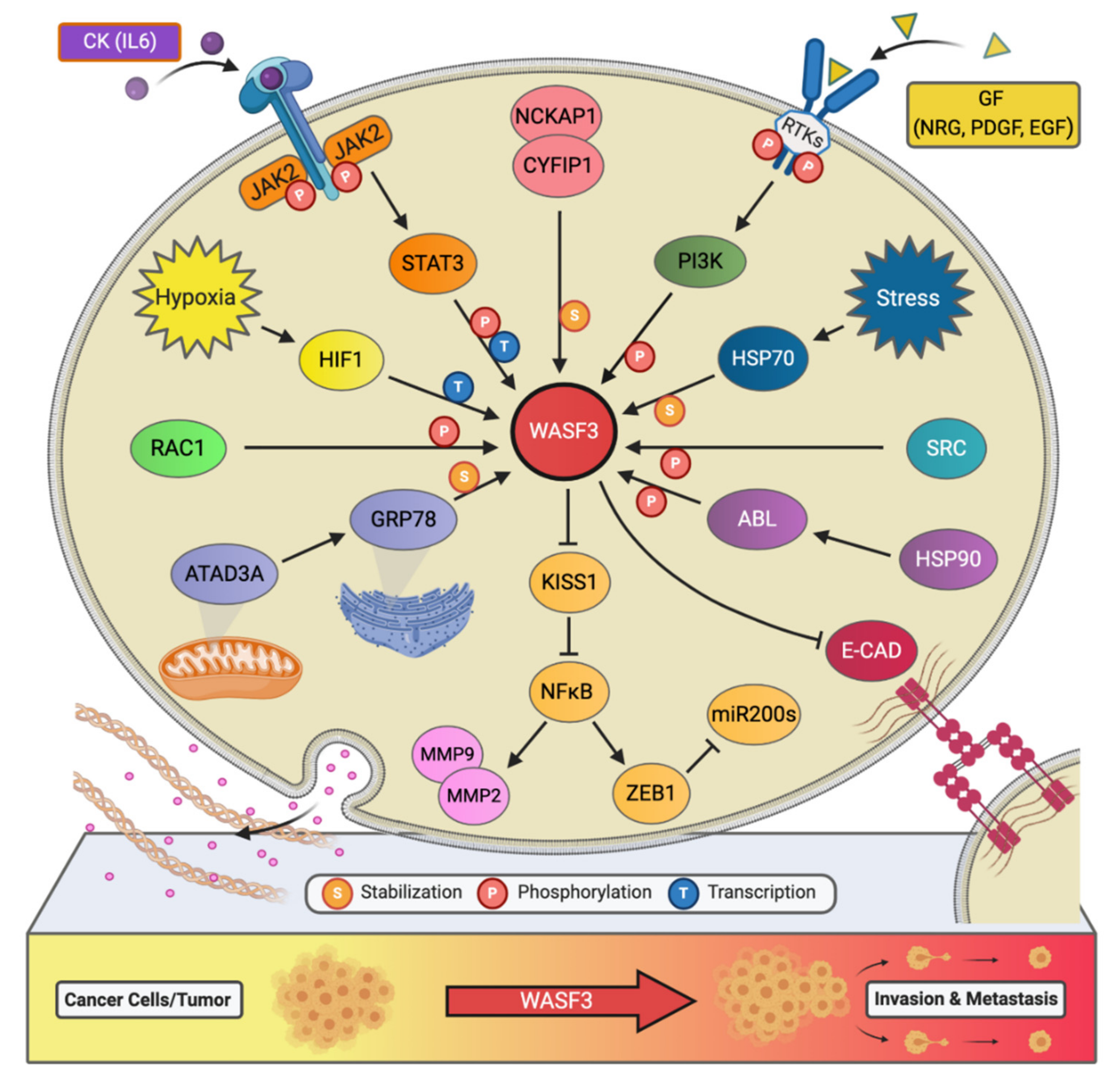
Several roads can contribute to WASF3 activation, here a recent paper :

Figure 2. The signaling network of WASF3 in cancer metastasis. The WASF3-dependent signaling pathways and related regulatory networks critical to controlling cancer metastasis are summarized here.

ijms-22-00836-g002.png

https://www.mdpi.com/1422-0067/22/2/836
 

datadragon

Senior Member
Messages
408
Location
USA
Last edited:

Tsukareta

Senior Member
Messages
150
Isn't tudca a fairly common supplement ? something to do with artificial bile, surely some of us would have taken it since we have various stomach related problems, personally I did not but I took digestive enzymes, promotility stuff, many antiviral or antibacterial / antifungal herbs, acid boosters, random vitamins etc.
 

datadragon

Senior Member
Messages
408
Location
USA
Another article today explaining the potential mechanism breakthrough:
To test the link between ER stress and WASF3, researchers treated human myoblasts with ER stress inducers and observed increased WASF3 protein. The level of WASF3 was inversely correlated with that of MTCO1, the last enzyme in the mitochondrial electron transport chain, which drives oxidative phosphorylation. This disruption leads to reduced mitochondrial oxygen consumption, providing a molecular explanation for symptoms like exercise intolerance and post-exertional malaise in patients with chronic fatigue. Muscle samples from ME/CFS patients also displayed higher WASF3 levels and lower levels of associated mitochondrial protein complexes.
https://medicalxpress.com/news/2023-08-chronic-fatigue-mechanisms-source-wasf3.html


In response to endoplasmic reticulum (ER) stress, increases in the GRP78 protein level leads to increased WASF3 protein levels. From that article: "Because WASF3 is regulated by BiP (GRP78), an endoplasmic reticulum (ER) protein that checks proteins for quality control, the researchers went upstream to see if BiP levels were reduced – and they were even more significantly altered than WASF3."

So I will add: Histone deacetylase (HDAC) inhibitors blocked GRP78 release by inducing its aggregation in the ER. https://www.nature.com/articles/srep30406 Butyrate inhibits histone deacetylase activity (HDAC) https://www.ncbi.nlm.nih.gov/pubmed/12840228 https://www.ncbi.nlm.nih.gov/pmc/articles/PMC6333934/ Butyrate enhances mitochondria https://www.nature.com/articles/s41398-017-0089-z Sodium butyrate reduces endoplasmic reticulum stress (posted above)

@LINE covered videos where butyrate is found low https://www.youtube.com/playlist?list=PLA9aaXOpxIE3QBETBfDtz545PrnROUkBo and in the fecal metabolome, the % butyrate was increased in the NoPEM group compared with both the PEM and control groups.

https://forums.phoenixrising.me/threads/an-acod1-genomics-question.90574/#post-2441131
 
Last edited:

SWAlexander

Senior Member
Messages
2,049
I was hesitant to post my short observation and theory I have sent to several scientists who were dealing foremost with C-19.

Now encouraged by Efthymios Kalafatis post (ME/CFS Research Foundation @MECFSResearch)
Maybe I´ll find a response or suggestion here. Comments and suggestions are welcome.

I gathered detailed information already in 2021 after a DVT, VWD and APS diagnosis.

As far as I know, none of the researchers have even tried to investigate P450.
This is what I sent out in Febr. 2022 and this year again.
1692536737073.png
 

Dude

Senior Member
Messages
210
@SWAlexander
This might be complete irrelevant and i just mention it, to help you find an answer, but fibrinogen is also mentioned in the hypothesis about how bc007 works.

More plausibly, the lasting effect of BC-007 may be related to an additional effect on the
inflammatory matrix of LC (which may possibly extend to CNS functions):
For one, the improved perfusion could induce a virtuous circle by abolishing the „drivers“ of
inflammation like tissue hypoxia and the associated mitochondrial dysfunction, but also by possibly
decreasing the disruption of the blood brain barrier (BBB), which has been shown to maintain neuroinflammation through a constant influx of albumin, fibrinogen and immune cells.
You find this document in the other thread with sources.
 

SWAlexander

Senior Member
Messages
2,049
This might be complete irrelevant and i just mention it, to help you find an answer, but fibrinogen is also mentioned in the hypothesis about how bc007 works.
I remember the struggle with BC007. Dr. Hohberger Erlangen and Bhupesh K Prusty Würzburg were one of the researchers I wrote to. There is no way, in my opinion, that clots entangled by fibrin, can be savly removed without dissolving the fibrin net.

Remember when Prof. Resia Pretorius mentioned in the early stage of C-19, that she found the micro clots but can't open them? I sent her a message to try PG or P450. No answer.
 
Last edited:

hapl808

Senior Member
Messages
2,325
Isn't tudca a fairly common supplement ? something to do with artificial bile, surely some of us would have taken it since we have various stomach related problems, personally I did not but I took digestive enzymes, promotility stuff, many antiviral or antibacterial / antifungal herbs, acid boosters, random vitamins etc.

I've taken TUDCA. Seems to improve digestion a bit. Not sure I've noticed any other major effects.
 

Judee

Psalm 46:1-3
Messages
4,584
Location
Great Lakes
So that Wiley Online Library paper says, "Under irremediable endoplasmic reticulum (ER) stress, hyperactivated inositol-requiring enzyme 1a (IRE1a) triggers the terminal untolded protein response (T-UPR), causing crucial cell dysfunction and apoptosis." (Simplistic question but I started taking Inositol recently. Does that mean Inositol will "feed" the mechanism that we don't want since the enzyme requires it?)

2nd simplistic question, the article mentions, "Doxycycline-inducible IRE1a overexpression or ER stress agents were used to induce IRE1a activation." (Again, does that mean Doxycycline is not a good idea either...that it could trigger this ER stress and induce IRE1a activation?

Again, asking because that is also something people here take from time to time esp for things like Lyme.

:(

My brain is just not scientific enough so that's why I'm asking and please if someone responds to my questions could you keep the answer to a level that someone who has had brain fog most of their life can understand...dumb it down for my sake if you would. Thank you. :)
 

Forummember9922

Senior Member
Messages
188
(One of those posts where you just sort of lawlessly paint a picture because it's what you want to see. And increasingly it is obvious that GPT will reliably paint any picture you want it to)

From: "Metabolic profiling of a myalgic encephalomyelitis/chronic fatigue syndrome discovery cohort reveals disturbances in fatty acid and lipid metabolism" (2018) https://www.ncbi.nlm.nih.gov/pmc/articles/PMC5365380/

Pathway analysis points to a few pathways with high impact and therefore potential disturbances in patients, mainly taurine metabolism and glycerophospholipid metabolism, combined with primary bile acid metabolism, as well as glyoxylate and dicarboxylate metabolism and a few other pathways, all involved broadly in fatty acid metabolism.
----

And then back to GPT "How does tudca effect taurine metabolism, glycerophospholipid metabolism, or primary bile acid metabolism?"
  1. Taurine Metabolism:
    • TUDCA and Taurine Synthesis: Taurine is an amino acid that plays various roles in the body, including in bile acid conjugation. Taurine conjugates with bile acids to form bile salts, which are essential for digestion and absorption of dietary fats. TUDCA has been shown to increase the synthesis of taurine in the liver. This can be beneficial for individuals with taurine deficiency or impaired taurine metabolism.
  2. Glycerophospholipid Metabolism:
    • TUDCA and Lipid Metabolism: Glycerophospholipids are important components of cell membranes and play a role in lipid metabolism. TUDCA has been studied for its potential to improve lipid metabolism and reduce lipid-related disorders, such as non-alcoholic fatty liver disease (NAFLD). TUDCA may help reduce lipid accumulation in the liver and improve insulin sensitivity, which can positively affect glycerophospholipid metabolism.
  3. Primary Bile Acid Metabolism:
    • TUDCA and Primary Bile Acids: Bile acids are synthesized in the liver from cholesterol and are essential for digestion and absorption of dietary fats. TUDCA is a secondary bile acid that is formed in the intestine by the gut microbiota from primary bile acids like chenodeoxycholic acid (CDCA). TUDCA has been shown to have cytoprotective and anti-apoptotic properties, which can help maintain the integrity of liver cells. It can also influence the balance between primary and secondary bile acids in the gut
 
Last edited:

datadragon

Senior Member
Messages
408
Location
USA
From Cort’s article, regarding the woman with elevated WASF3 in her muscles, “When they knocked down WASF3 levels in her cells her mitochondrial functioning improved.” Does that mean her energy levels and symptoms improved?

Not directly. Both S1 (the woman) and the people with ME/CFS had biochemical signatures of ER stress in their muscles, and treating S1’s cells in a dish with a drug that blocks ER stress lowered WASF3 levels and restored mitochondrial function. On the flipside, using toxins to artificially induce ER stress in cultured cells or in mice caused a rise in WASF3 levels,

Researchers have identified a protein that’s present at unusually high levels in the muscles of people with ME/CFS and that disrupts cells’ ability to generate energy. The researchers wondered whether WASF3 was interacting with mitochondria, cellular compartments responsible for energy generation that have been suggested to malfunction in people with ME/CFS. Sure enough, by changing levels of WASF3 inside cultured cells from S1 as well as in other human and mouse cells, the team found the protein could disrupt mitochondrial function. Specifically they found that high levels of WASF3 interfered with the assembly of mitochondrial proteins into molecular complexes that support normal energy production.

Hwang’s group next genetically engineered mice to produce elevated amounts of WASF3. These animals also had defects in their mitochondrial function and were only able to run about half as far on a treadmill as regular mice. Curious as to whether these results might be relevant to people formally diagnosed with ME/CFS, the researchers compared muscle samples from 14 people living with the illness with those of 10 healthy individuals. They found higher average levels of WASF3—and lower levels of the associated mitochondrial protein complexes—in people with the condition.
 

datadragon

Senior Member
Messages
408
Location
USA
Interesting that JAK2 increases WASF3. Could this fit with the Itaconate shunt hypothesis? A downstream effect?

My initial thought is that low butyrate is one connection to both higher WASF3 and the itaconate shunt which I mentioned in this thread that hopefully htester will look over. https://forums.phoenixrising.me/threads/an-acod1-genomics-question.90574/

Bioavailable copper (attached to ceruloplasmin) is necessary for the electron transport chain to operate and so when bioavailable copper is low mitochondria dysfunction also results. Another thought is that Zinc is also lowered through a reduction in Shank3 levels via NLRP3 over activation, and this can lead to reduction in Vitamin A and bioavailable (usable) copper. Most of the ATP in neurons is derived from oxidative metabolism, and cytochrome c oxidase (COX) is a critical energy-generating enzyme. It is an integral protein of the inner mitochondrial membrane, catalyzing the final step of oxidative metabolism. Cytochrome c oxidase, the terminal oxidase in the electron transport chain, is copper dependent https://www.ncbi.nlm.nih.gov/pmc/articles/PMC2775551/ https://pubmed.ncbi.nlm.nih.gov/9185337/

Copper also requires Zinc, Vitamin A, and Magnesium for ceruloplasmin) and why copper transport becomes dysregulated after prolonged inflammation, a loss of bioavailable copper for the body to utilize, and a buildup of unbound copper. Ceruloplasmin, a copper-containing acute phase plasma protein, has been shown to be regulated by 13-cis retinoic acid in rats. Ceruloplasmin activity was significantly increased within 24 h and remained elevated for at least 72 h after a single injection of 13-cis retinoic acid. https://pubmed.ncbi.nlm.nih.gov/3655940/ Ceruloplasmin secretion-based drug screening identified all-trans retinoic acid (ATRA) and other active Vitamin A metabolites as promising candidates for rescuing Ceruloplasmin secretion. ATRA also alleviated reactive oxygen species (ROS) production induced by lipid accumulation in Wilsons Disease-specific hepatocytes. https://www.biorxiv.org/content/10.1101/2021.08.10.455792v1

Until now, vitamin A (retinol) was solely regarded as a biochemical precursor for bioactive retinoids such as retinaldehyde and retinoic acid (RA), but recent results indicate that this compound has its own physiology. It functions as an electron carrier in mitochondria. By electronically coupling protein kinase Cδ (PCKδ) with cytochrome c, vitamin A enables the redox activation of this enzyme. https://www.ncbi.nlm.nih.gov/pmc/articles/PMC4739943/ Retinoic acid has also been shown to regulate mitochondrial transcription and function. Mitochondrial encoded genes have also been reported to be regulated by retinoic acid. These include NADH dehydrogenase subunit 5 (ND5;5), cytochrome c oxidase subunit I mRNA, and 16srRNA https://pubmed.ncbi.nlm.nih.gov/12440518/

Vitamin A is lowered from inflammation and infection https://forums.phoenixrising.me/thr...-fatigue-syndrome-patients.90588/post-2441323 A high level of copper from a lowering of zinc over time also will contribute to deplete vitamin a stores due to nutritional relationships and its requirement for ceruloplasmin.
 
Last edited:
Back