Phoenix Rising Team submitted a new blog post:
Ottawa 2011 Conference Reports Pt V: the Brain Studies
Posted by Cort Johnson
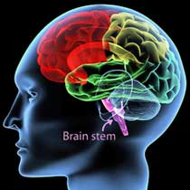 Many researchers think the problems in the brain or central nervous system probably play a key role in ME/CFS. Some of the most interesting research in the past couple of years has focused on the brain and the Ottawa conference was no exception.
At the conference we saw research findings begin to focus on specific areas of the brain, a spinal fluid study suggesting a brain injury is leaking proteins into the spinal fluid, a nasal study demonstrating more whacky autonomic nervous system results and a paper suggest that for some patients, it may all be in the nose......
PRESENTATIONS
The Seat of Fatigue in the Brain Identified?
Andrew Miller, Jones, Drake, Tian, Unger. Decreased basal ganglia activation in CFS subjects is associated with fatigue
The basal ganglia and related areas in the brain were news for a while. Several studies in the early 2000s appeared to be pointing an arrow straight at these areas, but, as happens so often in CFS, positive study results don’t necessarily mean more funding…and the studies stopped; but Andrew Miller, the Director of the Mind/Body Program at Emory University, in association with several CDC researchers has revived interest in this area.
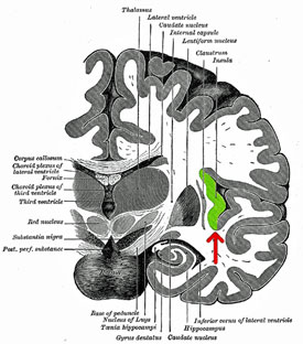
The study - In this study, the CFS patients showed reduced activity of two parts of the basal ganglia, the caudate and the globus pallidis. The fact that basal ganglia activity and fatigue levels were correlated in the ME/CFS patients but were not in the healthy controls highly suggested that the basal ganglia plays a role in producing fatigue in ME/CFS (as it appears to do in Parkinson’s disease, MS and other disorders.)
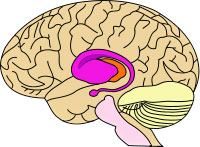
Focus on the Basal Ganglia - Sitting in the interior of the brain, the basal ganglia consists of six interconnected nuclei (including the amygdala) that provide a link with the limbic system and the hypothalamus.
A Deep Brain Focus - Several factors suggest that deep brain organs like the basal ganglia could play a role in CFS. These organs seem to excel in affecting both emotions and physical activity, both of which (at least in my case) have taken a hit in this disorder. The basal ganglia are hit hard in a number of highly fatiguing disorders (Parkinson’s disease, MS, stroke and AIDS). Dopamine activity in the basal ganglia plays an important role in mood, motivation, sleep/wake (arousal), cognition and motor activity. Study evidence suggests problems with dopamine metabolism may play an important role in ‘central pain/fatigue’ states such as Fibromyalgia.
Poor Planning? - Does just the thought of doing something overwhelm you sometimes? Basal ganglia problems could be a reason for that. Almost ten years ago two CFS researchers, Chaudhuri and Behan, proposed that the reduced self-motivation and increased fatigue seen in all central fatigue disorders (CFS, Parkinson’s, MS, etc.) is at least partly due to poor functioning in the basal ganglia; specifically its inability to effectively process the cues needed to do tasks. (This hearkens back to studies suggesting that problems with ‘motor planning,’ i.e. the planning needed to engage in movement, may make movement more effortful in ME/CFS. Apparently the brain conceptualizes movement before it occurs. …Both models touch on the idea that the ME/CFS brain has particular difficulty in ‘planning,’ which does have some intuitive appeal…)
Several small magnetic resonance spectroscopy (MRS) studies have found increased choline peaks in the basal ganglia of CFS patients, possibly due to membrane damage, perhaps due to infection (See Choline on the Brain?).
Miller proposed several reasons why the basal ganglia seem to be less effective in people with ME/CFS.
Increased pro-inflammatory cytokine activity. Pro-inflammatory cytokines have long been associated with fatigue. The cytokine/fatigue association was blown open when it was found that the administration of interferon alpha - a cytokine used to combat hepatitis - produced profound fatigue and flu-like states in many patients. Researchers later discovered that IFN-a also produces reduced glucose uptake (e.g. reduced activity) in yes, the basal ganglia.
Infection (and the ‘Good’ Fatigue) – Fatigue is believed to be an adaptive response initiated by the brain to slow the body down, assisting healing and inhibiting the spread of the pathogen. In a discussion during a break Miller related that he felt viruses played a key role in ME/CFS. He talked about a system that was on alert - and for a good reason. Miller sees ME/CFS from an evolutionary or ecological perspective. When, he asks, might having fatigue be better than having a high energy level? When the body needs to conserve its resources in order to heal itself, and having co-workers and friends visit would spread a pathogen to them.
 Infection Whacks Basal Ganglia Functioning - During the Q&A session, Dr. Miller noted that toxins associated with some bacteria (endotoxins) also have been shown to reduce basal ganglia functioning. These toxins apparently promote inflammation and pro-inflammatory cytokine production, and thereby produce fatigue and altered moods.
Interestingly, in some individuals these cytokines can cause ‘increased arousal’ - perhaps a more common state in ME/CFS than depression. (Dr. Baraniuk thinks the research community has been way off base with its focus on depression in ME/CFS….the much more common state, he believes, is ‘increased arousal’ caused by too much sensory information flooding the brain.)
Gene Silencing - Dr. Miller wasn’t sure, though, that whatever started the problem is still there, and he talked about an alternative culprit to pathogens. He said that when the body is attacked by a virus, or in the midst of some other traumatic event, the immune system revs up to fight off a possible attack. During the revving up process, things can go wrong, and he thinks a level of controls called ‘epigenetics’ may have gone awry in people with CFS. Studies have shown that immune activation can trigger the methylation process to turn genes on or off - permanently - causing what is called ‘epigenetic changes’.
(If you feel really different after coming down with CFS - if this theory is right - then it’s possible that you really are, genetically, somewhat different; that is, the genes you are born with are not acting in the same way.) In the case of CFS, he suggested that a methylation process triggered during infection could have altered the functioning of a main immune regulator, the glucocorticoid system.
He’s not the first to suggest this. Just last year, CDC researcher Falkenburg found evidence of increased methylation of a serotonin transporter in CFS (Neuromolecular Medicine, 2011 (13) 66-76) At the Ottawa conference, Falkenburg unsuccessfully looked for evidence that methylation had turned down the activity of genes that produce perforin in natural killer cells. Both Falkenburg and Miller are looking for physiological changes triggered by that initial infection (or whatever it was) which altered the playing field for people with ME/CFS.
Pressing the Reset Button - Some work is going into reversing these harmful epigenetic changes. Miller stated anti-methylation drugs have changed cowering lab rodents with epigenetic changes into exuberant, active ones and he related the story of a chemotherapy patient with documented epigenetic changes whose depression scores went way down when given an anti-methylation drug. He said it was like pressing a ‘reset button’. (‘Chemofog’ and similar CFS symptoms are common outcomes of cancer treatment.) Could people with CFS be displaying epigenetic changes caused by immune activation? Only time will tell.
Immune Modulation - Given his focus on the immune system, it wasn’t surprising that Dr. Miller is intrigued by the use of immune modulating drugs, not just to combat overt infection, but to affect ‘neuropsychiatric’ symptoms such as pain, fatigue and sleep. He noted that ‘cytokine antagonists’ (e.g. Enbrel, etc.) are helping to explain the contributions cytokines make to depression and pain (and fatigue). With some physicians now using them successfully to reduce pain in cancer patients, the role of immunomodulators and immune suppressants appears to be opening up. (A recent Miller paper argued for the use of more immune drugs to suppress pain in cancer.)
Direct immune modulation is becoming more and more an option for the right kind of CFS patients. Immune modulation can have serious consequences, but a pattern seems to building. Ampligen, of course, is an immunomodulator, and rituximab, a B-cell depressant, had excellent (if transitory) results in some patients. Dr. Klimas and other ME/CFS specialists appear to be exploring these powerful drugs for selected patients.
Dr. Miller had many intriguing ideas, but unfortunately his time with the CDC and CFS is over and he’s moving on.
The Mysterious CDC Insula Study
Interaction of Self- and Illness-related Cognitive Processing in the Right Anterior Insula of the CFS patients: an fMRI study. Jones, Rajendra, Drake, Miller, Unger, Tian, Pagnoni
Andrew Miller, an accomplished psychoneuroimmunologist who presented his own fMRI brain study, stated that he didn’t have a clue what this study was about.
Dr. Jones is surely close to ending his long stint on the CDC’s CFS research team and they have been giving him room to roam. Dr. Jones' first interoception paper in 2005 paper laid out his idea that abnormal behavior in several regions of the brain (insula, anterior/posterior cingulate, orbitofrontal cortex) might be associated with autonomic nervous system problems and increased symptoms.
He theorized that, depending on which part of the brain is affected, people with ME/CFS might respond well to pharmacological drugs, biofeedback, meditation or CBT. (Before his departure, Dr. Reeves stated a study was underway at the CDC that would match therapies to brain scan results; i.e. pharmacological drugs would be used to treat one type of brain dysfunction, meditation, another, etc.)
The insula is a hot topic in the neuro-endocrine-immune disorder field. Insula problems have been found in a number of ‘central sensitization’ disorders such as FM, IBS, TMJ and now the insula is showing up in ME/CFS.
Focus on the Insula
The insula is a fascinating organ in the brain that in some ways seems almost made to order for ‘central sensitization’ disorders. Got problems with bright lights, sharp noises or painful body sensations? It’s the insula that determines how bright the lights are or how painful your body sensations are. Got concentration problems? One recent paper suggests that the high levels of insula activity found in FM may actually impair their working memory as the insula ‘steals’ resources from other parts of the brain. (Several studies indicate that inability of CFS brains to turn off their attention to things like background noise is an energy drain as well. )
Exercise and Emotions - Interestingly, the insula is activated by two factors problematic for CFS patients - exercise and negative emotional experiences - and it regulates two systems at the core of ME/CFS symptoms - the autonomic nervous and immune systems. The insula helps control blood pressure and blood flows during and after exercise, affects gastric motility (IBS symptoms?) and even speech and coordination - all of which tend to falter after exercise in CFS.
(Might one ME/CFS study (Bogaerts et al. 2007), that showed a propensity for reduced CO2 levels when ME/CFS patients imagined negative experiences, also point to the insula as the cause? It suggested poor autonomic nervous system functioning (blood flows, blood pressure) in response to a negative emotional hit.)
In his study, Jones asked people with CFS a series of questions related to illness and ‘self’. The CFS patients had the same level of insula activity to questions asked about illness which were not related to the self, but an increased insula response to questions about illness related to themselves. The healthy controls did not exhibit increased activation in response to these questions.
Jones concluded that one of two things could explain the increased insula activity in people with CFS: physiological problems that underlie their symptoms, or aberrant interoceptive processes that cause fatigue and other symptoms to be experiences more strongly than usual.
We’ll learn more about the first Jones interoception study when the full paper comes out, but the conclusions - ME/CFS is caused either by physiological aberrations or by problems with interoception - didn’t on the face of it appear to add much to the field.
Assessment of regional cerebral blood flow in CFS using arterial spin labeling MRI; Dyke, Weiduschat, Mao, Pillemer, Murrough, Natelson, Mathew, Shungu
Dr. Shungu, the lead researcher in this study, has published two studies finding increased lactate levels in the brains of ME/CFS patients. A byproduct of anaerobic energy production, the increased levels of lactate in the brain suggested that some sort of reduced oxygen usage in the brain might be present, caused by reduced blood flows, mitochondrial dysfunction, high rates of oxidative stress, or all of the above.
In this study, he took a long look at the most prominent theory: that reduced blood flows in the brain are causing high rates of oxidative stress, which punch out the mitochondria causing high levels of lactate. The low blood flow theory works in so many ways; we know that blood volume is low in ME/CFS; plus, there is evidence of blood pooling in the lower parts of the body, which would reduce blood flows in the brain; plus, postural tachycardia syndrome (POTS) is synonymous with reduced blood flows to the brain. It sounds like it's gotta be happening.
Unfortunately, Dr. Shungu, using a new technique, didn’t find evidence of substantially reduced blood flows in the brain! Lower blood flows were found in CFS patients but they were only 4% smaller, and instead of being broadly spread across the brain, they were quite localized. Shungu expected a bigger decrease and a greater area of the brain with low blood flows.
Read more about Dr. Shungu’s study with Dr. Natelson and his finding of decreased glutathione levels in the brains of ME/CFS patients in Kim McCleary’s interview - http://www.research1st.com/2011/10/07/brainiacs/
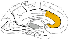
Focus on the Anterior Cingulate (ACC) - The place low blood flows were found, the anterior cingulate, however, was interesting. This is not the first time this part of the brain has popped up in ME/CFS research; it, like the basal ganglia, seems almost made to order for this disorder in some important ways.
Forming a kind of "collar" in the back lower part of the brain, the ACC regulates autonomic nervous system activity (heart rate, blood flows, etc.), pain sensitivity, ‘premotor’ (movement) control and what are called ‘rational cognitive functions’ such as decision making and reward anticipation. (For my part, difficulty making decisions should rank as one of the top cognitive problems in ME/CFS. It’s just pitiful sometimes… Of course, the movement problems for a formerly athletic person are also bizarre.)
Connections, Connections - Divided into two parts, a cognitive (rear - dorsal) and an emotional (front - ventral), the ACC is strongly connected with several areas of interest in ME/CFS such as the insula and the amygdala; the ACC has shown up in studies before. Schmaling (2003) found a marked ACC activation both in fatigued multiple sclerosis and in CFS patients. The ACC’s involvement in motor learning/planning and ‘attentional tasks’ suggests, as do the basal ganglia findings, that the planning process for movement may be impaired in ME/CFS. Interestingly, hepatitis treatment with interferon alpha also results in ACC activation. (A recent Miller paper suggested that the same processes occurring in IFN-a induced fatigue may be occurring in ME/CFS. Tying the fatigue processes in hepatitis C treatment and ME/CFS together would be a huge boost for the legitimization of ME/CFS.)
(Personal aside - long term planning has not been my strong suit after CFS. During a transfer factor treatment that temporarily boosted me out of my CFS-like state, however, I began thinking about and making plans for the future. It was like a circuit got reconnected….Once the bloom faded, I was back in my day to day grind. On a gut level, my guess is that all sorts of planning processes in the brain have taken a hit.)
Another study (Yamamoto, 2004) found reduced numbers of serotonin transporters in the front part of the ACC in CFS. A ‘feel good’ neurotransmitter that contributes to feelings of wellness and health, reduced levels of serotonin transmitters in the ACC could play a role in the pain and basically crappy feelings found in CFS. Falkenburg at the CDC recently found evidence of reduced transcription of serotonin in people with CFS.
CDC - Using brain scans, including magnetic resonance imaging (MRI) and positron emission tomography (PET), Emory scientists have found that IFN-alpha affects two parts of the brain, the basal ganglia and the dorsal anterior cingulate cortex (dACC). The basal ganglia play an important role in the regulation of motor activity and motivation as well as symptoms of fatigue. Indeed, IFN-alpha effects on the basal ganglia were linked with symptoms of fatigue. Ongoing studies at CDC are currently evaluating whether similar changes in the basal ganglia occur in patients with CFS. The dACC is a brain region associated with arousal and alarm, and changes in this brain region have been found in connection to anxiety.
POSTERS
Baraniuk Proteome Studies Suggest Brain Injury
Dr. Baranuik of Georgetown University had to unexpectedly leave the conference early but he was clearly happy about the results of his second proteome study. He presented more abstracts than any other individual and expects many papers to result.
Spinal fluid proteome analyses are pretty hot right now…The Schuster/Natelson study differentiating CFS patients from Lyme disease and healthy controls definitely got Dr. Natelson excited - something you don’t see very often. Baraniuk presented posters, not talks at the Conference - so our information is pretty scanty but things should get interesting soon.....
Distinct Clustering of Cerebropsinal Fluid Peptides and other Ion Peaks in Clusters of CFS and Healthy Subjects. J. Baraniuk, O Adewuyi, C Di Poto et. al.
Distinct Cerebrospinal Fluid Proteomic Patters in Clusters of CFS and Healthy Subjects
One study found evidence of four distinct subsets suggesting that ‘four distinctive pathophysiological mechanisms’ are present in the CFS population. Some of the significance factors =p<0.00001) were enormous, suggesting a very wide differentiation indeed. In his first brain proteome paper about five years ago Baraniuk presented evidence of a single proteomic signature in CFS and FM. Now, with his much larger and more complex study using better technology, he’s finding distinct subsets..
In most of these subsets he’s finding ME/CFS patients have higher levels of proteins in their spinal fluid. Where does he think these proteins are coming from? A brain injury that’s releasing proteins into the cerebrospinal fluid.
'Exercise' Test Reveals System Under Disarray
Blunted Nasal and Systemic Sympathetic Reflexes in Chronic Fatigue Syndrome. J Baraniuk, U. Le, K. Petrie et al.
Dr. Baraniuk, like many other researchers, has been fascinated by the role the autonomic nervous system plays in this disorder. Got a perennial case of sniffles, a clogged up nose or nasal drip? Dr. Baraniuk believes this may be caused by autonomic problems and he finds this kind of non-allergenic rhinitis in about 75% of people with ME/CFS.
In this study he had the participants clamp down hard on a hand grip while sticking what looked like a large horn into their nasal passages to measure nasal volume…..Since clamping down hard on the handgrip should stimulate the sympathetic nervous system to narrow the blood vessels in the nose (reducing nasal volume), increasing heart rate and blood pressure.
He found that all those things happened with the healthy controls but remained flat in the CFS group. One group had immediate increases in SNS measures which then decayed or even negative over time suggesting that their systems quickly pooped out quickly demonstrating in Dr. Baraniuk’s words “dysfunctional ‘on-demand’ sympathetic” activity.
Another Possible (and Treatable) Cause of ‘CFS’
Chronic Rhinosinuitis as an overlooked Chronic Fatigue Exclusionary condition
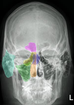
Dr. Alexander Chester is another Georgetown professor who has noticed sinus problems in people with ME/CFS. In his poster presentation Dr. Chester noted that not only is fatigue common in chronic rhinosinusitis (CRS) but that one study showed that ‘vitality scores’ in CRS patients were lower than in patients with congestive heart failure, chronic obstructive pulmonary disorder or chronic back pain.
Two studies, one which suggested surgery was more effective for severely fatigued (than less fatigued) people with CRS and one which found patients with FM and CRS responded better to surgery than patients with just CRS, suggested this approach might be effective for some with pain and fatigue producing disorders.
Chronic sinusitis or inflammation of the sinuses usually caused by a chronic viral or bacterial infection and Dr. Chester noted that it usually begins with an upper respiratory infection – much like ME/CFS does. It can cause stuffiness, head pains, headaches, post-nasal drip and fatigue. Cat Scans often do not pick up abnormalities in patients who later improve after surgery and there are no blood tests for it.

Continue reading the Original Blog Post
Ottawa 2011 Conference Reports Pt V: the Brain Studies
Posted by Cort Johnson
 Many researchers think the problems in the brain or central nervous system probably play a key role in ME/CFS. Some of the most interesting research in the past couple of years has focused on the brain and the Ottawa conference was no exception.
At the conference we saw research findings begin to focus on specific areas of the brain, a spinal fluid study suggesting a brain injury is leaking proteins into the spinal fluid, a nasal study demonstrating more whacky autonomic nervous system results and a paper suggest that for some patients, it may all be in the nose......
PRESENTATIONS
The Seat of Fatigue in the Brain Identified?
Andrew Miller, Jones, Drake, Tian, Unger. Decreased basal ganglia activation in CFS subjects is associated with fatigue
The basal ganglia and related areas in the brain were news for a while. Several studies in the early 2000s appeared to be pointing an arrow straight at these areas, but, as happens so often in CFS, positive study results don’t necessarily mean more funding…and the studies stopped; but Andrew Miller, the Director of the Mind/Body Program at Emory University, in association with several CDC researchers has revived interest in this area.
The study - In this study, the CFS patients showed reduced activity of two parts of the basal ganglia, the caudate and the globus pallidis. The fact that basal ganglia activity and fatigue levels were correlated in the ME/CFS patients but were not in the healthy controls highly suggested that the basal ganglia plays a role in producing fatigue in ME/CFS (as it appears to do in Parkinson’s disease, MS and other disorders.)
Focus on the Basal Ganglia - Sitting in the interior of the brain, the basal ganglia consists of six interconnected nuclei (including the amygdala) that provide a link with the limbic system and the hypothalamus.
A Deep Brain Focus - Several factors suggest that deep brain organs like the basal ganglia could play a role in CFS. These organs seem to excel in affecting both emotions and physical activity, both of which (at least in my case) have taken a hit in this disorder. The basal ganglia are hit hard in a number of highly fatiguing disorders (Parkinson’s disease, MS, stroke and AIDS). Dopamine activity in the basal ganglia plays an important role in mood, motivation, sleep/wake (arousal), cognition and motor activity. Study evidence suggests problems with dopamine metabolism may play an important role in ‘central pain/fatigue’ states such as Fibromyalgia.
Poor Planning? - Does just the thought of doing something overwhelm you sometimes? Basal ganglia problems could be a reason for that. Almost ten years ago two CFS researchers, Chaudhuri and Behan, proposed that the reduced self-motivation and increased fatigue seen in all central fatigue disorders (CFS, Parkinson’s, MS, etc.) is at least partly due to poor functioning in the basal ganglia; specifically its inability to effectively process the cues needed to do tasks. (This hearkens back to studies suggesting that problems with ‘motor planning,’ i.e. the planning needed to engage in movement, may make movement more effortful in ME/CFS. Apparently the brain conceptualizes movement before it occurs. …Both models touch on the idea that the ME/CFS brain has particular difficulty in ‘planning,’ which does have some intuitive appeal…)
Several small magnetic resonance spectroscopy (MRS) studies have found increased choline peaks in the basal ganglia of CFS patients, possibly due to membrane damage, perhaps due to infection (See Choline on the Brain?).
Miller proposed several reasons why the basal ganglia seem to be less effective in people with ME/CFS.
Increased pro-inflammatory cytokine activity. Pro-inflammatory cytokines have long been associated with fatigue. The cytokine/fatigue association was blown open when it was found that the administration of interferon alpha - a cytokine used to combat hepatitis - produced profound fatigue and flu-like states in many patients. Researchers later discovered that IFN-a also produces reduced glucose uptake (e.g. reduced activity) in yes, the basal ganglia.
Infection (and the ‘Good’ Fatigue) – Fatigue is believed to be an adaptive response initiated by the brain to slow the body down, assisting healing and inhibiting the spread of the pathogen. In a discussion during a break Miller related that he felt viruses played a key role in ME/CFS. He talked about a system that was on alert - and for a good reason. Miller sees ME/CFS from an evolutionary or ecological perspective. When, he asks, might having fatigue be better than having a high energy level? When the body needs to conserve its resources in order to heal itself, and having co-workers and friends visit would spread a pathogen to them.
 Infection Whacks Basal Ganglia Functioning - During the Q&A session, Dr. Miller noted that toxins associated with some bacteria (endotoxins) also have been shown to reduce basal ganglia functioning. These toxins apparently promote inflammation and pro-inflammatory cytokine production, and thereby produce fatigue and altered moods.
Interestingly, in some individuals these cytokines can cause ‘increased arousal’ - perhaps a more common state in ME/CFS than depression. (Dr. Baraniuk thinks the research community has been way off base with its focus on depression in ME/CFS….the much more common state, he believes, is ‘increased arousal’ caused by too much sensory information flooding the brain.)
Gene Silencing - Dr. Miller wasn’t sure, though, that whatever started the problem is still there, and he talked about an alternative culprit to pathogens. He said that when the body is attacked by a virus, or in the midst of some other traumatic event, the immune system revs up to fight off a possible attack. During the revving up process, things can go wrong, and he thinks a level of controls called ‘epigenetics’ may have gone awry in people with CFS. Studies have shown that immune activation can trigger the methylation process to turn genes on or off - permanently - causing what is called ‘epigenetic changes’.
(If you feel really different after coming down with CFS - if this theory is right - then it’s possible that you really are, genetically, somewhat different; that is, the genes you are born with are not acting in the same way.) In the case of CFS, he suggested that a methylation process triggered during infection could have altered the functioning of a main immune regulator, the glucocorticoid system.
He’s not the first to suggest this. Just last year, CDC researcher Falkenburg found evidence of increased methylation of a serotonin transporter in CFS (Neuromolecular Medicine, 2011 (13) 66-76) At the Ottawa conference, Falkenburg unsuccessfully looked for evidence that methylation had turned down the activity of genes that produce perforin in natural killer cells. Both Falkenburg and Miller are looking for physiological changes triggered by that initial infection (or whatever it was) which altered the playing field for people with ME/CFS.
- Dig Deeper! - The CFIDS Association of America recently launched a major study examining epigenetic changes across the genome.
Pressing the Reset Button - Some work is going into reversing these harmful epigenetic changes. Miller stated anti-methylation drugs have changed cowering lab rodents with epigenetic changes into exuberant, active ones and he related the story of a chemotherapy patient with documented epigenetic changes whose depression scores went way down when given an anti-methylation drug. He said it was like pressing a ‘reset button’. (‘Chemofog’ and similar CFS symptoms are common outcomes of cancer treatment.) Could people with CFS be displaying epigenetic changes caused by immune activation? Only time will tell.
Immune Modulation - Given his focus on the immune system, it wasn’t surprising that Dr. Miller is intrigued by the use of immune modulating drugs, not just to combat overt infection, but to affect ‘neuropsychiatric’ symptoms such as pain, fatigue and sleep. He noted that ‘cytokine antagonists’ (e.g. Enbrel, etc.) are helping to explain the contributions cytokines make to depression and pain (and fatigue). With some physicians now using them successfully to reduce pain in cancer patients, the role of immunomodulators and immune suppressants appears to be opening up. (A recent Miller paper argued for the use of more immune drugs to suppress pain in cancer.)
Direct immune modulation is becoming more and more an option for the right kind of CFS patients. Immune modulation can have serious consequences, but a pattern seems to building. Ampligen, of course, is an immunomodulator, and rituximab, a B-cell depressant, had excellent (if transitory) results in some patients. Dr. Klimas and other ME/CFS specialists appear to be exploring these powerful drugs for selected patients.
Dr. Miller had many intriguing ideas, but unfortunately his time with the CDC and CFS is over and he’s moving on.
The Mysterious CDC Insula Study
Interaction of Self- and Illness-related Cognitive Processing in the Right Anterior Insula of the CFS patients: an fMRI study. Jones, Rajendra, Drake, Miller, Unger, Tian, Pagnoni
“Something is happening here
But (we) don't know what it is
Do (we), Mister Jones?”
Bob Dylan “Ballad of a Thin Man”
Andrew Miller, an accomplished psychoneuroimmunologist who presented his own fMRI brain study, stated that he didn’t have a clue what this study was about.
Dr. Jones is surely close to ending his long stint on the CDC’s CFS research team and they have been giving him room to roam. Dr. Jones' first interoception paper in 2005 paper laid out his idea that abnormal behavior in several regions of the brain (insula, anterior/posterior cingulate, orbitofrontal cortex) might be associated with autonomic nervous system problems and increased symptoms.
He theorized that, depending on which part of the brain is affected, people with ME/CFS might respond well to pharmacological drugs, biofeedback, meditation or CBT. (Before his departure, Dr. Reeves stated a study was underway at the CDC that would match therapies to brain scan results; i.e. pharmacological drugs would be used to treat one type of brain dysfunction, meditation, another, etc.)
The insula is a hot topic in the neuro-endocrine-immune disorder field. Insula problems have been found in a number of ‘central sensitization’ disorders such as FM, IBS, TMJ and now the insula is showing up in ME/CFS.
Focus on the Insula
The insula is a fascinating organ in the brain that in some ways seems almost made to order for ‘central sensitization’ disorders. Got problems with bright lights, sharp noises or painful body sensations? It’s the insula that determines how bright the lights are or how painful your body sensations are. Got concentration problems? One recent paper suggests that the high levels of insula activity found in FM may actually impair their working memory as the insula ‘steals’ resources from other parts of the brain. (Several studies indicate that inability of CFS brains to turn off their attention to things like background noise is an energy drain as well. )
Exercise and Emotions - Interestingly, the insula is activated by two factors problematic for CFS patients - exercise and negative emotional experiences - and it regulates two systems at the core of ME/CFS symptoms - the autonomic nervous and immune systems. The insula helps control blood pressure and blood flows during and after exercise, affects gastric motility (IBS symptoms?) and even speech and coordination - all of which tend to falter after exercise in CFS.
(Might one ME/CFS study (Bogaerts et al. 2007), that showed a propensity for reduced CO2 levels when ME/CFS patients imagined negative experiences, also point to the insula as the cause? It suggested poor autonomic nervous system functioning (blood flows, blood pressure) in response to a negative emotional hit.)
In his study, Jones asked people with CFS a series of questions related to illness and ‘self’. The CFS patients had the same level of insula activity to questions asked about illness which were not related to the self, but an increased insula response to questions about illness related to themselves. The healthy controls did not exhibit increased activation in response to these questions.
Jones concluded that one of two things could explain the increased insula activity in people with CFS: physiological problems that underlie their symptoms, or aberrant interoceptive processes that cause fatigue and other symptoms to be experiences more strongly than usual.
We’ll learn more about the first Jones interoception study when the full paper comes out, but the conclusions - ME/CFS is caused either by physiological aberrations or by problems with interoception - didn’t on the face of it appear to add much to the field.
Assessment of regional cerebral blood flow in CFS using arterial spin labeling MRI; Dyke, Weiduschat, Mao, Pillemer, Murrough, Natelson, Mathew, Shungu
Dr. Shungu, the lead researcher in this study, has published two studies finding increased lactate levels in the brains of ME/CFS patients. A byproduct of anaerobic energy production, the increased levels of lactate in the brain suggested that some sort of reduced oxygen usage in the brain might be present, caused by reduced blood flows, mitochondrial dysfunction, high rates of oxidative stress, or all of the above.
In this study, he took a long look at the most prominent theory: that reduced blood flows in the brain are causing high rates of oxidative stress, which punch out the mitochondria causing high levels of lactate. The low blood flow theory works in so many ways; we know that blood volume is low in ME/CFS; plus, there is evidence of blood pooling in the lower parts of the body, which would reduce blood flows in the brain; plus, postural tachycardia syndrome (POTS) is synonymous with reduced blood flows to the brain. It sounds like it's gotta be happening.
Unfortunately, Dr. Shungu, using a new technique, didn’t find evidence of substantially reduced blood flows in the brain! Lower blood flows were found in CFS patients but they were only 4% smaller, and instead of being broadly spread across the brain, they were quite localized. Shungu expected a bigger decrease and a greater area of the brain with low blood flows.
Read more about Dr. Shungu’s study with Dr. Natelson and his finding of decreased glutathione levels in the brains of ME/CFS patients in Kim McCleary’s interview - http://www.research1st.com/2011/10/07/brainiacs/
Focus on the Anterior Cingulate (ACC) - The place low blood flows were found, the anterior cingulate, however, was interesting. This is not the first time this part of the brain has popped up in ME/CFS research; it, like the basal ganglia, seems almost made to order for this disorder in some important ways.
Forming a kind of "collar" in the back lower part of the brain, the ACC regulates autonomic nervous system activity (heart rate, blood flows, etc.), pain sensitivity, ‘premotor’ (movement) control and what are called ‘rational cognitive functions’ such as decision making and reward anticipation. (For my part, difficulty making decisions should rank as one of the top cognitive problems in ME/CFS. It’s just pitiful sometimes… Of course, the movement problems for a formerly athletic person are also bizarre.)
Connections, Connections - Divided into two parts, a cognitive (rear - dorsal) and an emotional (front - ventral), the ACC is strongly connected with several areas of interest in ME/CFS such as the insula and the amygdala; the ACC has shown up in studies before. Schmaling (2003) found a marked ACC activation both in fatigued multiple sclerosis and in CFS patients. The ACC’s involvement in motor learning/planning and ‘attentional tasks’ suggests, as do the basal ganglia findings, that the planning process for movement may be impaired in ME/CFS. Interestingly, hepatitis treatment with interferon alpha also results in ACC activation. (A recent Miller paper suggested that the same processes occurring in IFN-a induced fatigue may be occurring in ME/CFS. Tying the fatigue processes in hepatitis C treatment and ME/CFS together would be a huge boost for the legitimization of ME/CFS.)
(Personal aside - long term planning has not been my strong suit after CFS. During a transfer factor treatment that temporarily boosted me out of my CFS-like state, however, I began thinking about and making plans for the future. It was like a circuit got reconnected….Once the bloom faded, I was back in my day to day grind. On a gut level, my guess is that all sorts of planning processes in the brain have taken a hit.)
Another study (Yamamoto, 2004) found reduced numbers of serotonin transporters in the front part of the ACC in CFS. A ‘feel good’ neurotransmitter that contributes to feelings of wellness and health, reduced levels of serotonin transmitters in the ACC could play a role in the pain and basically crappy feelings found in CFS. Falkenburg at the CDC recently found evidence of reduced transcription of serotonin in people with CFS.
CDC - Using brain scans, including magnetic resonance imaging (MRI) and positron emission tomography (PET), Emory scientists have found that IFN-alpha affects two parts of the brain, the basal ganglia and the dorsal anterior cingulate cortex (dACC). The basal ganglia play an important role in the regulation of motor activity and motivation as well as symptoms of fatigue. Indeed, IFN-alpha effects on the basal ganglia were linked with symptoms of fatigue. Ongoing studies at CDC are currently evaluating whether similar changes in the basal ganglia occur in patients with CFS. The dACC is a brain region associated with arousal and alarm, and changes in this brain region have been found in connection to anxiety.
POSTERS
Baraniuk Proteome Studies Suggest Brain Injury
Dr. Baranuik of Georgetown University had to unexpectedly leave the conference early but he was clearly happy about the results of his second proteome study. He presented more abstracts than any other individual and expects many papers to result.
Spinal fluid proteome analyses are pretty hot right now…The Schuster/Natelson study differentiating CFS patients from Lyme disease and healthy controls definitely got Dr. Natelson excited - something you don’t see very often. Baraniuk presented posters, not talks at the Conference - so our information is pretty scanty but things should get interesting soon.....
Distinct Clustering of Cerebropsinal Fluid Peptides and other Ion Peaks in Clusters of CFS and Healthy Subjects. J. Baraniuk, O Adewuyi, C Di Poto et. al.
Distinct Cerebrospinal Fluid Proteomic Patters in Clusters of CFS and Healthy Subjects
One study found evidence of four distinct subsets suggesting that ‘four distinctive pathophysiological mechanisms’ are present in the CFS population. Some of the significance factors
In most of these subsets he’s finding ME/CFS patients have higher levels of proteins in their spinal fluid. Where does he think these proteins are coming from? A brain injury that’s releasing proteins into the cerebrospinal fluid.
'Exercise' Test Reveals System Under Disarray
Blunted Nasal and Systemic Sympathetic Reflexes in Chronic Fatigue Syndrome. J Baraniuk, U. Le, K. Petrie et al.
Dr. Baraniuk, like many other researchers, has been fascinated by the role the autonomic nervous system plays in this disorder. Got a perennial case of sniffles, a clogged up nose or nasal drip? Dr. Baraniuk believes this may be caused by autonomic problems and he finds this kind of non-allergenic rhinitis in about 75% of people with ME/CFS.
In this study he had the participants clamp down hard on a hand grip while sticking what looked like a large horn into their nasal passages to measure nasal volume…..Since clamping down hard on the handgrip should stimulate the sympathetic nervous system to narrow the blood vessels in the nose (reducing nasal volume), increasing heart rate and blood pressure.
He found that all those things happened with the healthy controls but remained flat in the CFS group. One group had immediate increases in SNS measures which then decayed or even negative over time suggesting that their systems quickly pooped out quickly demonstrating in Dr. Baraniuk’s words “dysfunctional ‘on-demand’ sympathetic” activity.
Another Possible (and Treatable) Cause of ‘CFS’
Chronic Rhinosinuitis as an overlooked Chronic Fatigue Exclusionary condition
Dr. Alexander Chester is another Georgetown professor who has noticed sinus problems in people with ME/CFS. In his poster presentation Dr. Chester noted that not only is fatigue common in chronic rhinosinusitis (CRS) but that one study showed that ‘vitality scores’ in CRS patients were lower than in patients with congestive heart failure, chronic obstructive pulmonary disorder or chronic back pain.
Two studies, one which suggested surgery was more effective for severely fatigued (than less fatigued) people with CRS and one which found patients with FM and CRS responded better to surgery than patients with just CRS, suggested this approach might be effective for some with pain and fatigue producing disorders.
Chronic sinusitis or inflammation of the sinuses usually caused by a chronic viral or bacterial infection and Dr. Chester noted that it usually begins with an upper respiratory infection – much like ME/CFS does. It can cause stuffiness, head pains, headaches, post-nasal drip and fatigue. Cat Scans often do not pick up abnormalities in patients who later improve after surgery and there are no blood tests for it.
Please Support Phoenix Rising
(Why not support Ph0enix Rising with a $5 , $10, $15, $20 or more monthly 'subscription')
(Click on the image and look on the left hand side of the page for Recurring Donation Options)

Continue reading the Original Blog Post

