• Welcome to Phoenix Rising!

    Created in 2008, Phoenix Rising is the largest and oldest forum dedicated to furthering the understanding of, and finding treatments for, complex chronic illnesses such as chronic fatigue syndrome (ME/CFS), fibromyalgia, long COVID, postural orthostatic tachycardia syndrome (POTS), mast cell activation syndrome (MCAS), and allied diseases.

    To become a member, simply click the Register button at the top right.

Type 2 Deiodinase Disruption in Astrocytes Results in Anxiety-Depressive-Like Behavior

pattismith

Senior Member
Messages
3,946
Type 2 Deiodinase Disruption in Astrocytes Results in Anxiety-Depressive-Like Behavior in Male Mice
AC Bianco and col
2016
Abstract

Millions of levothyroxine-treated hypothyroid patients complain of impaired cognition despite normal TSH serum levels.
This could reflect abnormalities in the type 2 deiodinase (D2)-mediated T4-to-T3 conversion, given their much greater dependence on the D2 pathway for T3 production.
T3 normally reaches the brain directly from the circulation or is produced locally by D2 in astrocytes.

Here we report that mice with astrocyte-specific Dio2 inactivation (Astro-D2KO) have normal serum T3 but exhibit anxiety-depression-like behavior as found in open field and elevated plus maze studies and when tested for depression using the tail-suspension and the forced-swimming tests.

Remarkably, 4 weeks of daily treadmill exercise sessions eliminated this phenotype. Microarray gene expression profiling of the Astro-D2KO hippocampi identified an enrichment of three gene sets related to inflammation and impoverishment of three gene sets related to mitochondrial function and response to oxidative stress.
Despite normal neurogenesis, the Astro-D2KO hippocampi exhibited decreased expression of four of six known to be positively regulated genes by T3, ie, Mbp (∼43%), Mag (∼34%), Hr (∼49%), and Aldh1a1 (∼61%) and increased expression of 3 of 12 genes negatively regulated by T3, ie, Dgkg (∼17%), Syce2 (∼26%), and Col6a1 (∼3-fold) by quantitative real-time PCR.
Notably, in Astro-D2KO animals, there was also a reduction in mRNA levels of genes known to be affected in classical animal models of depression, ie, Bdnf (∼18%), Ntf3 (∼43%), Nmdar (∼26%), and GR (∼20%), which were also normalized by daily exercise sessions.

These findings suggest that defects in Dio2 expression in the brain could result in mood and behavioral disorders.
.....

Astro-D2KO animals exhibited 70%–95% reduction in Dio2 mRNA levels and D2 activity in the cerebral cortex and hippocampus (Figure 1, A–D) when compared with GFAP-Cre (control) animals. D2 activity was preserved in the pituitary gland (Figure 1E) and hypothalamus (18);

@Hip
 
Last edited:

Hip

Senior Member
Messages
17,874
I don't know much about deiodinase, but in chronic coxsackievirus B infection of the brain (as occurs in ME/CFS), it is the astrocytes that get chronic infected. Ref: 1 Thus some of the symptoms of ME/CFS might be due to an astrocyte infection, that impairs astrocyte functioning.
 

duncan

Senior Member
Messages
2,240
Here we report that mice with astrocyte-specific Dio2 inactivation (Astro-D2KO) have normal serum T3 but exhibit anxiety-depression-like behavior as found in open field and elevated plus maze studies and when tested for depression using the tail-suspension and the forced-swimming tests
Forced swimming tests. Anxiety? Try fear for their lives.

Remarkably, 4 weeks of daily treadmill exercise sessions eliminated this phenotype.
Thank God for the treadmill. :rolleyes:

My takeaway: Fear of drowning eases in mice once they are pulled out of the water. They remain less fearful even when placed on a treadmill.

If they really wanted to test for anxiety/depression, perhaps they should look into how many packs of cigarettes the mice smoked.

What ever happened to humanity and common sense?
 

Hip

Senior Member
Messages
17,874
Forced swimming tests. Anxiety? Try fear for their lives.

To properly criticize the forced swim test, you'd first need to spend some time reading about it and understanding it. Otherwise you are criticizing from a position of ignorance.
 

duncan

Senior Member
Messages
2,240
To properly criticize the forced swim test, you'd first need to spend some time reading about it and understanding it.
@Hip, have you ever written research reports? Spun their results for press releases?

I have.

I also have ended up in deep ocean water where I didn't want to be and was afraid of drowning.

I frequently can also distinguish fear from anxiety/depression.

Regardless, the kicker for me - and perhaps for some other readers - is the treadmill offered up as curative. Exercise, the panacea.

Thanks, but no thanks.
 

pattismith

Senior Member
Messages
3,946
@Hip
Regardless, the kicker for me - and perhaps for some other readers - is the treadmill offered up as curative. Exercise, the panacea.

Thanks, but no thanks.

Duncan, this is really not the point I wanted you to focus on, because nothing can say if this part of the experiment is relevant to any other condition where astrocytes deiodinase type 2 down-regulation could occur.

What is amazing in this study, (and Hip already pointed it), is that we can easily imagine that an infected astrocytes (by an enterovirus for example) may have it's D2 disrupted, which could lead to an hypothyroid state in astrocytes and in some parts of the brain, leading to hypometabolism and behavioral symptoms.
This kind of impairment couldn't be detected, because nothing would show up in blood as it is the case in these mice.
 

duncan

Senior Member
Messages
2,240
What is amazing in this study, (and Hip already pointed it), is that we can easily imagine that an infected astrocytes (by an enterovirus for example) may have it's D2 disrupted, which could lead to an hypothyroid state in astrocytes and in some parts of the brain, leading to hypometabolism and behavioral symptoms.'

I can appreciate that, @pattismith . I can imagine other antigens inducing the same thing, e.g. borrelia. I get that possibility, and I embrace it, at least in theory.

But what I am trying to point out is the methodology/assumptions of this study leaves me a little wanting, and if the methodology/assumptions are suspect, then to prove your suspicians and @Hip's and by extension maybe even some of mine, someone may need to build a better mouse trap.

And btw, isn't everyone getting sick and tired of researchers throwing mice into water to try to generate ME/CFS or similar conditions in those creatures? Not that this is an ME/CFS study, but you get my drift.
 
Last edited:

pattismith

Senior Member
Messages
3,946
And btw, isn't everyone getting sick and tired of researchers throwing mice into water to try to generate ME/CFS or similar conditions in those creatures? Not that this is an ME/CFS study, but you get my drift.

Yes of course, I agree and I would prefer those poor mice not to suffer. But it is the way research goes, and I don't know if we can avoid it totally.
On the other hand I wouldn't want human to become the guinea pigs, if mice had to be spared.
So the debate is very big, and this is not my willing to go into this.
 

Hip

Senior Member
Messages
17,874
And btw, isn't everyone getting sick and tired of researchers throwing mice into water to try to generate ME/CFS or similar conditions in those creatures?

I think we can all agree that the usual mouse model of ME/CFS — in which they first exhaust mice with forced exercise and then equate that exhaustion to ME/CFS — would almost certainly not be anywhere near an accurate model of ME/CFS.

However, as for the techniques used in this study to measure anxiety and depressive behaviors, I myself am not in any position to comment on whether these methods are valid or not, as I know nothing about them, and would have to spend hours if not days reading about these methods to come any conclusions.
 

duncan

Senior Member
Messages
2,240
However, as for the techniques used in this study to measure anxiety and depressive behaviors
Do you mean anxiety and depression? :)

You have to admit this is at least one very large step removed from that.
 

Hip

Senior Member
Messages
17,874
Do you mean anxiety and depression?

No, there is no way to directly measure anxiety and depression in mice; you can't ask them how they feel; so you can only measure or observe behaviors that are suggestive of anxiety and depression.
 

duncan

Senior Member
Messages
2,240
Precisely. Inference is a messy business, riddled with risk. And you no doubt remember how I regard that entire sickness behavior silliness.
 
Last edited:

Hip

Senior Member
Messages
17,874
Precisely. Inference is a messy business.

But you would not know the details, because I suspect you have not looked into it at all. Criticism without knowing the background details is no criticism at all. It's just random moaning, which takes someone's thread off track.
 

duncan

Senior Member
Messages
2,240
Based on the abstract, it would seem they are inferring depression and anxiety in mice after throwing them in water. I take exception to that logic and methodology; for me it brings into question other aspects of the study. Not that their conclusions are necessarily wrong. How is that random? Why must I read the entire study? You think most clinicians or even researchers will? There is a reason abstracts exist, just as there are reasons some abstracts are better than others, @Hip.

But point taken about derailing the thread, so I will stop.
 
Last edited:

pattismith

Senior Member
Messages
3,946
What are the ways viruses could disrupt D2?

-Lowering expression of DIO2 (which means less produced D2)
-Upregulation of D2 ubiquitination (which means increasing D2 clearance)


Lot's of scientific papers are about virus hijacking the cells' ubiquitination pathway, so it could be involved; Viruses can also code for some ubiquitin- conjugating ligases (enzyme that ubiquitinate proteins in the cells)
 

wastwater

Senior Member
Messages
1,271
Location
uk
I take 75mcg for hypothyroidism not otherwise specified and the effect is very subtle ie not much better but it gets my labs back to normal
Col6a1 is a gene I’ve heard of
 

pattismith

Senior Member
Messages
3,946
What are the ways viruses could disrupt D2?

-Lowering expression of DIO2 (which means less produced D2)
-Upregulation of D2 ubiquitination (which means increasing D2 clearance)


Lot's of scientific papers are about virus hijacking the cells' ubiquitination pathway, so it could be involved; Viruses can also code for some ubiquitin- conjugating ligases (enzyme that ubiquitinate proteins in the cells)
@Hip ,

I checked how Melatonin is affecting D2, it seems that by inhibiting D2 ubiquitination, Melatonin upregulates brain's cells' D2.
This action could rescue a pathologic pathway in which D2 would be knocked down (like the AstroD2KO mice)
It may explain why some people need a high amount of Melatonin, if their brain D2 has been knocked down by a triggering agent.(your case)

On the contrary, people with a pituitary highly sensitive to T3 have probably an already upregulated pituitary D2, so are very sensitive to the Melatonin activity. (my case)

Here some more explanations in this paper
 

pattismith

Senior Member
Messages
3,946
Dr Bianco article is really helpful to understand how D2 is regulated in cells, it could explain how ubiquitination/de-ubiquitination can modulate T3 availability in cells.


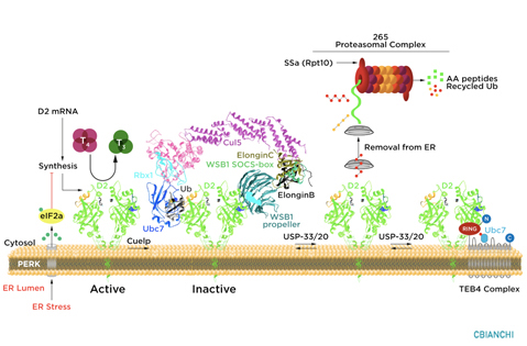
"The thyroid hormone activating deiodinase, D2, is a type I endoplasmic-reticulum (ER)-resident thioredoxin fold-containing selenoprotein. It is anchored to the ER membrane in close proximity to the cell nucleus through a single transmembrane domain. The D2 globular domain containing the catalytic active site is in the cytosol whereas only a few amino acids exist in the ER lumen. D2 is structured as a homodimer, D2:D2, and monomers are inactive.

D2 has a short half-life that can be stabilized depending on whether its natural substrate T4 is available. In the presence of T4, D2 is inactivated with an approximately 20 min half-life whereas in the absence of T4 its half-life is prolonged to hours. This provides a mechanism through which the production of T3, the biologically active thyroid hormone, can be regulated according to the availability of T4. For example, an accumulation of D2 in cells increases the fractional conversion of T4 to T3 when serum T4 levels are low, such as in the case of iodine deficiency or hypothyroidism. In contrast, serum levels of T4 are relatively high in patients maintained with levothyroxine replacement therapy, and the fractional conversion of T4 to T3 is consequently reduced.

D2 ubiquitination is the molecular mechanism underlying these changes in D2 half-life, i.e. the covalent attachment of multiple ubiquitin molecules to D2, which both inactivates the enzyme and targets it to degradation in the proteasomes. Ubiquitination is thought to inactivate D2 by disrupting the conformation of the D2:D2 dimer, critical for enzyme activity. A unique 18-amino acid loop confers intrinsic metabolic instability to D2, facilitating binding to proteins involved in the ubiquitination process.

The ubiquitin activating enzymes UBC6 and UBC7 are critical in the process of D2 ubiquitination, as well as two ubiquitin ligases, the hedgehog-inducible WSB-1, and TEB4, a ligase involved in the degradation of proteins in the endoplasmic reticulum. Ubiquitinated D2 (UbD2) is not immediately taken up by the proteasomes. Instead, UbD2 can be reactivated by de-ubiquitination, a process catalyzed by two USP-class D2-interacting de-ubiquitinases (DUBs), USP-20 and USP-33. The other two deiodinases, D1 and D3, are not known to be ubiquitinated or undergo post-translational modifications.

D2 ubiquitination occurs via K48-linked ubiquitin chains and exposure to its natural substrate, T4, accelerates UbD2 formation. UbD2 is taken up by proteasomes located in the cytoplasm. D2 retrotranslocation to the cytoplasm occurs via interaction with the p97-ATPase complex. D2 retrotranslocation also includes deubiquitination by the p97-associated DUB Ataxin-3. Once in the cytosol, D2 is delivery to the proteasomes as evidenced by coprecipitation with 19S proteasome subunit S5a and increased co-localization with the 20S proteasome.

D2 ubiquitination may occur at different rates depending on the cell where D2 is expressed.
As opposed to the rest of the body, D2 in the hypothalamus is poorly ubiquitinated (or greatly de-ubiquitinated) sustaining conversion of T4 to T3 despite interaction with T4. As a result, TRH and TSH secretion are highly sensitive to serum T4. This also explains why serum TSH is normalized in many levothyroxine-treated patients even as serum T3 levels remain below the normal range."
Deiodinases-Thyroid-Hormone-Homeostasis-1.jpg
 
Last edited:

pattismith

Senior Member
Messages
3,946
Ubiquitination of D2 in the brain is less sensitive to T4 inhibition than in the other tissues;
This means that D2 is more active in the brain, and that more T3 is produced in the brain, so the brain sensitivity to TH is greater than the other tissues.
What could make the brain more resistant to TH:

-decreased entry of TH in brain cells (decreased transporters activity)
-increased ubiquitination of D2
-increased activity of D3 (deactivate T3 and T4)

D2 ubiquitination may occur at different rates depending on the cell where D2 is expressed.
As opposed to the rest of the body, D2 in the hypothalamus is poorly ubiquitinated (or greatly de-ubiquitinated) sustaining conversion of T4 to T3 despite interaction with T4. As a result, TRH and TSH secretion are highly sensitive to serum T4. This also explains why serum TSH is normalized in many levothyroxine-treated patients even as serum T3 levels remain below the normal range."

and from this other article by Bianco:

"In the hypothalamus-pituitary dyad as well as the rest of the brain, the majority of T3 present is generated locally by T4 deiodination via the type 2 deiodinase (D2); this pathway is self-limited by ubiquitination of D2 by the ubiquitin ligase WSB-1.
Here, we determined that tissue-specific differences in D2 ubiquitination account for the high T4/T3 serum ratio in adult thyroidectomized (Tx) rats chronically implanted with subcutaneous L-T4 pellets.

While L-T4 administration decreased whole-body D2-dependent T4 conversion to T3, D2 activity in the hypothalamus was only minimally affected by L-T4.

In vivo studies in mice harboring an astrocyte-specific Wsb1 deletion as well as in vitro analysis of D2 ubiquitination driven by different tissue extracts indicated that D2 ubiquitination in the hypothalamus is relatively less. As a result, in contrast to other D2-expressing tissues, the hypothalamus is wired to have increased sensitivity to T4. These studies reveal that tissue-specific differences in D2 ubiquitination are an inherent property of the TRH/TSH feedback mechanism and indicate that only constant delivery of L-T4 and L-T3 fully normalizes T3-dependent metabolic markers and gene expression profiles in Tx rats.