This is a new pilot study funded by the ME Association Ramsay Research Fund that introduced a relatively new technique and provided for some intriguing results.
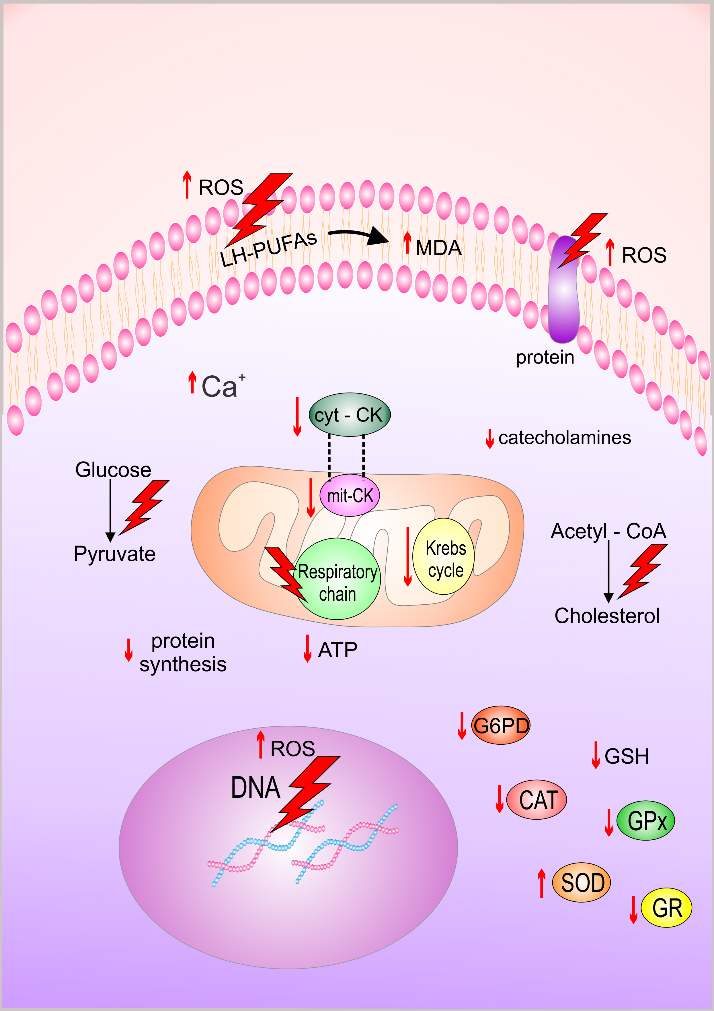
“It is becoming clear that metabolic/energetic dysfunction plays a role in ME/CFS. More information is required to determine if these differences are driving the illness or are a consequence of having ME/CFS.
“Single Cell Raman Spectroscopy is an exciting new tool which can give a readout on aspects of intracellular metabolism. Live cells/tissue are not required, which if the approach is successful, will be a major benefit in developing a diagnostic test.”
Dr Karl Morten
About the study
Dr Morten and Prof Wei Huang (Department of Engineering) from Oxford University, attempted to link mitochondrial dysfunction and ME/CFS pathogenesis by comparing the ‘fingerprint’ of a cell model containing no mitochondrial DNA (known as ‘ρ0’) to the ‘fingerprint’ of molecules from the blood cells of ME/CFS patients.
The study involved the use of a cell imaging method called single-cell Raman micro-spectroscopy (SCRM). A light (usually from a laser) shining on a cell results in changed frequencies of photons – due to the energy exchange between the incident light and vibrations of biomolecules in cells – which are then detected and observed in the form of a Raman spectrum, named after Indian Physicist Sir C. V. Raman who earned the 1930 Nobel Prize for the discovery.
Each biomolecule has a unique ‘fingerprint’ on the Raman spectrum (shown as different length bands) and the sum of all biomolecular fingerprints in a cell can be used as a phenotype of the single cell. These fingerprints can be used to indicate changes in cellular metabolism and identify disease- related biomarkers.
This non-invasive biochemical analysis technique has an advantage over other biomarker-identifying methods as it can be performed on living cells. Also, since it is a label-free technique, the cells do not need to be radioactively labelled or stained with a dye to be imaged, so they are much closer to their natural bodily state; reflecting the intrinsic biochemical profiles of the cells with less manipulation.
However, this technique is not widely used in clinical practice at this time.
Phenylalanine – an amino acid
The researchers found that both the cells with no mitochondria and the ME/CFS patients’ blood cells had high ‘bands’ (or markers) associated with phenylalanine-like compounds, whereas the controls did not.
Phenylalanine is an amino acid (a building-block employed by the body to make important molecules) readily detectible by Raman. It is used to make many neurotransmitters, such as adrenaline (involved in the fight/flight response for example).
Although this initial exploratory study only involved 5 ME/CFS patients, it will be exciting to see where this goes when tested on a larger cohort from the ME/CFS Biobank. These findings could support a possible metabolic defect, or perhaps even a mitochondrial defect, with implications for diagnostic assessment and, ultimately, for treatment.
“As similar changes were observed in the ρ 0 cell model with a known deficiency in the mitochondrial respiratory chain as well as in CFS patients,
our results suggest that the increase in cellular phenylalanine may relate to mitochondrial/energetic dysfunction in both systems.
“Interestingly, phenylalanine can be used as a potential biomarker for diagnosis of CFS by SCRM [Single-cell Raman Spectroscopy].
“A machine learning classification model achieved an accuracy rate of 98% correctly assigning Raman spectra to either the CFS group or the control group.
“SCRM combined with machine learning algorithm therefore has the potential to become a diagnostic tool for CFS.”
From: A new approach to find biomarkers in CFS/ME by single-cell Raman micro- spectroscopy (Xu, et al. 2018)
For more on the amino acid phenylalanine in ME/CFS see also for example:
- Fluge et al. (2016) Metabolic profiling indicates impaired pyruvate dehydrogenase function in myalgic encephalopathy/chronic fatigue syndrome
- Niblett et al. (2007) Hematologic and urinary excretion anomalies in patients with chronic fatigue syndrome
For more information on the research being conducted by Dr Morten’s team in Oxford (funded by the ME Association Ramsay Research Fund), see:
• MEA research update: Metabolomics and ME/CFS | 13 August 2018

")