
Scientific Findings Distinguish Between Myalgic Encephalomyelitis and Chronic Fatigue Syndrome
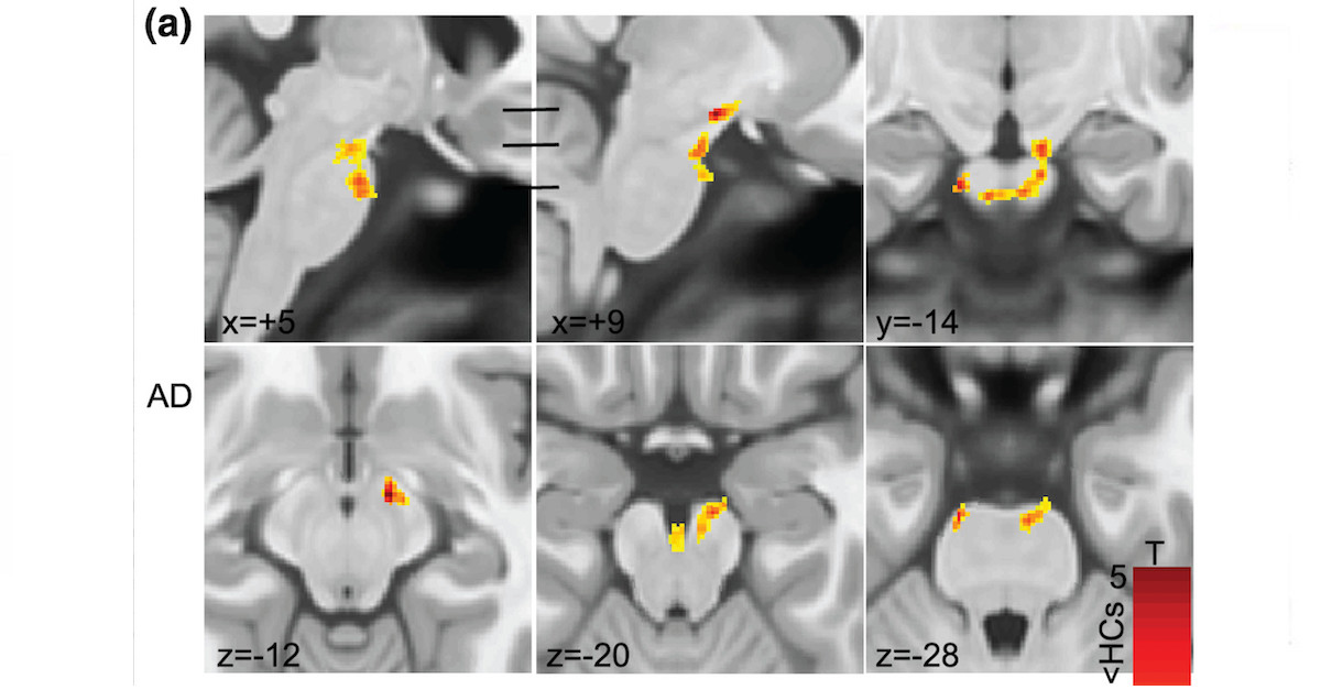 Dr. Kiran Thapaliya and colleagues used an advanced form of medical imaging to spot significant abnormalities in the brainstem of patients diagnosed with myalgic encephalomyelitis. However, no significant differences were found in patients only diagnosed with chronic fatigue syndrome. Image is Figure 1a from Thapaliya, K., Marshall-Gradisnik, S., Staines, D., & Barnden, L. (2021). Diffusion tensor imaging reveals neuronal microstructural changes in myalgic encephalomyelitis/chronic fatigue syndrome. European Journal of Neuroscience, 54(6), 6214–6228. Licensed CC-BY-NC-4.0.

By Bronc
People living with myalgic encephalomyelitis/ chronic fatigue syndrome (ME/CFS) are definitely not “just a little tired.” But they are indeed tired of some things — such as dealing with medical ignorance, the very slow pace of research, and seemingly endless discussions over terminology and definitions.But in science, precise terminology and precise definitions are absolutely essential in order to produce exact, repeatable results. Neuroscientist Dr. Kiran Thapaliya knows this all too well.
In the last few years, Dr. Thapaliya has produced three key papers researching ME/CFS. Two of these dramatically show the difference between using the modern definition of myalgic encephalomyelitis produced by an international group of experts, and using the 30-year-old definition of chronic fatigue syndrome produced by the U.S. CDC.

Dr. Kiran Thapaliya is a neuroscientist at the National Centre for Neuroimmunology and Emerging Diseases (NCNED) in Australia. Image courtesy of Dr. Kiran Thapaliya.
In 2020, Dr. Thapaliya published a review of the known neuroimaging findings in ME/CFS.
He followed this up with a 2021 publication that used a technique called “diffusion tensor imaging” to follow the nerve pathways in the brains of patients.
Then, in a 2022 publication, he looked at a key part of the brain known as the hippocampus.
Dr. Thapaliya took some time out from his busy work schedule to talk to us about his research and these three recent publications.
How did you get involved in the field of ME/CFS research?
Dr. Kiran Thapaliya:
I got involved in ME/CFS research a few years ago. At the end of 2019, I was about to submit my Ph.D. thesis on the development of neuroimaging methods to study tissue microstructure.
Then, I was offered a job with the National Centre for Neuroimmunology and Emerging Diseases (NCNED), at the Menzies Health Institute of Griffith University to undertake Magnetic Resonance Imaging (MRI) research on ME/CFS.
When I heard that ME/CFS has unknown aetiology, it motivated me to implement my MRI knowledge to understand the pathophysiology of ME/CFS.
Phoenix Rising:
Can you tell us about your current research projects?
Dr. Kiran Thapaliya:
Currently, we are studying brain dysfunction in a large cohort of ME/CFS patients using an ultra-high-field 7T MRI scanner that yields better tissue contrast. This project is funded by ME Research UK (MERUK). We are also using multi-modal MRI imaging to study the pathophysiology of ME/CFS.
Phoenix Rising:
In 2020 you co-authored a study which reviewed 55 papers related to the structural and functional neurological changes in ME/CFS patients, as measured by neuroimaging techniques.
After assessing the findings of these papers regarding neurological or cognitive differences in adult ME/CFS patients, compared with healthy controls, you noticed several outcomes.
Can you describe the results of your systematic review of these 55 papers? What conclusions did you draw?
Dr. Kiran Thapaliya:
Across the 55 papers included in the neuroimaging systematic review, evidence of widespread disruption of the nervous system in ME/CFS patients — when compared to healthy controls — was found using various neuroimaging techniques.
Findings included changes in: structural morphology in parts of the brain such as the cingulate cortex, cerebral blood flow, gray matter, and white matter volume.
Evidence of neuroinflammation in regions such as the hippocampus, cognitive dissonance, and alteration in functional connectivity networks were also observed.
These findings, however, were at times unconfirmed and at times not consistent. Although neurological impairments were present, further studies are required to understand their contribution to the pathology of ME/CFS.
2021: Diffusion tensor imaging reveals neuronal microstructural changes in ME/CFS
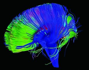
Diffusion Tensor Imaging is a scientific technique that can show the nerve fiber pathways in the brain. Image by NICHD NIH, licensed under CC BY 2.0.
Phoenix Rising:
What does the imaging technique called diffusion tensor imaging show?
Dr. Kiran Thapaliya:Diffusion tensor imaging (DTI) is sensitive to cellular structure that measures the diffusion of water molecules.
It provides information on the white matter nerve fibre tracts connecting the different regions of the brain.
Phoenix Rising:What significant differences did you find between healthy controls and patients diagnosed with the modern definition of myalgic encephalomyelitis?
Dr. Kiran Thapaliya:We performed voxel-based analysis on DTI metrics like fractional anisotropy (FA), axial diffusivity (AD), mean diffusivity (MD), radial diffusivity (RD), eigenvalues (λ2, and λ3), and mode of anisotropy (MO).
We found significant clusters with decreased AD, MD, and λ2 in the brainstem of ME/CFS patients — as well as in the superior longitudinal fasciculus region for MO.
Phoenix Rising:
What significant differences did you find between healthy controls and patients diagnosed with only the 30-year-old definition of chronic fatigue syndrome?
Dr. Kiran Thapaliya:We performed a voxel-based analysis for all DTI metrics with the 30-year-old definition of chronic fatigue syndrome. We did not find any significant clusters.
Phoenix Rising:Did you find any correlation with symptom severity?
Dr. Kiran Thapaliya:We could not perform a correlation between DTI metrics and symptom severity. In our future study, we will correlate DTI findings with symptom severity.
2022: Volumetric differences in hippocampal subfields and associations with clinical measures in ME/CFS
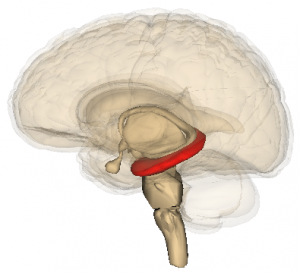
The hippocampus, shown in red, is a part of the brain that is essential for forming memories. Image adapted from one by Life Science Databases (LSDB), licensed CC-BY-SA-2.1-jp.
Phoenix Rising:
In March of this year you had a new paper published, which noted that people with ME/CFS suffer from cognitive and memory dysfunction.
Since the hippocampus part of the brain plays a key role in both cognition and memory, you and your colleagues tested for size differences (volumetric differences) in specific parts (subfields) of the hippocampus in ME/CFS.
Can you explain the role of the hippocampus, what are subfields of the hippocampus and why you examined hippocampal volume reduction?
Dr. Kiran Thapaliya:The hippocampus is an extension of the temporal lobe of the cerebral cortex and plays an important role in cognitive functions such as memory, executive processing, and reward processing.
It is an extremely active region, both biochemically and electrically, and therefore it is ultra-sensitive to many factors which influence brain functioning.
Anatomically, the hippocampus is a complex structure consisting of specific parts called subfields (dentate gyrus, subiculum, parasubiculum, entorhinal cortex, and the three cornu ammonis [CA1, CA3, CA4]), each with distinct memory functions.
These subfields of the hippocampus may be involved in selective neurocognitive processes in health and disease.
Recent research has shown that patients with a defective hippocampus also have an impaired ability to imagine new experiences, to generate predictions about the future, and to elaborate rules to guide their behaviour.
We know that ME/CFS patients have difficulty in processing information (slowed thought and impaired concentration) and short-term memory loss (recalling information and poor working memory).
This motivated us to investigate the hippocampus regions mostly involved in cognitive function.
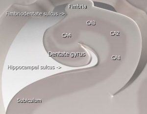
A cross-sectional view of the hippocampus shows specific parts of the hippocampus, known as subfields. Image adapted from one by Frank Gaillard, licensed GFDL.
Did your study reveal any correlations between the size of subfields and symptom severity?
Dr. Kiran Thapaliya:Unfortunately, we could not perform a correlation between subfield volumes and symptom severity.
However, we did perform a correlation analysis between subfield volumes and the symptoms of pain and fatigue — which showed that an increase in the pain and fatigue level is correlated with decreased subfield volumes.
In our future study, we will correlate the MRI findings with symptom severity.
Phoenix Rising:What conclusions did you draw from the data you analysed?
Dr Kiran Thapaliya:Our study revealed more subfield volumes were different in ME/CFS patients meeting the International Consensus Criteria for myalgic encephalomyelitis (ICC) than in ME/CFS patients only meeting the Fukuda criteria for chronic fatigue syndrome, when compared with healthy controls.
Therefore, volumetric changes in the hippocampus depend on patient selection criteria and demonstrate the importance of strict case definitions in ME/CFS research.
Unlike in other neurodegenerative diseases where subfield volumes were reduced, some subfield volumes — like the left subiculum, pre-subiculum head, and fimbria volumes — were significantly larger in ME/CFS patients who met ICC criteria.
Therefore, changes in hippocampus volume may be associated with cognitive impairment in ME/CFS patients — and increased volumes may reflect a compensatory effect for deficits elsewhere.
Phoenix Rising:How might this research into neurological impairments help in the development of treatments or biomarkers for the illness?
Dr. Kiran Thapaliya:The overall volumetric difference in the hippocampus may serve as an indicator for neurological impairments in ME/CFS patients. Of course, additional research needs to validate this as a potential biomarker for this illness.
Further research is also needed to confirm the cause of cognitive impairment in ME/CFS, but these results are promising.
Maksoud R, du Preez S, Eaton-Fitch N, Thapaliya K, Barnden L, et al. (2020) A systematic review of neurological impairments in myalgic encephalomyelitis/ chronic fatigue syndrome using neuroimaging techniques. PLOS ONE 15(4): e0232475.
Thapaliya, K., Marshall-Gradisnik, S., Staines, D., & Barnden, L. (2021). Diffusion tensor imaging reveals neuronal microstructural changes in myalgic encephalomyelitis/chronic fatigue syndrome. European Journal of Neuroscience, 54(6), 6214–6228.
Thapaliya, K., Staines, D., Marshall-Gradisnik, S., Su, J. & Barnden, L. (2022). Volumetric differences in hippocampal subfields and associations with clinical measures in myalgic encephalomyelitis/chronic fatigue syndrome. Journal of Neuroscience Research, 100, 1476–1486.
Bronc is a former historian who is active in his local ME support group. He enjoys interviewing scientists involved in ME research to help himself and others better understand their illness.
Acknowledgment: The author would like to thank Jody Smith and Eric Pyrrhus for help with editing and preparing this article
Last edited by a moderator:
