sb4
Senior Member
- Messages
- 1,907
- Location
- United Kingdom
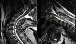
Hope it's okay to dump my images on this thread also.

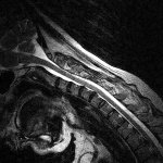
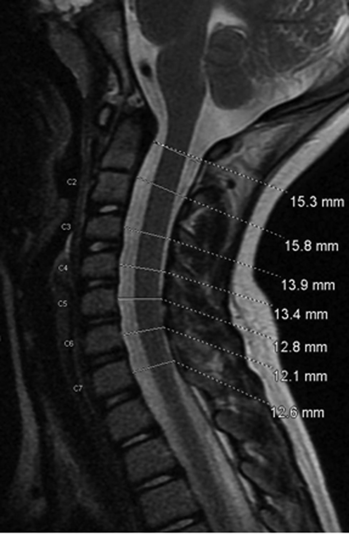
From my extremely amateur view it looks like the most potentially significant thing is disk herneation / bulging at C5/6 and a little at C4/5. It looks similar to the CFS study where the three patients had disk surgery and got better.
The top of the brainstem seems to turn at a bit of a sharp angle and this is backed up by borderline CXA.
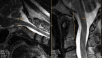
One thing I have noticed is that looking at other peoples MRIs who have Dx they seem to have areas where the CSF seems super thin thanks to herneiation etc but with me I seem to have quite a lot of white CFS around the core the majority of the time.
I don't suffer from much neck pain. My neck gets a bit achey during the evening and this usually coincides with me feeling better. So it could be just from being up right all day but I think something else is at play.
Not done it yet. However I think its possible that I could be the opposite way around as I noticed sometimes my fingers would get wrinkly for no reason and touching things would be wierd. This only happened when I was more ill. I know my fingers do get wrinkly after whilst in the bath however it would be a case of seeing how long it takes I suppose.@sb4 , autonomic function is probably at play, you need to test it.
How do you react to the finger wrinckling test?
One thing I have noticed is that looking at other peoples MRIs who have Dx they seem to have areas where the CSF seems super thin thanks to herneiation etc but with me I seem to have quite a lot of white CFS around the core the majority of the time.
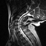
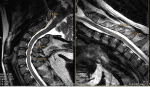
Although there are parts where the spinal cord seems to be bent a bit awkwardly it is never pinched.

I think spinal stenosis is 90% in older people, so yes it is very common. As @valentinelynx said, the symptoms are decisive. The problem is that some symptoms are not taken into account by neurologists like dizziness, tinnitus, brain fog, autonomic symptoms, etc...So symptomatic people may be more than it is currently admitted.Also anyone have any idea on the rates of cervical disk degeneration in relatively young people? I am 28 and would like to know how likely I would have mild disk bulge by chance at my age. I found a study suggesting I think 25% of people below 50 have stenosis which seems pretty high and you would imagine the vast majority of these cases it causes no problems.
yes it is the more simple test we can find for autonomic system.Not done it yet. However I think its possible that I could be the opposite way around as I noticed sometimes my fingers would get wrinkly for no reason and touching things would be wierd. This only happened when I was more ill. I know my fingers do get wrinkly after whilst in the bath however it would be a case of seeing how long it takes I suppose.
Am I right in saying water is around 40*C and after 15mins fingers should be wrinkly?
You have any idea whether the mild stenosis / bulging could be impacting the sympathetic nervous that lead to the heart as you see this is the level they occur at.
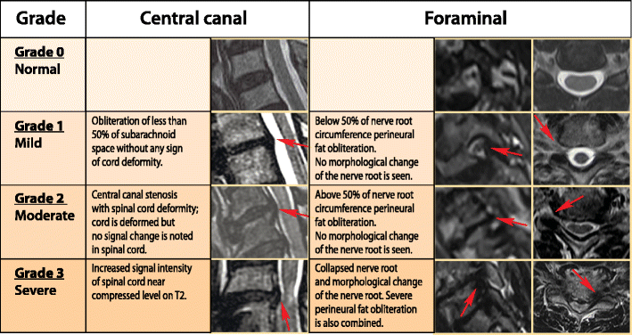
Not sure I understand this. Grade 0 is no disk bulging, right?To answer my own question. It seems that even grade 0 and 1 could be symptomatic
Yes I had the tilt table test and my heart rate went above 120 and increased by over 30 within 10 minutes. This was when I was near bed bound. Now I can stand for quite a long time, maybe an hour on a good day without much increase in symptoms. I can walk moderate distances. So I am not even sure I would qualify for POTS anymore with those metrics however my body is still significantly messed up.Did you had tilt table test? Do you have change in your heart rate and blood pressure when standing?
Not sure I understand this. Grade 0 is no disk bulging, right?