• Welcome to Phoenix Rising!

    Created in 2008, Phoenix Rising is the largest and oldest forum dedicated to furthering the understanding of, and finding treatments for, complex chronic illnesses such as chronic fatigue syndrome (ME/CFS), fibromyalgia, long COVID, postural orthostatic tachycardia syndrome (POTS), mast cell activation syndrome (MCAS), and allied diseases.

    To become a member, simply click the Register button at the top right.

Autopsy evidence of chronic EV infection Dr Chia Oct 2016

Countrygirl

Senior Member
Messages
5,452
Location
UK
https://www.facebook.com/notes/jerr...tient-presented-by-dr-john-c/1267675596637350

Autopsy Evidence of Chronic EV Infection in ME Patient Presented by Dr. John Chia at IACFSME Conference October 2016

Poster 13
Chronic enterovirus (EV) infection in a patient with myalgic encephalomyelitis / chronic fatigue syndrome (ME/CFS) – Clinical, Virologic and Pathological Analysis

John Chia, David Wang, Andrew Chia, Rabiha El-Habbal. EV Med Research. Lomita, CA
Objectives: A 23 y/o Caucasian male developed prolonged, recurrent gastrointestinal symptoms, followed by onset of severe ME/CFS (CDC criteria, ICC).

At initial evaluation, Echovirus 11 antibody titer was >1:640 (normal <1:10); IgG and IgM antibody for EBV and HHV6 were negative, CMV IgG was positive. He failed to respond to combination of alpha and gamma interferon; and debilitating symptoms of the stomach and central nervous system were minimally alleviated by SSRI, benzodiazepine and acid-suppressant.

Repeated MRI scans of brain and spinal cords showed normal results. The patient committed suicide 6 years after the onset of symptoms. Brain was harvested and frozen within 24 hours of death for evaluation of chronic viral infection.

Method: Using EV- and dsRNA-specific monoclonal antibody (5D8/1 and J2 mAb), stomach and colon biopsies obtained 5 months after onset of illness were stained for viral capsid protein (VP1) and dsRNA by immunoperoxidase technique. Blood drawn in Paxgene tube 3 years after illness was screened for enterovirus RNA by RT-PCR. ~1 cm3 sample was taken from the ponto-medullary junction (PM), medial temporal lobe (MT), frontal lobe (FL), occipital lobe (OL), cerebellum (CL) and midbrain / hypothalamus area (MB) of brain.
The brain samples were homogenized in 10 ml of serum-free medium. Aliquots were processed for viral cultures. Trizol-LS reagent was used for RNA and protein extraction, as well as other lysis agents.
'
Tris-Glycine and MES gels, wet and semi-dry transfer, then western blot was performed with Ibind (Life technology) using EV-, CMV- and HHV6-specific mAbs, patient’s own serum and control serum samples. Viral culture was performed in WI-38 and BGMK-DAF cell lines. RT-PCR for conserved highly-conserved sequences of 5’ end and 3D polymerase sequence were performed on extracted RNA.

Results: Stomach and colon biopsies stained positive for EV VP1 soon after initial infection documenting the initial viral infection; dsRNA was detected in the stomach biopsies. EV RNA was not detected in blood 3 years after illness.
Initial culture of brain samples did not grow virus; 5’ EV RNA sequence was not detected by RT-PCR. Using 5 D8/1 mAb, western blot revealed 37-42K and 46K protein bands in the brain samples, which corresponded to viral protein and creatine kinase b extracted from infected stomach biopsies, but not in brain biopsy samples taking from patients with brain tuberculoma and lymphoma. 3D pol gene was amplified from the DNase-treated RNA extracted from PM, MT and FL. 5’ RNA sequence was in one of the FL specimens.
'
Conclusion: The analysis of the second brain specimen taken from ME/CFS patient replicated the British findings published in 1994 (Ann. IM). The finding of viral protein and RNA in the brain specimens 6 years after documented acute enterovirus infection of the gastrointestinal tract is consistent with a chronic, persistent infection of the brain causing debilitating symptoms.

'EV is clearly one of the causes of ME/CFS, and antiviral therapy should be developed for chronic EV infection.

John Chia MD, 25332 Narbonne Ave. # 170, Lomita, CA 90717. evmed@sbcglobal.net
 
Last edited:

M Paine

Senior Member
Messages
341
Location
Auckland, New Zealand
My thoughts and gratitude go out to this patient, may they rest in peace. I'm truly thankful for their contribution, and I know first hand how devastating this situation is for friends and family. I hope the family will find small comfort in the large contribution and selfless act this person was able to make in progressing research.
 

ash0787

Senior Member
Messages
308
" EV VP1 " enterovirus ? anyone know what this means and what the test results show in terms of how the infection
behaved ?
 

Countrygirl

Senior Member
Messages
5,452
Location
UK
@ash0787
I recall way back in 1986 when I was shipped into a nursing home with severe ME Prof Mowbury (I don't think I've used the correct spelling) of London visited the home to take some of our thigh muscle fibres for the VP1 test to check for chronic enteroviral infection.

When I was diagnosed I was told ME was chronic coxsackie infection.
 
Last edited:

M Paine

Senior Member
Messages
341
Location
Auckland, New Zealand
" EV VP1 " enterovirus ? anyone know what this means and what the test results show in terms of how the infection
behaved ?

I have some Virology training. Just enough to be dangerous anyway. There are some interesting findings here.

EV in this context means Echovirus 11. VP1 is a capsid protein (outer shell of viral particle)... I think it stands for something imaginative like Viral Protein 1. I've seen EV refer to enteroviruses in general, so it can be a confusing term. More than Echovirus 11 expresses VP1, so we are already starting to get confusing in terminology.

They report detecting high titer of Echovirus 11 VP1 antibodies in this patient shortly after initially seeing this patient. The also reported detection of IgG for CMV, so a secondary immune response, probably not a recent infection.

Using EV- and dsRNA-specific monoclonal antibody (5D8/1 and J2 mAb), stomach and colon biopsies obtained 5 months after onset of illness were stained for viral capsid protein (VP1) and dsRNA by immunoperoxidase technique

Stomach and colon biopsies stained positive for EV VP1 soon after initial infection documenting the initial viral infection; dsRNA was detected in the stomach biopsies.

So here they confirmed the serum findings of VP1 antibodies related to an infection of the GI tract, and further documented that EV VP1 was present in tissue samples of the stomach and colon.

EV RNA was not detected in blood 3 years after illness.

This is an interesting finding, I'm not sure exactly how this should be interpreted. They didn't mention testing for EV RNA at any point prior to this. If this means that the virus was being suppressed effectively by the patients immune system or not is hard to know.


Now to the more recent history of this patient...


Initial culture of brain samples did not grow virus;

I'm really glad they at least attempted doing cell culture. It was the first thing I looked for.

5’ EV RNA sequence was not detected by RT-PCR

OK, no EV RNA detected via PCR in the brain tissue samples initially...

3D pol gene was amplified from the DNase-treated RNA extracted from PM, MT and FL. 5’ RNA sequence was in one of the FL specimens.

I'm a bit rusty here... but I think 3D pol is probably a genetically conserved region of a viral rna polymerase. So what they did here was changed tact and looked for a different viral sequence instead of the capsid protein. And they did get a hit on the brain tissue sample of the frontal lobe.

Using 5 D8/1 mAb, western blot revealed 37-42K and 46K protein bands in the brain samples, which corresponded to viral protein and creatine kinase b extracted from infected stomach biopsies

So they pulled out some more classical virology here, which is nice. And they appear to have semi-convincing evidence that at least viral proteins are present in one or more of the brain samples. They said 'samples' so I assume they meant all three.

It's no secret that tissue culture is very difficult, and it's also entirely possible that there is no viable virus in brain tissue here. Viral protein and RNA is a few steps short of verifiable infection of grey matter. Healthy skepticism aside, it's a very interesting finding worth serious attention.
 

alex3619

Senior Member
Messages
13,810
Location
Logan, Queensland, Australia
It would be nice if we had technology that can test living patient brains without damaging them. A small number of isolated cases will not convince sceptics.

I am interested that, despite widespread brain infection, the virus was not found in the blood. I wonder how much of that is due to the issue that its complete viral particles that are in the blood in a typical infection, and that alternative enterovirus lifecycles might result in incomplete viral particles, and in particular mostly only RNA etc. which standard viral blood testing might not pick up.
 

charles shepherd

Senior Member
Messages
2,239
Some preliminary results (as published in the Journal of Neurological Sciences) from the UK post mortem research group:

Pathology of Chronic Fatigue Syndrome: Pilot Study of Four Autopsy Cases
, the position may become more clear.
DG O’Donovan1, 2, T Harrower3, S Cader2, LJ Findley2, C Shepherd4, A Chaudhuri2
1Addenbrooke’s Hospital Cambridge UK
2Queen’s Hospital Romford Essex UK
3Royal Devon & Exeter Hospitals UK
4Honorary Medical Advisor to ME Association UK

Chronic Fatigue Syndrome / Myalgic Encephalomyelitis is a disorder characterised by chronic exercise induced fatigue, cognitive dysfunction, sensory disturbances and often pain. The aetiology and pathogenesis are not understood.

We report the post mortem pathology of four cases of CFS diagnosed by specialists.

The causes of death were all unnatural and included: suicidal overdose, renal failure due to lack of food and water, assisted suicide and probable poisoning.

Selected portions of tissue were made available by the various Coroners in the UK and with the assent of the persons in a qualifying relationship.

The cases were 1 male, and 3 female. Ages (years) M32, F32, F43 & F31.

One case showed a vast excess of corpora amylacea in spinal cord and brain of unknown significance but Polyglucosan Body Disease was not supported by clinicopathologial review. No ganglionitis was identified.
One case showed a marked dorsal root ganglionitis and two other cases showed mild excess of lymphocytes with nodules of nageotte in the dorsal root ganglia.

This raises the hypothesis that dysfunction of the sensory and probably also the autonomic nervous system may lead to abnormal neural activity eg hyperalgesia & allodynia rather than anaesthesia and may explain some of the symptoms of CFS / ME such as pain, hypotension, hyperacusis and photophobia. However, the syndrome may be heterogeneous.

Nevertheless, the precise relationship of fatigue, which may be either peripheral or central, to abnormalities in the peripheral nervous system (PNS) needs to be studied.

The differential diagnosis of ganglionitis should be investigated in CFS / ME patients hence Varicella Zoster, Lyme disease, HIV, Sjogren’s disease, paraneoplastic sensory ganglionopathy should be excluded by appropriate history and tests.

Thorough histopathological study of cases coming to autopsy may help to confirm or refute the hypothesis, that CFS is a disease process, and whether the symptomatology may be explained by inflammation of the sensory and autonomic divisions of the PNS.

A specific CFS / ME brain and tissue bank in the UK is proposed.

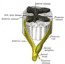
Anatomy of a dorsal root ganglion - which is part of the peripheral nervous system:

250px-Gray675.png
 

CFS_for_19_years

Hoarder of biscuits
Messages
2,396
Location
USA
" EV VP1 " enterovirus ? anyone know what this means and what the test results show in terms of how the infection
behaved ?
Taken from the article:
Chronic enterovirus (EV) infection in a patient with myalgic encephalomyelitis....[..]
[......]....were stained for viral capsid protein (VP1)
So EV VP1 means enterovirus viral capsid protein.

Abbreviations are always explained when they are first used in a paper, and one should go back to the first part of the article to understand their meaning.
 

IreneF

Senior Member
Messages
1,552
Location
San Francisco
Putting on my sceptic's hat: We don't have researchers going around trying to grow viruses from normal people's brains, do we? We're all exposed to pathogens. It could be that some of them leave traces of themselves but are effectively suppressed in the living person. Kind of like shingles, which is reactivated chicken pox. Not everyone who had chicken pox gets shingles.

Nevertheless interesting.
 

Hip

Senior Member
Messages
17,852
Thanks for posting this, @Countrygirl. You might consider adding a blank line to separate each paragraph: you know the trouble us people with enterovirus-filled brains have with reading large blocks of text!


Putting on my sceptic's hat: We don't have researchers going around trying to grow viruses from normal people's brains, do we?

Two previous ME/CFS brain autopsies are detailed in this post (along with Dr Chia's autopsy); in the Mcgarry, Gow and Behan brain autopsy of one deceased ME/CFS patient, they also tested the brains of 8 controls, and all these 8 were found to be enterovirus negative.
 

Hip

Senior Member
Messages
17,852
It's no secret that tissue culture is very difficult, and it's also entirely possible that there is no viable virus in brain tissue here.

It is quite possible that the infection in the brain was a pure enteroviral RNA infection, with no viral particles (virions) being synthesized.

Enterovirus is known to exist in the form of a pure RNA infection: this form of the virus is termed a non-cytolytic enterovirus infection, and it lives inside cells as a chronic intracellular infection, comprising stands of enteroviral ssRNA and dsRNA which can replicate without needing to create viral particles.

This situation of detecting enteroviral RNA in the tissues, but finding no virus that you can cultivate, occurs in chronic coxsackievirus B infection of the adult heart muscle (myocarditis). This is because the heart muscle infection is primarily a non-cytolytic enterovirus infection, with very few viral particles being synthesized. See Invest in ME - Human Enteroviruses and Chronic Infectious Disease.
 
Last edited:

Countrygirl

Senior Member
Messages
5,452
Location
UK
Thanks for posting this, @Countrygirl. You might consider adding a blank line to separate each paragraph: you know the trouble us people with enterovirus-filled brains have with reading large blocks of text!

When I returned to read the comments, I found it a problem to read my own post for that very reason. I usually ensure the text is broken up. The letters clearly conspired against me when my back was turned.

I've brought them to heel now. Apologies!
 

ash0787

Senior Member
Messages
308
So would it be fair to say this test was inconclusive ? or did the evidence prove that the infection was still going strong within the stomach ?

I dont know what the difference is between an enterovirus and a normal virus, how could an infection persist for years without the immune system removing it ?

I know that viruses are debatably considered to be not alive but rather just a piece of 'information', if thats so then how can they deactivate themselves ?
 

halcyon

Senior Member
Messages
2,482
It's no secret that tissue culture is very difficult, and it's also entirely possible that there is no viable virus in brain tissue here. Viral protein and RNA is a few steps short of verifiable infection of grey matter.
It has been found that enteroviruses can pass between cells without triggering cell lysis or even encapsidating into normal virion. If viral proteins are present that means that translation is occurring and the viral RNA is viable. Any cell containing viral protein likely has normal mRNA translation suppressed and will not function normally but also won't die. Even the RNA alone is immunogenic.
 
Last edited:

halcyon

Senior Member
Messages
2,482
I dont know what the difference is between an enterovirus and a normal virus, how could an infection persist for years without the immune system removing it ?
Many viruses have evolved countermeasures that allow them to evade the immune response. If an infection becomes persistent and is antigenically stimulating enough it will trigger T cell exhaustion and the immune system will actually happily leave the virus alone for the most part.
 

barbc56

Senior Member
Messages
3,657
I wonder if patients with other health conditions might have the same result of this patient. Maybe people without health conditions? Who knows? This is patient presentation and a poster at the conference.

Are there any blinded studies? At this point it's a hypothesis, an interesting hypothesis, but not proven one way or another.

It might make a good study for the future.
 

Daffodil

Senior Member
Messages
5,875
since many of us get CFS after other infections, I wonder if the common denominator is how the infections affect gut flora. an enterovirus would affect gut flora for sure..maybe somehow that weakens the body so that the infection cannot be controlled.?

eh. more questions than answers as usual
 

ash0787

Senior Member
Messages
308
it is scary realizing that we could have a virus in our brain tissue while it is not detectable anywhere else.

I'd say more annoying than scary, even if there is some silly virus with a stealth mechanism which is playing games with our brain cells, we would still need to find a way to explain how that could affect other areas of the body