
WoolPippi
Senior Member
- Messages
- 556
- Location
- Netherlands
Yesterday I visited a practitioner who does Live Blood Analysis. They take a small drop of blood with a really gentle finger prick and put it under a dark field microscope which is attached to a screen.
The analysis-part is controversial so lets not talk about that but seeing my red blood cells move about was fascinating!
This picture is from the internet -please ignore the texts on it- and it's pretty similar to what I got to see from my own blood:
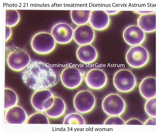
Those are nice round red blood cells. They were slowly moving about, tumbling. Some of mine were misformed or jagged.
The grainy cell is a white blood cell. Mine looked like this one and that's a good one. It has more than one structure within.
The bit of "fluff" on the cell at the left above the white one was called "debris" in my case. Stuff that should have been evacuated from the body. (??)
There are 3 or 4 little star like dots in this picture, close to cells. Mine had many more, floating amongst the cells and they were actively vibrating and moving. She said they were nutrients like minerals (??) They looked magical!
With me there was also a structure the size of 5 nutrient-dots and she said it was a parasite, a bacterium. A common one, not a spirally one because those would have indicated Lyme, something-else-I-forget or Syphilis. (??)
As I said, I'm not prepared to defend the interpretation/analysis-part of this practise. But seeing the shape of the cells and the ratio between red and white cells gives information. Lots of white blood cells indicate an inflammation somewhere in the body.
I think my drop of blood had about 300 red blood cells and 20 white blood cells. I had about 200 vibrating nutrient-dots. 25 bits of debris-fluff. 20 bacteria.
Some of my red blood cells had a pointy bit. This happens when the specimen gets older (we're talking minutes here) but also when there's need of B12. This would coincide with my situation but now we're in analysis-territory.
It was really cool to see.
The analysis-part is controversial so lets not talk about that but seeing my red blood cells move about was fascinating!
This picture is from the internet -please ignore the texts on it- and it's pretty similar to what I got to see from my own blood:
Those are nice round red blood cells. They were slowly moving about, tumbling. Some of mine were misformed or jagged.
The grainy cell is a white blood cell. Mine looked like this one and that's a good one. It has more than one structure within.
The bit of "fluff" on the cell at the left above the white one was called "debris" in my case. Stuff that should have been evacuated from the body. (??)
There are 3 or 4 little star like dots in this picture, close to cells. Mine had many more, floating amongst the cells and they were actively vibrating and moving. She said they were nutrients like minerals (??) They looked magical!
With me there was also a structure the size of 5 nutrient-dots and she said it was a parasite, a bacterium. A common one, not a spirally one because those would have indicated Lyme, something-else-I-forget or Syphilis. (??)
As I said, I'm not prepared to defend the interpretation/analysis-part of this practise. But seeing the shape of the cells and the ratio between red and white cells gives information. Lots of white blood cells indicate an inflammation somewhere in the body.
I think my drop of blood had about 300 red blood cells and 20 white blood cells. I had about 200 vibrating nutrient-dots. 25 bits of debris-fluff. 20 bacteria.
Some of my red blood cells had a pointy bit. This happens when the specimen gets older (we're talking minutes here) but also when there's need of B12. This would coincide with my situation but now we're in analysis-territory.
It was really cool to see.
") Thanks for sharing!
Thanks for sharing!